
Abstract
BACKGROUND:
HCC as the 6th most common tumor entity with the fourth highest mortality and an increasing prevalence especially due to today’s lifestyle acquires a high attention in the clinical setting. Beside CECT and CEMRI, CEUS depicts a dynamic, low-risk and radiation free imaging method that finds its use mainly in screening and active surveillance programs.
PURPOSE:
The aim of the retrospective study was to evaluate the diagnostic value of CEUS in correlation to pathologic findings.
MATERIALS AND METHODS:
Between 2004 and 2018 a total number of 119 patients were included in this retrospective single-center study. Every patient underwent CEUS in addition to a native B-mode and Color-Doppler scan. After given informed consent SonoVue® (Bracco, Milan, Italy), a second-generation blood-pool agent, was used as contrast medium. Every examination was performed and interpreted by a single experienced radiologist (EFSUMB level 3). A low mechanical index (MI) of <0,2 was chosen to obtain a good imaging quality.
RESULTS:
All 119 included patients received CEUS followed by a liver biopsy for inter-modality comparison. In correlation to the pathology results, CEUS showed a diagnostic sensitivity of 96,6%, a specificity of 63,9%, a PPV of 86,7% and a NPV of 88,5% by detecting liver lesions suspicious for HCC. According to the Cohen’s Kappa coefficient (k = 0,659) CEUS shows a strong inter-modality agreement in comparison to the histopathological finding.
CONCLUSION:
With a high sensitivity and a strong cross-modality comparability to histopathology, the CEUS is highly effective in the detection of suspicious HCC lesions.
Keywords
Introduction
According to the International Agency for Research on Cancer (IARC), liver cancer represented the sixth most common cancer with the fourth highest mortality in 2018 worldwide. Subdivided between men and women, the mortality was 2nd for men and 6th for women. The hepatocellular carcinoma (HCC) reflects the most common de novo liver malignancy in patients arising in a cirrhotic liver [1, 2]. Approximately 85% of primary liver tumors are HCC [3–5] with chronic alcohol consumption, hepatitis B, hepatitis C and non-alcoholic fatty liver disease (NAFLD) as their major risk factors [2, 6]. NAFLD describes an entity that, due to its association with today’s western lifestyle problems such as obesity, diabetes and metabolic syndrome, has increased in incidence in Western nations in recent decades and is expected to continue to increase in the future [2, 7]. In addition, the development of HCC can also be associated with hereditary diseases such as hemochromatosis, Wilson’s disease and α-1-antitrypsin deficiency [8]. Hepatocarcinogenesis is based on a multi-step process which develops in the cirrhotic liver from hypertrophic regenerative nodules and low grade dysplastic nodules to high grade dysplastic nodules and finally to HCC [1]. The HCC itself is further subdivided into well-, moderately- and poor-differentiated HCC [1, 9].
The primary goal is to prevent the development of an HCC lesion or to detect even small lesions with a high specificity and sensitivity by monitoring risk patients, e.g. patients with liver cirrhosis or patients with hepatitis B but without detectable cirrhosis [5]. Within CEUS, the pathognomonic imaging features are an early arterial hyper-perfusion, followed by wash-out during the delayed venous phase [10–17].
Within the various leading societies including the American Association for the Study of Liver Diseases (AASLD), the European Association for the Study of the Liver (EASL) and the Asian Pacific Association for the Study of the Liver (APASL), there is broad agreement that clinical monitoring of high-risk patients at six month intervals should be carried out using imaging procedures whereas ultrasound (US) being the recommended modality with an sensitivity up to 80% and a specificity of more than 90%. The additive determination of special biomarkers, e.g. AFP, is still a matter of recent debate by having possibly a lower benefit than initially expected due to suboptimal cost-effectiveness for routine surveillance of early HCC [3, 18–22]. In case of suspicious or unclear liver lesions, diagnostic imaging should be expanded. In this case, multiphase contrast-enhanced computed tomography (CE-CT) and contrast-enhanced magnetic resonance imaging (CE-MRI) are recommended as well as performing a biopsy followed by histopathological analysis. The histopathological analysis has its high value especially in unclear cases either to confirm or to reject the diagnosis of suspicion and nowadays for further detection of immunohistochemical and molecular characteristics [3, 23–25, 3, 23–25].
Materials and methods
The performed single-center study was approved by our institutional ethical committee and all data were gained according to the principles expressed in the Declaration of Helsinki/Edinburgh 2002.
All CEUS examinations were performed and subsequently analyzed by a single well experienced radiologist (EFSUMB level 3). The included patients were examined with high-end up-to-date ultrasound systems by using adequate CEUS protocols (GE Healthcare LOGIQ L9, Milwaukee, Wisconsin, USA; Siemens Ultrasound Sequoia, ACUSON Sequoia, Mountain View, California, USA; Philips Ultrasound iU22, EPIQ 7, Seattle, Washington, USA). As contrast medium we used SonoVue® (Bracco, Milan, Italy), a second generation blood-pool contrast agent with an only intravascular distribution pattern. To avoid an early destruction of the injected microbubbles the examinations were performed with a low mechanical index (<0,2). Over a peripheral venous access, a total volume of 1,2–1,5 ml SonoVue® followed by 5 to 10 ml of sterile 0,9% sodium chloride solution were applied.
In this present study 119 patients were included with a mean age of 62 and a range between 20 and 88 years. The period of investigation was between 2004 and 2018. Before performing B-mode, Color Doppler and CEUS scan, oral and written informed consent were obtained by every patient. No adverse side effects were registered due to the applied contrast medium and sufficient imaging quality was acquired in every single examination. The patient files were stored in the local archiving system of our institution to allow further analyses and a precise interpretation of the gained data. After performing CEUS the patients underwent a liver biopsy followed by an extensive histological interpretation of the collected material. CEUS and pathological data were retrieved from the in-house Picture Archiving and Communication System (PACS) and were correlated to the histopathological report written by pathologists of our institution.
To evaluate the inter-modality agreement the Cohen’s kappa statistic was calculated. In this scale values less than 0.2 indicate a positive but only poor agreement, values of 0.2–0.4 indicate a weak agreement whereas values between 0.4–0.6 indicate a clear, 0.6–0.8 a strong and values greater than 0.8 an excellent agreement (Fig. 1).
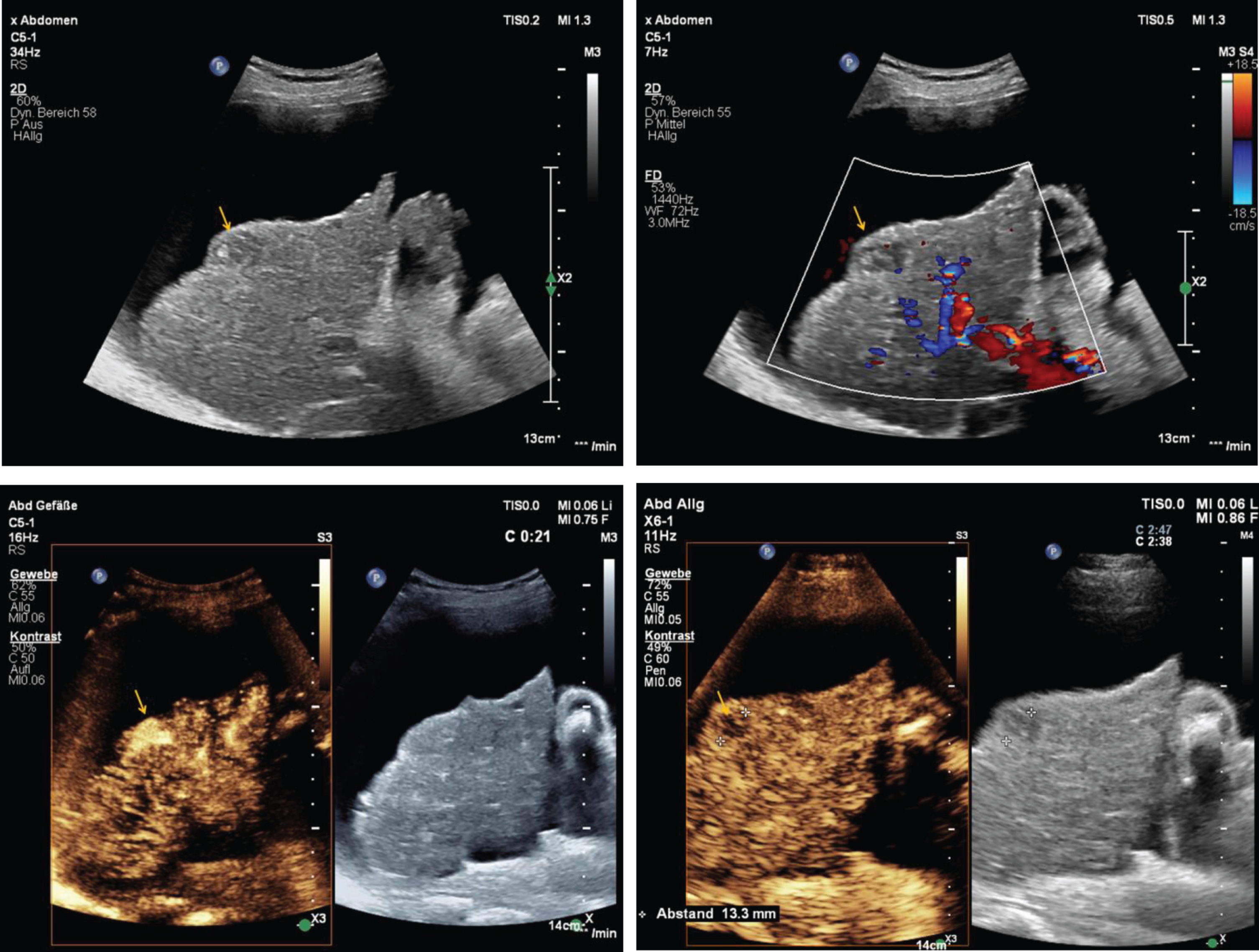
Sonomorphological appearance of a histopathologically verified hepatocellular carcinoma.
A 49-year old patient with hepatic cirrhosis shows a suspicious subcapuslar hypoechoic lesion in native B-mode (a). The lesion does not feature hypervascularization in Color Doppler sonography (b). Early arterial contrast enhancement (c) and venous wash-out (d) registered during CEUS.
Between 2004 and 2018 a total number of 119 patients with a suspected HCC lesion underwent 124 CEUS examinations followed by a liver biopsy. The patient population is subdivided into 84 male (70,6%) and 35 female patients (19,4%) with a mean age of 62 and a range between 20 – 88 years.
In detecting HCC-suspicious lesions CEUS showed a sensitivity of 96,6% and a specificity of 63,9%. In the underlying patient population, the positive predictive value (PPV) of CEUS was 86,7% with a negative predictive value (NPV) of 88,5%. Kappa coefficient between CEUS and the pathology showed a value of 0,659 with a significance (p) of < 0,001.
Discussion
The aim of the present study was to evaluate the diagnostic value of CEUS in comparison with corresponding histopathological results. Besides CE-CT and CE-MRI, few studies had already described CEUS as an effective and efficient non-invasive diagnostic tool for the detection and evaluation of the intra-tumoral microperfusion [9, 26–28]. As a result US is being implemented by leading professional societies, including EASL or AASLD, as the diagnostic tool of choice in screening and surveillance of patients at high risk for developing HCC [20–22, 29].
While sonography can make a statistically significant statement regarding the differentiation between HCC, focal nodular hyperplasia (FNH), hepatic adenoma (HA) or metastatic liver cancer (MLC) [30–32], it has some limitations for example in differentiating HCC from intrahepatic cholangiocarcinoma (ICC) due to overlapping sono-morphological features [33–35], in the context of hemodynamic changes in cirrhotic patients, in NAFLD or non-alcoholic steatohepatitis (NASH) patients, in the lack of a large field of view or for assessing liver lesions that are located at great depths [13, 36–39].
By comparing the morphological findings of CEUS with the histopathological results, Cohen’s kappa coefficient was 0,659 (p > 0,001) which indicates a strong inter-modality reliability and underlines the effectiveness of CEUS in detecting HCC. Additional studies had previously shown that CEUS may help to differentiate between poor, moderate and well differentiated HCC subtypes [9, 40–42]. Furthermore, CEUS showed a diagnostic sensitivity of 96,6%, a specificity of 63,9%, a PPV of 86,7% and a NPV of 88,5%. In comparison to our results a previous study described that CEUS showed high sensitivity of 93,5% for assessing even small HCC lesions of less than 2,0 cm size [43]. Besides the good correlation between CEUS and the histopathological results it has also a huge benefit in the examination of children where the use of US contrast agent has recently been approved by the U.S. Food and Drugs Administration (FDA) [44, 45]. While the patient is exposed to radiation during CT-scans and the use of CT or MRI contrast medium with its associated potential risks to the kidney function and the thyroid gland is almost indispensable to achieve sufficient diagnostic results, contrast media used for CEUS feature an excellent safety profile. First studies in small cohorts could already demonstrate safe and feasible application of CEUS during pregnancy for assessing unknown hepatic lesions [46, 47].
Conclusion
In addition to the excellent safety profile, CEUS offers a strong inter-modality reliability with histopathology and should therefore be included as a non-invasive examination method in the diagnostic clarification of unclear liver lesions.