



Abstract
The pig is the most widely used large animal model in Europe, with cardiovascular research being one of the main areas of application. Adequate refinement of interventional studies in this field, meeting the requirements of Russel and Burchs’ 3 R concept, can only be performed if blood-contacting medical devices are hemocompatible. Because most medical devices for cardiovascular interventional procedures are developed for humans they are tested mostly for compatibility with human blood. The aim of this study was therefore to determine whether there are differences in behavior of porcine and human platelets when they come into contact with glass, which was used as an exemplary thrombogenic material. For this purpose changes of platelet count, platelet volume and platelet expression of the activation markers CD61, CD62P and CD63 were measured using a modified chandler loop-system simulating the fluidic effects of the blood flow. Minipig and human platelets showed significant differences in number and volume, but not in activation after 4–8 min exposure to glass.
Keywords
Introduction
Sheep and pigs are important large animal models for interventional cardiovascular studies [1]. In 2019, 6,292 pigs were used in trials in Germany, of which 534 (8.5%) were used for cardiovascular studies and studies on the blood and lymphatic system (Federal Ministry of Food and Agriculture).
The study was prompted by the fact that these studies can involve the contact of medical devices with animal blood, many of which have only been tested for hemocompatibility with human blood. Recently, sheep platelets were shown to differ from human platelets in activability after 4–8 minutes of exposure to glass as a thrombogenic material, although these differences were small [6]. This indicates that for test refinement to meet the requirements of the 3R concept of Russel and Burch [2], it should be tested whether the hemocompatibility of these medical devices is also given for porcine blood.
For this reason, a study was set up to investigate in pigs (Göttingen minipigs) platelet number, volume, and activation changes in comparison to human platelets after glass contact for 4–8 min. The study was performed under dynamic testing conditions in a modified chandler loop-system [3, 4].
Materials and methods
Humans and animals
Human blood samples were collected from healthy adults (2 male, 1 female) who consented to blood collection on a voluntary basis. For each participant, a recovery period of 1 week was observed between each donation.
Blood sampling from the animals was approved by the Lower Saxony State Office for Consumer Protection and Food Safety (approval number 33.8-42502-05-20A513) and was performed with two adult and healthy Göttingen minipigs (both female, 11 years old). The animals were housed under conventional holoxenic hygiene conditions and cared for according to the guidelines of the European Societies of Laboratory Animal Sciences. Animals were without clinical signs of disease and monitored by whole blood analysis and general examination by an experienced veterinarian on each day of testing.
For each day of measurement, two platelet rich plasma (PRP) samples (silicone and silicone + glass) were available from pig or human. Each of these PRP samples was measured three times, before circulation and after passing through a modified chandler loop-system 20 and 40 times, respectively.
Blood sampling and dynamic test model
For each test at most 83 ml blood were sourced from the external jugular vein using an 18 G sized cannula and nine blood collection systems, eight filled with 1 ml of 0.106 molaric tri-sodium citrate each (S-Monovette® 10 ml 9NC) and one filled with 1.6 mg/ml blood EDTA (S-Monovette® 1.6 ml K3 EDTA) to prevent spontaneous coagulation. In addition, serum monovettes were collected (S-Monovette® Serum 4.9 ml) for determination of control parameters. There was a minimum of seven days per individual (human, minipig) between blood samplings and blood was sourced from each individual six times in total.
Within 30 minutes, the citrate-anticoagulated whole blood was centrifuged at 120 G for 15 min and PRP was carefully removed and placed in plastic test tubes. For the tests a sterilized modified chandler loop-system [4] was filled with the PRP for fluorescence-activated cell sorting (FACS) analysis of the platelets, and for platelet number and platelet volume analysis. In brief, the modified chandler loop-system consisted of two separate medical-grade silicone tubes with a non-thrombogenic inner surface, which were mounted to a closed loop using two three-way valves made of polyvinylidene fluoride (PVDF). The closed tube was filled with the PRP via one of both three-way valves. The second three-way valve ensured that the air in the system could escape. After insertion of the pump tube into a roller pump a defined flow was maintained (7.5 ml/h). Filled only with PRP the system was regarded as the negative-reference-system (non-thrombogenic control). For tests under thrombogenic conditions the tube system was partly filled with glass beads (OD 0.75–1.0 mm). The glass beads (n = 1084; calculated total surface 2,221 mm2) were prevented to follow the blood by a medical-grade V2A-steel-based mesh (mesh width 0.5 mm), which was integrated in the tube.
Directly after the initial filling of the tube system with PRP was completed, baseline values were determined. PRP volumes removed were always replaced isovolemically by autologous PRP. The PRP was analyzed in a total of 40 consecutive test cycles. Platelet analysis was performed after the PRP within the closed tubing had circulated through the total tube system 20 or 40 times. This corresponded to a circulation time of 4 or 8 min.
Platelet count and mean platelet volume
The platelets that were free to move in the tube system and that did not become adherent to inner surfaces of the tube system respectively were analyzed in terms of total number and mean platelet volume (as parameter for platelet aggregation). A double measurement was performed on all samples. These parameters were measured using an automated hematology analyzer (ProCyte Dx®, IDEXX, Germany).
FACS analysis
Platelet activation was evaluated using flow cytometry (MACSQuant® Analyzer, Miltenyi, Germany). Platelets were defined as CD42a (=GPIX) positive cells within the PRP-sample. To assess the platelet activation state the platelet markers CD61 (=GPIIIa), CD62P (=P-selectin), and CD63 (=GP53) were used.
In preliminary concentration tests the antibodies used (recombinant human IgG1 antibodies; PE-conjugated anti-CD42a, FITC-conjugated anti-CD61, APC-conjugated anti-CD62P, PE-Vio 771-conjugated anti-CD63; Miltenyi, Germany) were titrated to the endpoint so that measurements could always be conducted at saturation level, thereby excluding any dependence between the fluorescent intensity and the concentration of the antibodies.
Between 50,000 and 100,000 particles were collected in all flow cytometric measurements to ensure reliable analysis.
Statistics
Data analysis was performed with SPSS Statistics 26 using descriptive statistic parameters (means and standard deviations) and t-test for comparison of paired samples.
Results
In human and porcine PRP, platelet counts decreased significantly within 40 test cycles or 8 minutes when there was no contact with the glass (see Fig. 1). In this regard, the decrease in platelet count was greater for human PRP (–6.1%) than for porcine PRP (–2.4%). Moreover, platelet count in human PRP was already decreased after the first 20 test cycles (p < 0.05).
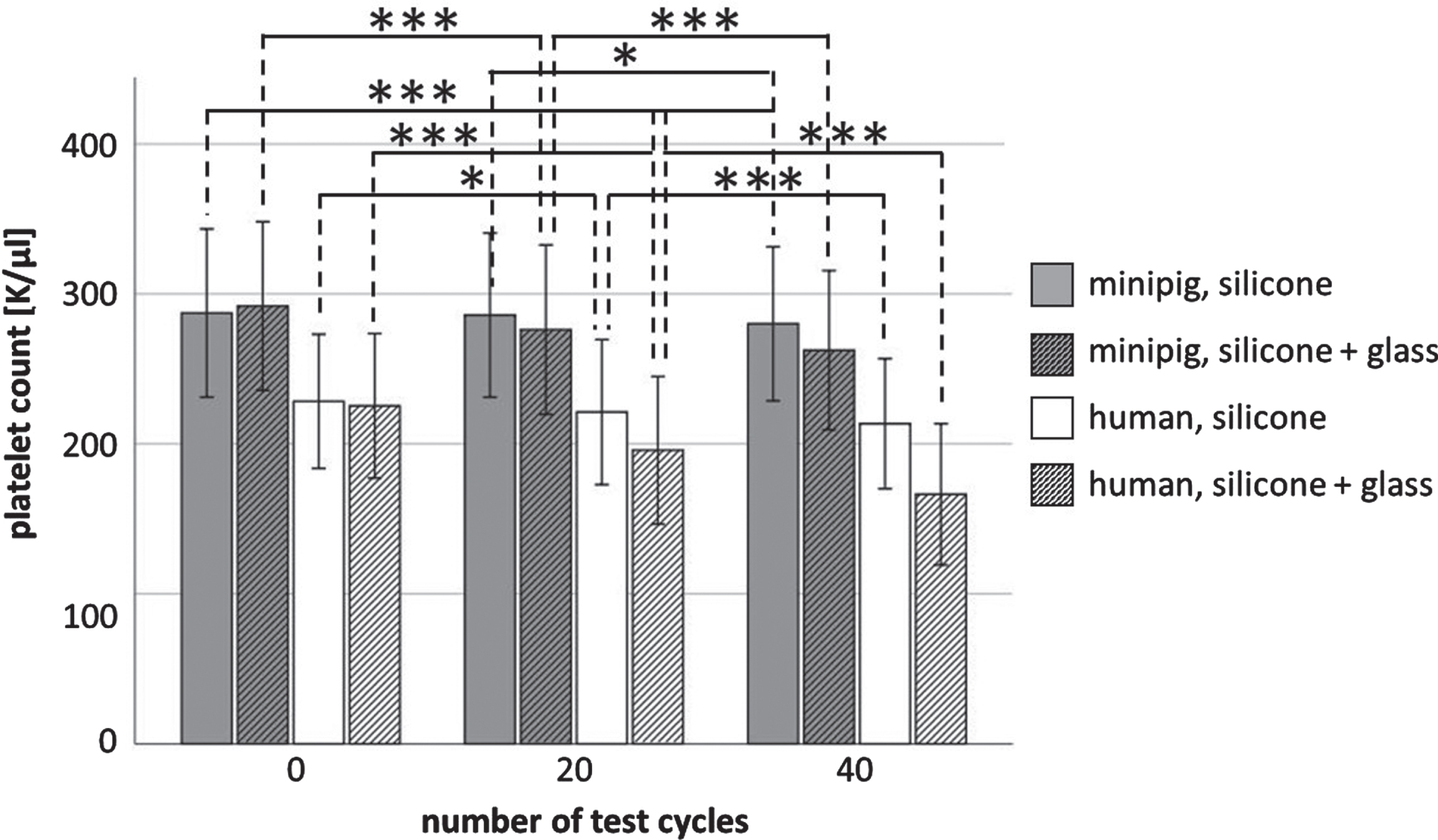
Total number of non-adherent platelets from platelet rich plasma (PRP) of adult Göttingen minipigs (n = 2) and humans (n = 3); each PRP sample was measured three times, before and when it has passed 20 or 40 times through a modified chandler loop-system filled with glass beads; means and standard deviation; *p < 0.05, ***p < 0.001.
Glass exposure also caused a decrease in platelet count in PRP of both species over time (p < 0.001), whereby again the decrease in platelet count was also greater in human PRP (–26.2%) than in minipig PRP (–10.3%).
No significant change in mean platelet volume (MPV) was measurable in human PRP after 20 or 40 test cycles without glass exposure (see Fig. 2). In contrast in minipig PRP there was an increase of the MPV within 40 test cycles (2.9%, p < 0.01).

Volume of non-adherent platelets from platelet rich plasma (PRP) of adult Göttingen minipigs (n = 2) and humans (n = 3); each PRP sample was measured three times, before and when it has passed 20 or 40 times through a modified chandler loop-system filled with glass beads; means and standard deviation; *p < 0.05, **p < 0.01, ***p < 0.001.
With glass exposure, in human and minipig PRP MPV increased over time. In human PRP, after 40 test cycles the MPV was increased by 1.6% (p < 0.001), in minipig PRP by 0.7% (p < 0.05).
The proportion of platelets (CD42a+ cells) within the PRP samples that were also positive for CD61 remained relative constant over the time of 20 or 40 test cycles regardless of whether the PRP was exposed to glass or not (see Fig. 3). In addition, platelet activation after glass contact did not differ between porcine and human platelets over time and was comparable to the control (tube system without glass beads). In human and porcine PRP only a proportion of less than 26% of the platelets (CD61+ and CD42a+ cells) was shown to be positive for CD62P and CD63 after glass contact for 4 or 8 min.
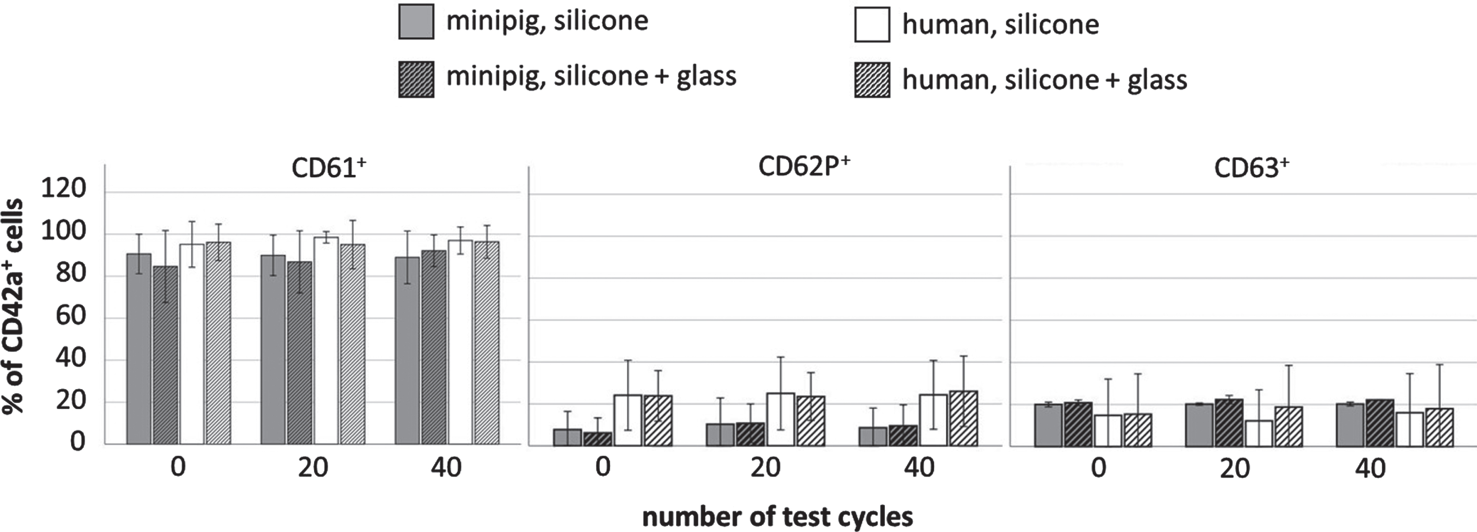
Percentage of CD42+/CD61+, CD42+/CD62+ and CD42+/CD63+ cells in platelet rich plasma (PRP) of adult Göttingen minipigs (n = 2) and humans (n = 3); each PRP sample was measured three times, before and when it has passed 20 or 40 times through a modified chandler loop-system filled with glass beads; means and standard deviation.
The study was performed to investigate how the behavior of porcine platelets after glass contact differs from the behavior of human platelets in terms of number, volume and activation. An established modified chandler loop-system was used for testing, which allows the behavior of platelets to be studied ex vivo in a dynamic mode.
The test results showed partly significant differences between the platelets of the two species. In porcine and human PRP, glass contact resulted in a decrease in platelet count, which was more pronounced in human than in minipig PRP. In the case of the MPV, there were also changes in both species due to glass contact, but these were minor. This could suggest that human platelets show stronger adherence after biomaterial contact than those of minipigs. Goodman et al. [5] found that human and porcine platelets showed comparable adherence to biomaterials. Using a dynamic test model, they studied the adherence behavior of platelets from three pigs (juvenile, Yorkshire cross), three sheep (two Suffolk sheep, one crossbred between a Suffolk sheep and a Dourset sheep), and adult humans on different polymers, including silicone. Interestingly, in Goodman’s tests, the pig platelets showed much greater adherence to silicone than the sheep platelets. This contrasts with results of our previous study, which showed a significant 33.5% decrease in ovine platelet count after only 4 min (20 test cycles) of silicone contact (w/o glass exposure) compared to our minipigs with a decrease of 0.4% [6]. The differences from the results of Goodman et al. [5] could be traced, at least in part, to the different assay approaches. Goodman’s group exposed platelets to the biomaterials for 45 min [5], whereas in the study presented here the maximum exposure time was 8 min. In addition, Goodman et al. [5] used a static model, whereas our results are based on tests with a dynamic model. In this model, shear forces become effective, which might have influenced platelet adhesion [7]. Furthermore, the different roughness of the foreign surfaces used may also play a role [8–10]. Hecker and Scandrett [9] examined how the thrombogenicity is affected by the roughness of polyvinyl chloride. They observed a positive correlation between roughness and thrombus formation after insertion of tubings with varying roughness (OD 3.0 mm) into the great saphenous vein and aortic artery of 40 adult Merino sheep.
Platelet behavior may also have been influenced by the mechanical effects of the roller pump, because experiments with a comparable chandler loop-model found that CD62P+ platelets can increase due to the physical effects of the roller pump, even without samples being introduced into the tubing system [3].
Species-specific differences in platelet behavior must also be considered. Pelagalli et al. [11] compared the adherence of sheep, porcine, and human platelets to autologous fibrinogen and found that sheep platelets could not adhere to autologous fibrinogen without pre-activation by adenosine 5-phosphate, whereas human and porcine platelets could, and the ability of human platelets to adhere to autologous fibrinogen was more pronounced than that of porcine platelets. It was suggested that these differences were due to structural differences in platelets and to differences in the availability of the fibrinogen (GPIIb/IIIa) receptor on the platelet surface. Species-specific differences in the ability to adhere to materials were also found by Grabowski et al. [12]. Using a dynamic circulation model, they compared platelets from different animal species with those from humans in terms of their ability to adhere to a cellulose-based hemodialysis membrane. They found that after 10 minutes of blood flow (shear rate 986/sec) fewer than 100 platelets/mm2 from sheep, pigs, and humans adhered, whereas in the test with platelets from dogs and rabbits, the number of adherent cells was much higher (dog 27,400±4,600; rabbit 78,400±6,400).
The MPV of the platelets in the human and minipig PRP increased over the total test period of 40 test cycles (8 min) with glass contact. This increase was slight, but more prominent in human PRP. This in turn could mean that platelets from minipig aggregate less than those from human during glass contact.
With regard to porcine platelet activation, it was also found that only a small proportion of platelets were activated by glass contact. In this respect, the results were comparable to those obtained with sheep and human blood [6]. Regardless, however, sheep platelets appeared to be more activatable than porcine platelets, as the proportion of CD62P-positive cells in sheep platelets increased significantly 8 minutes after glass exposure [6], whereas no significant increase in activation marker expression was observed in porcine platelets after glass contact.
Conclusion
The study showed that there are differences in the behavior of platelets from humans and pigs after exposure to glass. It became clear that exposure to glass has an effect on the number of platelets in the blood and on the mean platelet volume, but not on the activation status of platelets.
Footnotes
Acknowledgments
We thank Dr. Alexandra von Altrock, Dr. Theresa Maria Punsmann, Klaus Schlotter, and Thorsten Waßmann, staff members of the Clinic for Swine and Small Ruminants, Forensic Medicine and Ambulatory Service, University of Veterinary Medicine Hannover, Foundation, Hannover, Germany, for performing the blood collections, housing, and care of the animals.