
Abstract
BACKGROUND:
Over the years, diphtheria was known as contagious fatal infection caused by Corynebacterium diphtheria that affects upper respiratory system. The spread of diphtheria epidemic disease is best prevented by vaccination with diphtheria toxoid vaccine. Aluminum adjuvants were reported to stimulate the immune responses to killed and subunit vaccines.
OBJECTIVE:
Our study aimed to minimize adjuvant particles size, to gain insight of resulting immunity titer and impact on immune response antibody subtypes.
METHODS:
Aluminum salts and calcium phosphate adjuvants were prepared, followed by micro/nanoparticle adjuvants preparation. After formulation of diphtheria vaccine from diphtheria toxoid and developed adjuvants, we evaluated efficacy of these prepared vaccines based on their impact on immune response via measuring antibodies titer, antibodies isotyping and cytokines profile in immunized mice.
RESULTS:
A noteworthy increase in immunological parameters was observed; antibodies titer was higher in serum of mice injected with nanoparticle adjuvants-containing vaccine than mice injected with standard adjuvant-containing vaccine and commercial vaccine. Aluminum compounds adjuvants (nanoparticles and microparticles formulation) and microparticles calcium phosphate adjuvant induce TH2 response, while nanoparticles calcium phosphate and microparticles aluminum compounds adjuvants stimulate TH1 response.
CONCLUSIONS:
Different treatments to our adjuvant preparations (nanoparticles and microparticles formulation) had a considerable impact on vaccine immunogenicity.
Introduction
Corynebacterium diphtheria causes an acute infection in upper respiratory tract called diphtheria. This disease starts with diphtheria toxin produced by Corynebacterium diphtheria alongside a bacteriophage infection that is responsible for providing this pathogenic microbe with the gene encoding toxin production. The symptoms of this infection are adherent pseudomembrane of the pharynx and tonsil, low grade fever, and sore throat. The mortality rate associated with this illness ranges between 5 and 10%, with children under 5 years reported to have higher (up to 20%) rates followed by adults over 40 years [1, 2].
Vaccination is the only way to obtain immunity against diphtheria. After about eight decades, the diphtheria prevalence around the world was considerably reduced by the extensive application of DTP vaccines [3]. Although outbreaks of diphtheria were noticeably reduced worldwide [4], occasional outbreaks were reported in both developed and developing countries [5, 6, 7, 8]. A booster dose of anti-diphtheria vaccine is given to adults every 10 years as recommended by US Centers for Disease Control (CDC) to achieve prevention of infection [2]. Vaccine for diphtheria depends mostly on toxoid, which is a modified bacterial toxin prepared by detoxification with formalin [9, 10]. This deactivated toxoid produces antitoxin via stimulating antibodies production specially IgG. Adjuvants such as alum salts are needed to adsorb diphtheria toxoid resulting in effective formulation of diphtheria vaccine [11]. So far, diphtheria toxoid is the safest vaccine available [6, 12, 13]. Formulation of the vaccine antigen with the proper adjuvant is vital to accomplish effective immune response. Adjuvant enhances the adaptive immune response, also it has strong role in activation of innate immunity [14].
Recently, adjuvants with biodegradable nano particulation was developed by numerous procedures [15, 16, 17, 18], to direct immune response and achieve increase in surface area of adjuvant and vaccine antigen loading. These advantages boost necessary immune response represented in immunoglobulin type and subtypes depending on the helper T lymphocyte [15, 19]. Also, some types of adjuvants specifically enhance antibody dependent immune responses at mucosal surfaces [20, 21]. Biodegradable nanoparticles that have entrapped vaccine antigens, for instance peptides and proteins, possess significant potential as vaccine delivery systems [22]. Li et al. reported that protein antigens adjuvanted with alum nanoparticles induce more durable and stronger specific antibody responses than that induced by the same amount of antigens adjuvanted with the standard alum microparticles [23]. Akagi et al. suggested that nano- and microparticles biodistribution and the particle-related immune response can be regulated by controlling the size of the particles [22]. Vaccination of nanoparticles mostly elicited a higher serum IgG response than that obtained with the vaccination of normal-sized particles [22, 24]. Consequently, controlling the size of adjuvants nanoparticles is vital for efficient vaccine delivery and specific immune response stimulation.
The current study aimed to prepare diphtheria vaccines formulated with nanoparticle salt adjuvants then comparing it with internationally corresponding vaccines regarding their immunogenicity and in vivo potency.
Materials and methods
Salts for adjuvants preparation
Calcium chloride and dibasic sodium phosphate were purchased from BDH chemicals (London, UK). Trisodium phosphate was obtained from Techno Pharm Chem manufacturers (Bahadurgarh, Haryana, India). Aluminum chloride, sodium acetate, and sodium citrate were purchased from Alfa Aesar (Tewksbury, USA).
Alum salts and calcium phosphate adjuvants preparation
Aluminum hydroxide and aluminum phosphate adjuvants were prepared by exposing an aqueous solution of aluminum chloride to alkaline conditions in a well-defined controlled chemical environment. Aluminum hydroxide adjuvant was prepared by mixing aluminum chloride (AlCl
Aluminum phosphate adjuvant was prepared by mixing two solution, aluminum chloride solution (0.03 M) and trisodium phosphate solution (0.02 M). Trisodium phosphate solution was added to aluminum chloride solution with good stirring until a pH of 5 was reached, and then mixture was centrifuged. The pellet was dissolved in distilled water, then 0.36% NaCl was added [25].
Calcium phosphate adjuvant preparation was dependent on reaction between an aqueous solution of dibasic sodium phosphate and an aqueous solution of calcium chloride with good stirring and controlling pH to 7.4 until gel precipitated [26].
Preparation of microparticle adjuvants by microwave
To prepare micro-particles of adjuvant, a suspension of each adjuvant was exposed to microwave for 10 to 15 min, microwave was stopped after each boiling of solution for 30 s, and then restarted again until 15 min ended, while keeping continuous shaking. After microwave exposure ended, pH was adjusted for each adjuvant as it was originally before this treatment [23, 27].
Preparation of nanoparticle adjuvants by ultrasonication
To prepare nanoparticles, all prepared adjuvants were thoroughly mixed prior to ultrasonication. Ultrasonication was performed at room temperature, and then transferred to a water bath ultrasonicator (Branson, Emerson, USA). A 15/10 s on/off time ultrasonication and cooling cycles was applied for solutions; this procedure was continued for 30 min with constant gentle mixing to break down the particle size. The pH was monitored and adjusted for each adjuvant as it was originally before this treatment [23, 28].
Characterization of adjuvants by electron microscopy
The size of nanoparticles and microparticles formulation was measured by scanning electron microscopy (Quanta FEG 450 SEM, Thermo Fisher Scientific, USA) at Electron Microscopy Unit in King Abdulaziz University (Jeddah, Saudi Arabia). To prepare samples for SEM, all samples were diluted with 1x PBS to the same concentration, then totally dried to prevent any damage in samples while low pressure vaccum through SEM work [29].
Adjuvant-toxoid mixing and protein loading evaluation
To prepare diphtheria vaccine, diphtheria toxoid was mixed with prepared adjuvants with continuous stirring at room temperature for 3 days, and then stored at 4
Mice immunization
Fifty five female mice of strain BALB/c, 7 weeks old weighing 18-20 gram were purchased from ani-mal house, king Fahd Medical Research Centre, Jeddah, Saudi Arabia. After obtaining mice from animal house, they were restrained for at least 7 days at room temperature. All procedures were approved by Animal House and Department of Training and Development at King Fahd Medical Research Centre. Mice were divided into 11 groups; groups 1, 2, 3, 4, 5, 6, 7, 8, 9, 10, and 11 immunized intramuscular with vaccines prepared from untreated calcium phosphate, microwave treated calcium phosphate, ultrasonication treated calcium phosphate, untreated aluminum hydroxide, microwave treated aluminum hydroxide, ultrasonication treated aluminum hydroxide, untreated aluminum phosphate, microwave treated aluminum phosphate, ultrasonication treated aluminum phosphate, and standard adjuvant Alum 2%, and commercial vaccine TETAVAX (SANOFI PASTEUR S.A., France), respectively. After 21 days, blood was collected and centrifuged to obtain polyclonal serum [31].
Immune response evaluation
We evaluated the efficacy of prepared vaccines based on their impact on immune response through measuring three parameters of immunity; antibodies titer, antibodies isotyping and cytokines profile. Different kits were purchased to evaluate immune response, mouse immunoglobulin isotyping ELISA kit from BD Biosciences (New Jersy, USA) and multi-analyte ELISA array kit for detection of mouse Th1/Th2/Th17 cytokines from QIAGEN (Hilden, Germany).
Statistical analysis
Differences between the variants were tested using Student’s t test and McNemar’s test [32]. P values of less than 0.05 were considered statistically significant.
Results
Structure and morphology of prepared adjuvants
Aluminum hydroxide adjuvant characterization
Aluminum hydroxide adjuvant has amorphous structure. The images taken by SEM for alum hydroxide adjuvant with 2500 x and 5000 x magnification showed an amorphous structure composed of nanoparticles varying in diameter from 1.969
Structure and morphology of prepared adjuvants examined by SEM. (A) Aluminum hydroxide adjuvant at 2500 x magnification. (B) Aluminum hydroxide adjuvant at 5000 x magnification. (C) Calcium phosphate adjuvant at 2500 x magnification. (D) Calcium phosphate adjuvant at 10.000 x magnification. (E) Aluminum phosphate adjuvant at 2500 x magnification. (F) Aluminum phosphate adjuvant at 10.000 x magnification.
Calcium phosphate adjuvant has spherical crys-talline structure. The images taken by SEM for calcium phosphate adjuvant with 2500 x and 10.000 x magnification showed a spherical structure composed of small microparticles varying in diameter from 5.113
Aluminum phosphate adjuvant characterization
Aluminum phosphate adjuvant has completely am-orphous structure. The images taken by SEM for alum phosphate adjuvant with 2500 x and 10.000 x magnification showed amorphous structure composed of nanoparticles, varying in diameter from 1
Alum 2% standard adjuvant characterization
Alum 2% adjuvant consists of crystalline magnesium hydroxide and amorphous aluminum hydroxyl carbonate. The images taken by SEM for alum 2% adjuvant with 2500 x and 10.000 x magnification showed amorphous like crystalline structure composed of aggregated small microparticles varying in diameter, at magnification 10000 x (Fig. 2A and B).
Evaluation of protein loading for all prepared adjuvants
Evaluation of protein loading for all prepared adjuvants
Characterization of Alum 2% standard adjuvant and commercial vaccine TETAVAX by SEM. (A) Alum 2% adjuvant at 2500 x magnification. (B) Alum 2% adjuvant at 10.000 x magnification. (C) TETAVAX at 2500 x magnification. (D) TETAVAX at 12.000 x magnification.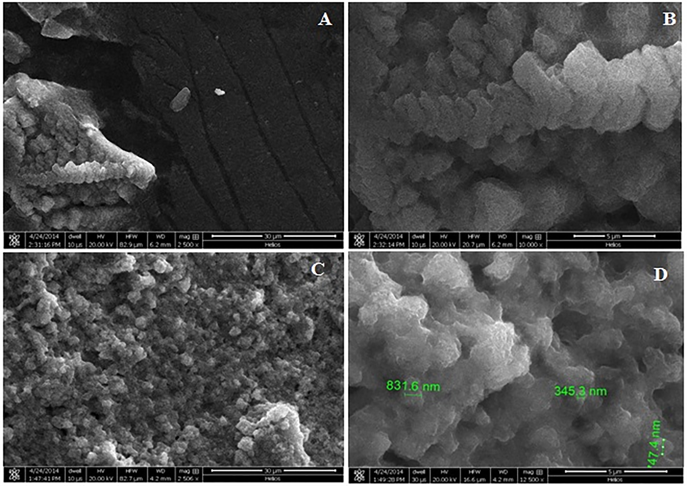
This commercial vaccine consists of tetanus toxoid and alum salts and has amorphous structure. The images taken by SEM for TETAVAX with 2500 x and 12.000 x magnification showed amorphous structure composed of nanoparticles varying in diameter from 345.3 nm to 831.6 nm at 12.000 x magnification (Fig. 2C and D).
Immune response to different prepared anti-diphtheria vaccines in immunized mice.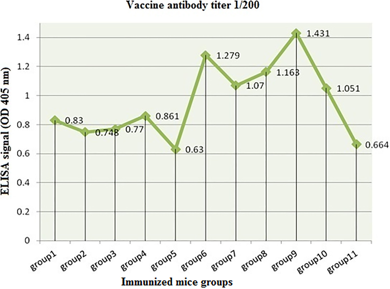
Incubation of adjuvants and diphtheria toxoid after 24 h had no significant effect on adjuvant-toxoid interaction, as the majority of their attraction and subsequently loading of diphtheria toxoid protein occurred within 24 h incubation for all prepared adjuvants. Thus, 24 h preparations have been used for mice immunization.
Evaluation of adjuvant adsorption ability and quantity of adsorbed antigen was performed by Bradford assay. After all adjuvant preparations were incubated for 24 h with protein toxoid, protein in vaccine preparations was measured by spectrophotometry at 595 nm. Results revealed that adjuvants treated by ultrasonication and microwave had significantly (
Anti-diphtheria vaccine antibody titration
We found that the highest optical density at 1/200 dilution was in serum of mice group 9 injected with vaccine containing ultrasonic treated aluminum phosphate, followed by group 6 injected with vaccine containing ultrasonic treated aluminum hydroxide (Fig. 3). In group 9, optical density was highest at 1/200–1/1600 dilution, and then gradually decreased till reaching 1/204800 dilution (the highest antibody titer among all mice groups). The antibody titer in mice groups immunized with vaccines prepared from untreated calcium phosphate, microwave treated calcium phosphate, ultrasonic treated calcium phosphate, untreated aluminum hydroxide, microwave treated aluminum hydroxide, ultrasonic treated aluminum hydroxide, untreated aluminum phosphate, microwave treated aluminum phosphate, ultrasonic treated aluminum phosphate, and standard adjuvant Alum 2%, and TETAVAX were 1/12800, 1/12800, 1/12800, 1/25600, 1/12800, 1/102400, 1/51200, 1/51200, 1/204800, 1/51200, and 1/12800, respectively. From these data, we concluded that diphtheria vaccine containing aluminum phosphate adjuvant treated by ultrasonication is the optimal vaccine for producing a strong antibodies immune response, followed by vaccine containing ultrasonic treated aluminum hydroxide.
Anti-diphtheria vaccine antibodies isotyping
All vaccine preparations as well as Alum 2% vaccine, and TETAVAX could induce mainly IgG1, IgG2b, IgM, and Ig
TH1 type of immune response includes antibodies istotypes (IgG2 and IgG3), while TH2 includes antibodies istotypes (IgG1 and IgA). The results demonstrated that TH2 type of immune response antibodies istotypes was typically distributed in mice groups 2, 8, 9, and 11 which were immunized with calcium phosphate treated with microwave, aluminum phosphate microwave treated, aluminum phosphate ultrasonication treated, and TETAVAX vaccines, respectively (Table 2).
Anti-diphtheria vaccines antibodies isotyping (results represented as mean antibody OD at 405 nm
standard deviation)
Anti-diphtheria vaccines antibodies isotyping (results represented as mean antibody OD at 405 nm
The vaccine best induced TH2 cytokines was that prepared from aluminum hydroxide adjuvant treated with ultrasonication (Fig. 3.5). In serum of mice injected with vaccine prepared from aluminum hydroxide treated with ultrasonication, we detected the highest concentrations of cytokines of TH2 immunity (IL5, IL6, IL10 and IL13) (Fig. 3.5) and no detectable TH1 cytokines (IL12 or INF
Cytokines profile in different immunized groups.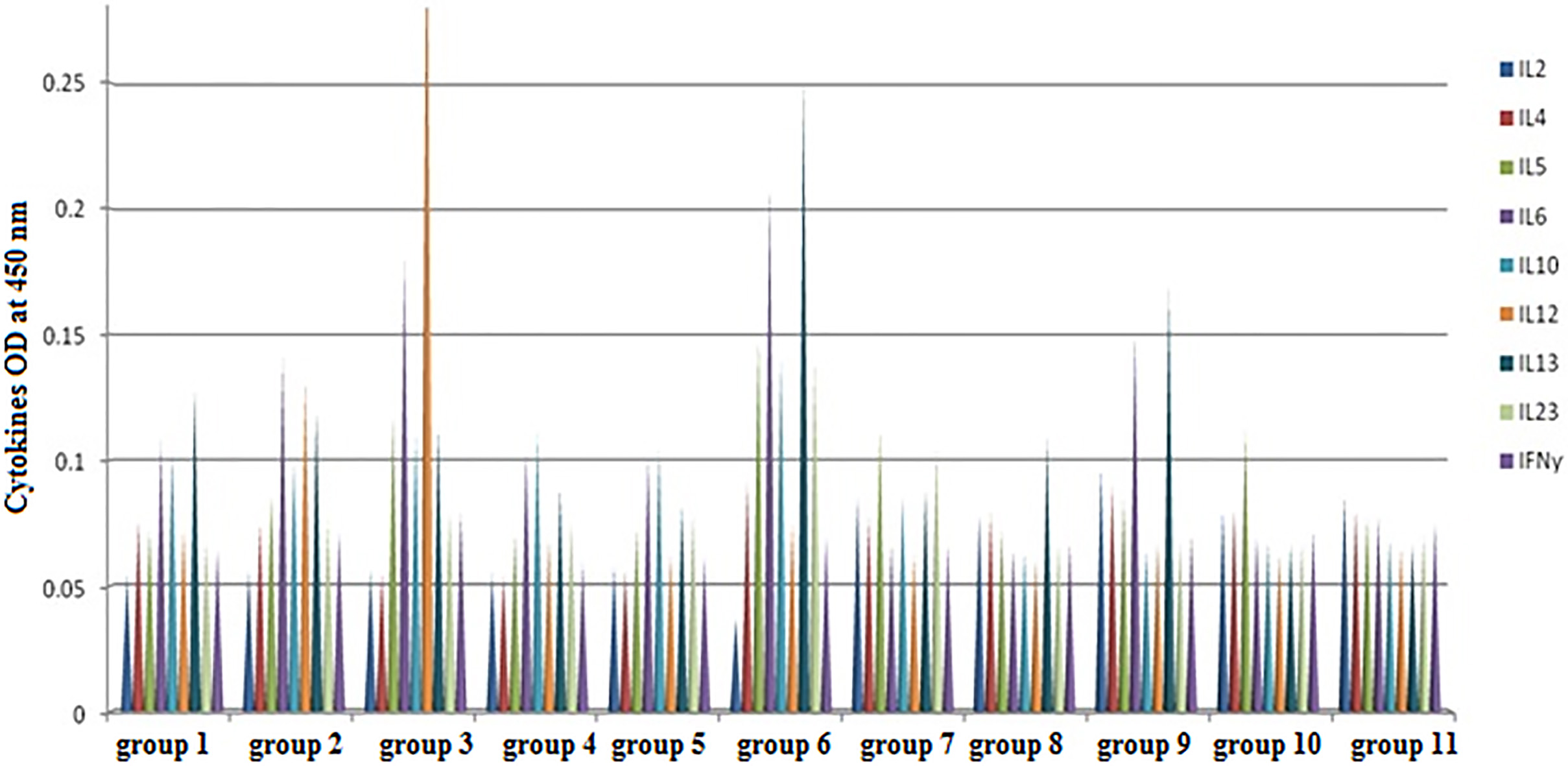
Vaccine prepared from calcium phosphate adjuvant treated with ultrasonication was the best for inducing TH1 cytokines (IL12), while TH2 immune response (IL13) against this vaccine was significantly (
The current study aimed to develop different adjuvant preparations (nanoparticles and microparticles formulation) to potentiate the immune response against diphtheria toxoid. We performed in vivo experiments to gain insight of resulting immunity against vaccine preparations from these adjuvants represented by antibodies titer, antibodies isotyping and cytokines profile.
Basically, vaccination targets stimulation of a protective, strong, and most importantly long lasting immune response to the administered antigen. In order to accomplish these targets, potent adjuvants and innovative vaccine approaches are required to induce a potent immune response [33]. To evaluate potency of a certain vaccine, vaccine components including the adjuvants need critical assessment. We evaluated our prepared adjuvants throughout this study, starting with estimating quantity of diphtheria antigen that was adsorbed on each adjuvant preparation, and finally in vivo analysis of resulting immunity against vaccine preparations from these adjuvants represented by antibodies titer, antibodies isotyping and cytokines profile. In the present study, three types of adjuvants (aluminum hydroxide, calcium phosphate, and aluminum phosphate) with different formulation, microparticles and nanoparticles were developed. The size of nanoparticles and microparticles formulation was measured by scanning electron microscopy. The obtained size values of these adjuvants particles were comparable to the size of microorganisms and were adequate for uptake by phagocytosis by antigen-presenting cells (APC). The adjuvant effect of micro/nanoparticles depends mainly on their uptake into APC. More importantly, particulate vaccines were proved to be more effective than soluble one for immune responses induction [34, 35].
Images taken by scanning electron microscope for adjuvants showed the differences in particles size between different preparations of adjuvant. For aluminum hydroxide, SEM showed nanoparticles of diameter ranging from 1.969
Adsorption is vital for the immunostimulatory effect of aluminum adjuvants. An important factor influencing adjuvant capacity of adsorption is the specific surface area. This parameter takes into account adjuvant particles size and shape in order to determine the real surface in contrast with the apparent surface [37]. As the particle size decreases, the specific surface area along with adsorption capacity increases. Nanoparticles have the highest specific surface area due to its very small size. The particulate nature of adsorbed antigens enables its uptake by APC through phagocytosis. It was confirmed that tetanus toxoid adsorbed on aluminum was easier to be uptaken by APC than soluble toxoid [38]. Also, adsorbed antigens are more slowly released from the injection site [39]. All these reasons and differences in preparations procedures explain why each adjuvant differs from other adjuvants with regard to morphology and capacity of adsorption. Antigens are simply adsorbed onto the alum due to strong electrostatic interaction between antigen and alum, which enhances antigen uptake and presentation by APC [40]. In current study, the adsorption capacity of the prepared adjuvants was evaluated by mixing diphtheria toxoid and different quantities of adjuvants, then detection of adsorbed and non-adsorbed toxoid protein by Bradford assay. The obtained data revealed that aluminum hydroxide and aluminum phosphate have the least quantities of free protein that didn’t adsorb onto adjuvant and highest percentage (85–94%) of antigen toxoid adsorbed. On the other hand, calcium phosphate has the highest free protein concentration and least percentage of adsorbed protein (46.67–50%).
In vivo experiments revealed that diphtheria vaccine containing aluminum phosphate adjuvant treated by ultrasonication is the optimal vaccine for producing a strong antibodies immune response (the antibody titer was 1/204800), followed by vaccine containing ultrasonic treated aluminum hydroxide (the antibody titer was 1/102400). These results were in agreement with those described by Akagi et al. “For a review, see [22]” and Li et al. “For a review, see [23]” who reported that protein antigens adjuvanted with alum nanoparticles induce more durable and stronger specific antibody responses than that induced by the same amount of antigens adjuvanted with the standard alum microparticles. Additionally, aluminum compounds adjuvants (nanoparticles and microparticles formulation) and microparticles calcium phosphate induce TH2 response (IgG1 and IgA), while nanoparticles calcium phosphate and microparticles aluminum compounds adjuvants stimulate TH1 response (IgG2 and IgG3). Aluminum phosphate adjuvant with nanoparticles size stimulates humoral immune response; IgM, IgG1, and IgG2b, after that comes aluminum hydroxide adjuvant with nanoparticles size that stimulates humoral immune response; IgM, and IgG1, therefore, both are effective adjuvant preparations for vaccine formulation that enhance immune response and provide protection against Corynebacterium diphtheria.
These results were in agreement with previous results reported by Gupta and Siber “For a review, see [41]”, who found that aluminum phosphate adjuvant gave the highest antibody responses for tetanus toxoid injected in mice when compared with calcium phosphate and stearyl tyrosine adjuvants. Also, they agree with results obtained by Issa et al. “For a review, see [42]”, who revealed that, alum and calcium phosphate adjuvants enhanced the immune response against vaccine adsorbed on them. But it was higher in case of alum than in calcium phosphate nanoparticles. Previous studies demonstrated that production of IgG1a and 1b was lower with calcium phosphate compared to aluminum hydroxide and phosphate but calcium phosphate has the advantage of not stimulating specific IgE response [43]. Anyway, although animal model results are divergent depending on antigen and adjuvant concentrations, human data suggested that calcium phosphate is an effective adjuvant and potentially more effective than alum after the booster [44]. It has a reasonable capacity to adsorb antigens, induces high levels of IgG antibodies, and does not increase IgE production [26, 36, 37, 44].
The presence of IL-12 and/or IFN-
In conclusion, the results of current study revealed that aluminum salts especially aluminum phosphate treated with ultrasonication (nanoparticle formulation) was the most potent adjuvant against diphtheria toxoid. Nanoparticle-based vaccine approaches can reduce the frequency of vaccine dosage and will increase patient compliance. In the near future, they can be applied in treatment of many infectious diseases or cancers.
Footnotes
Acknowledgments
The current work is a part of the Master degree of Ms. Fatimah M. Alshanqiti (Department of Biology, Faculty of Science, King Abdulaziz University). This work was supported in part by King Abdulaziz City for Scientific Research and Technology (KACST, AT-35-155), Saudi Arabia.
Conflict of interest
No conflicts of interest.