
Abstract
BACKGROUND:
The Lewis (b) blood group antigen-Binding Adhesion2 (BabA2) has been reported to mediate the attachment of H. pylori to human.
AIM:
assessment the diagnostic potential of detection of (BabA2) gene compared with immunostaining of Lewis (b) by specific mouse monoclonal antibodies in gastric biopsies from Egyptian Patients as a diagnostic maker for Helicobacter pylori infection.
MATERIALS AND METHODS:
Fifty untreated patients suffering from dyspeptic complaints were enrolled in this study and underwent for upper gastro-duodenal endoscopy. Biopsies were taken for histological examination by (H&E) and immunohistochemical analysis for Lewis b by specific mouse monoclonal antibodies, and scoring of Lewis b expression in gastric tissue biopsy as well as molecular detection of BabA2 gene of H. pylori by PCR. Biochemical analysis was performed to detect the presence of H. pylori urease activity using Rapid Urease Test (RUT).
RESULTS
: Out of 50 gastric biopsies, 41 biopsies were positive for histological, Immunostaining for Lewis b expression and urease activity test (RUT) for H pylori. RUT showed a sensitivity of 87.8%, specificity 88.9%, positive predictive value (PPV) 97.2%, and negative predictive value (NPV) 61.5%. BabA2 gene results revealed that, out of 41 positive biopsied cases, 39 (95.1%) were positive by the PCR test for BabA2 gene. And all 9 negative biopsies (100%) for H pylori negative for BabA2gene so the sensitivity and specificity of BabA2 gene detection in gastric biopsies by PCR were 95.1% and 100%; respectively.
CONCLUSION
: BabA2 gene detection in gastric tissue biopsies could be suggested as a diagnostic biomarker to be included among the other biomarkers routinely performed for clinical diagnosis of H. pylori infection.
Keywords
Introduction
Helicobacter pylori, is a causative agent of various severe gastroduodenal diseases, including gastric cancer [1, 2]. To succeed in these long-term associations, H. pylori has developed a unique set of virulence factors, which allow survival in a unique and hostile ecological niche – the human stomach. Several studies identified genes that are consistently present in all H. pylori strains analyzed. The core genomes described in these studies have ranged from 1091 to 1281 genes and contained two copies of the 16S, 23S, and 5S rRNA genes. Many strains carry one or more cryptic plasmids, which do not seem to carry antibiotic resistance genes or virulence genes [3, 4]. Blood group antigen adhesion (BabA), a 75 kDa adhesion molecule is one of the major adhesins considered to contribute to the pathogenicity of H. pylori encoded by the BabA2 gene. It has been identified as the adhesions responsible for H. pylori binding to the fucosylated blood group antigens Lewis b [5, 6]. The BabA2 gene encodes the blood group antigen binding adhesion, which binds to the fucosylated Lewis b antigen present on the surface of gastric epithelial cells. BabA facilitates colonization, the persistence of infection and release of virulence factors of the bacterium. Infection with BabA2-positive H. pylori has been associated with gastric ulcer, duodenal ulcer, and gastric adenocarcinoma and is related to increased risk of severe disease when it coexists with the cagA gene and the vacA s1 allele [7]. Several studies showed that BabA-mediated adherence leads to increased mucosal damage of gastric mucosa [8, 9]. Patients infected with strains harboring the BabA2 gene had a higher degree of granulocytic infiltration than patients infected with strains lacking BabA2 [10]. An important chemotactic factor for granulocytes is interleukin 8 (IL-8) which is mainly secreted by epithelial cells in response to H. pylori infection, and the highest levels of IL-8 were detected in patients infected with BabA2 positive strains. So, the suggested sequences of events were leading to higher pathogenicity of BabA2 positive strains [11]. Currently, there are several popular methods for detecting the presence of H. pylori infection, each having its own advantages, disadvantages, and limitations. Basically, the tests available for diagnosis can be separated according to whether or not an endoscopic biopsy is necessary. Histological evaluation, culture, polymerase chain reaction (PCR), and rapid urease tests are typically performed on tissue obtained at endoscopy [12]. Alternatively, simple breath tests, serology, and stool assays are sometimes used, and trials investigating PCR amplification of saliva, feces, and dental plaque to detect the presence of H. pylori are ongoing [13]. The available tests are generally divided into noninvasive tests, based on peripheral samples, such as blood, breath samples, stools, urine, or saliva for detection of antibodies, bacterial antigens, or urease activity, and invasive tests, based on gastric specimens for histology, culture, or other methods such as Polymerase Chain Reaction [3, 14, 15]. The main objective of the current study was the detection of BabA2 gene to estimate its prevalence in out-clinic patients admitted to Medical Research Institute hospital, Alexandria University, Egypt as one of the major virulence factors associated with H. pylori and contribute to the different clinical outcomes. Also to evaluate the efficiency of using conventional PCR technique as a powerful technique for its molecular detection in gastric tissue biopsies obtained during endoscopy from patients infected by H. pylori. Evaluation of the clinical outcomes due to infection by BabA2 positive strains, detection of the Lewis b expression status, and its relation to H. pylori colonization density and subsequent disease outcome were also done.
Materials and methods
Samples
Fifty untreated patients suffering from dyspeptic complaints were consecutively enrolled in this study and underwent upper gastro-duodenal endoscopy. This study was carried out at the hospital of Medical Research Institute (MRI), Alexandria University, Egypt. All patients were informed and signed written consent and then the Ethics committee of MRI agreed for the ethical approval for the study.
Biochemical analysis histopathological examination
Routine upper gastro-duodenal endoscopy with re- cordings of all pathological signs found and 6–8 gastric tissue biopsies were taken for detection of H. pylori infection in gastric tissue biopsy using biochemical analysis, histological examination and molecular detection. Biochemical analysis was performed to detect the presence of H. pylori urease activity using Rapid Urease Test (RUT).
Immunostaining of lewis b with specific mouse monoclonal antibodies
Gastric biopsies were fixed in 10% formalin, embedded in paraffin wax and then sections from tissue blocks were examined using routine Hematoxylin and Eosin (H&E) stain. Determination of H. pylori colonization densities (if present), and scoring of H. pylori infection as a gold standard were also carried out. Histopathological examination of H&E stained sections was carried out for examining gastric-duodenal pathology to detect the severity of gastritis, atrophy, ulceration, or intestinal metaplasia. The paraffin sections from tissue blocks were also stained using immunohistochemical analysis for Lewis b, and scoring of Lewis b expression in gastric tissue biopsy. Prediluted mouse anti-human Lewis b monoclonal antibody (Cat. # MS-379, Thermo Fisher Scientific Fremont, CA 94538, USA) and UltraVisondetection System (Anti-polyvalent, HRP/DAB Plus, Cat. # TP-004–HD, Thermo Fisher Scientific Fremont, Ca 94538, USA) kit was used for the immunohistochemical study.
Detection of BabA2 gene of H. pylori was carried out by polymerase chain reaction
BabA2 gene of H. pylori was carried out by conventional PCR technique [15, 16] using the thermal cycler (Little Genius, Bioer). DNA extraction from gastric tissue biopsy was performed out using Genomic DNA purification kit (Cat. # K0512, Fermentas, Lithuania). All PCR reagents used in this study were purchased from Fermentas, Lithuania except the primers which were synthesized by MetaBion, Germany (Pavlov et al., 2004). Primers were reconstituted with sterile distilled water to a final concentration of 100 pmol/
BabA2 – Sense (Forward): 5’-AATCCAAAAAG GAGAAAAAGTATGAAA-3’. BabA2 – Anti-Sense (Reverse): 5’-TGTTAGTG ATTTCGGTGTAGGACA-3’.
The PCR products (832 bp) were visualized on 2% agarose gels.
All data were expressed as mean
Sensitivity
Specificity
Predictive value for a positive result (PV
PV
Predictive value for a negative result (PV
PV
Diagnostic values of detection of BabA2 gene expression for diagnosis of H pylori infection compared with histological examination with Relation between BabA2 gene and H. pylori according to histological examination (A) and between BabA2 gene versus histological findings (B)
Diagnostic values of detection of BabA2 gene expression for diagnosis of H pylori infection compared with histological examination with Relation between BabA2 gene and H. pylori according to histological examination (A) and between BabA2 gene versus histological findings (B)
Relation between BabA2 gene and Lewis b expression (A) and BabA2 gene colonization density and Lewis b expression (B)
Immunostaining of Lewis B using specific monoclonal antibody. A: Case of mild chronic superficial gastritis of H. pylori etiology showing Lewis-b expression (X40), B: Moderate chronic superficial gastritis of H. pylori etiology showing strong expression of Lewis-b (X 400), C: Moderate/severe chronic gastritis of H. pylori etiology showing strong homogenous expression of Lewis-b (X400).
Detection of Blood Group Antigen-Binding Adhesion2 (BabA2) gene of H. pylori by conventional PCR technique. Ethidium bromide stained agarose gel showing bands of amplified PCR products of BabA2 gene of H. pylori from gastric biopsies at 832 bp: Lane (1) DNA molecular weight marker, lane (6) a negative case, while lane (14) a negative control, Lanes (2–5, 7–13, 15) positive cases.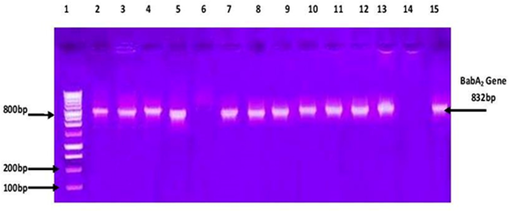
Diagnostic values of detection of BabA2 gene expression for diagnosis of H pylori infection compared with histological examination.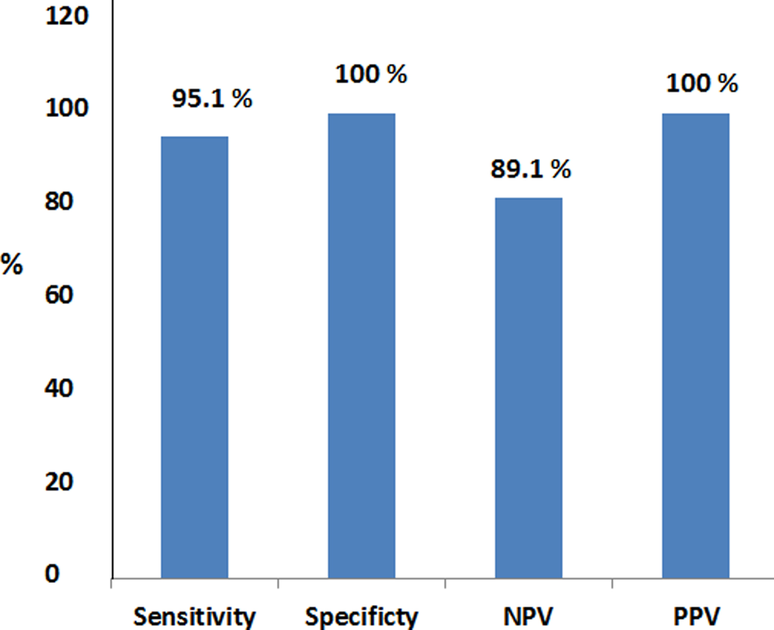
Biochemical findings of Rapid Urease test showed that 37 out of 50 (74%) cases were positive for RUT while 13 cases were negative. Histological grading of H. pylori infection as a gold standard showed that 41 out of 50 patients (82%) were positive (Table 1A and B). The histopathological examination of H&E stained sections showed that 35 out of 50 patients (70%) have chronic superficial gastritis. The relation between Immunohistochemical scoring of Lewis b expression (Fig. 1) and histopathological grading showed that 42% have grade 1. However, compared with histological examination of gastric biopsies, the measures of diagnostic test accuracy of the RUT showed a sensitivity of 87.8%, specificity 88.9%, positive predictive value (PPV) 97.2%, and negative predictive value (NPV) 61.5%.
DNA was extracted from gastric tissue biopsies and BabA2 gene was detected using PCR (Fig. 2). Out of 50 samples (39 DNA samples 74%) were positive for BabA2 gene. The results revealed that out of 41 positive cases by histological examination, 39 (95.12%) were positive for BabA2 gene expression by the PCR test. On the other hand, all the 9 negative cases by histological examination were also negative BabA2 gene expression by the PCR test (Table 1).
Compared with histological examination as a gold standard, it was found that BabA2 gene expression detection by PCR showed sensitivity, specificity, a positive predictive value (PPV), and a negative predictive value (NPV) of 95.1%, 100%, 100%, and 81.9% respectively (Fig. 3). Moreover, Table 2B showed the relation between BabA2 gene according to BabA2 gene expression and disease progression as regard to histopathological findings.
Table 2A illustrated the relation between BabA2 gene and colonization density in regard to the Lewis b expression grade detected using immunohistochemistry. Table 2B showed the relation between BabA2 gene, colonization density of H. pylori and expression grade of Lewis b. Studying the relation between BabA2 gene according to PCR results and disease outcomes regarding histopathological findings revealed that severity of disease outcome and BabA2 gene was found to be statistically significant (
Discussion
In the present study, out of 50 patients suffering from dyspepsia who’s their ages ranged between 20 to 69 years, undergoing upper gastrointestinal endoscopy, 41 patients were infected with H. pylori, giving a prevalence of 82%. It appears high, corresponding to the trend in developing countries, and confirming that infection in developing countries is seen in middle age people [17]. High prevalence was also reported in developing countries with up to 80% of the population in India [18], in Taiwan and China [19, 20]. On the other hand, lower prevalence ranging from 49.5% to 66.5% was reported by Raghunath et al. [21] and in the western world, the prevalence reported ranges from 25% to 50% [22]. The Rapid Urease Test (RUT) is the most frequently assayused for the diagnosis of H. pylori infection in routine gastrointestinal endoscopy practice [23, 24]. It is extremely valuable because it gives a result for H. pylori infection before the patient leaves the endoscopic suite, providing indirect identification of infection within a few hours of endoscopy [25]. In the current study, RUT was used to determine the H. pylori infection of studied cases. Compared with histological examination of gastric biopsies, the measures of diagnostic test accuracy of the RUT showed a sensitivity of 87.8%, specificity 88.9%, positive predictive value (PPV) 97.2%, and negative predictive value (NPV) 61.5%. These results are comparable to results reported by Monterio et al. [17], Yakoob et al. [25], and Lim et al. [26] who reported that the sensitivity of RUTwas 86.4%, 90% and 87.5% respectively. Higher sensitivity and specificity 94% and 99%; respectively of RUT was recorded in another study [27]. On the other hand, Adensay et al. recorded a lower sensitivity of 72%. The accuracy of the test is dependent on the number of tissue specimens tested, the location of biopsy sites, bacterial load, and previous usage of antibiotics and PPI, as well as the prevalence of H. pylori in the tested population [28].
Detection of BabA2 gene in gastric tissue biopsies in the present study using conventional PCR technique revealed that out of 41 cases positive for H. pylori by histological methods, 39 cases were BabA2 positive, giving a prevalence of about 95%. This result was in accordance with the reports from Asian countries, but not with European and some Latin American results. The PCR-based genotyping of Japanese isolates showed a higher prevalence (96.8%) than Costa Rica (73.7%) as reported by Con et al. [29]. The prevalence of BabA2 in Thai dyspeptic patients was 92% as reported by Chomvarin et al. [30]. Analysis of H. pyloriBabA2 from Brazilian population show prevalence of 69.3% as observed by Mattar et al. [27], H. pylori isolates exhibited a high frequency 82.3% of BabA2 as shown by Torres et al. [31]. In contrast, the western countries showed lower prevalence, Olfatet al. [32] recorded that the prevalence was 45%, 45%, 34% and 60% in Germany, Sweden, Portugal and Finland; respectively. To evaluate the efficiency of using conventional PCR technique to detect BabA2 gene in H. pylori-infected patients, the results of PCR were compared with those from histological findings of biopsy specimens obtained from the same patients as a gold standard. Based on these results, the sensitivity, specificity, positive predictive value (PPV), and negative predictive value (NPV) of PCR were determined and were found to be 95.1%, 100%, 100%, and 81.9%; respectively. However, Babi et al. [33] reported that the detection of H. pyloriBabA2 and other virulence genes in the gastric biopsies by PCR is highly sensitive and specific. Several studies suggested that there is no significant correlation between the BabA2 gene and clinical outcome, especially in countries with higher prevalence like Japan, Taiwan, Thailand, China, and Iran. This finding could be found for BabA2 because of its predominance, as opposed to the frequent association of BabA2 with severe diseases in other reports, especially in western countries with lower prevalence of BabA2 [19]. The discrepancy between the sensitivity of this detection of BabA2 in the current study and others may be related to primers used in PCR or the severity of infection of H pylori or other unknown factors. Immunostaining analysis showed that the pattern of Lewis b antigen expression distribution in the foveolar epithelium in infected patients with dense colonization of gastric mucosa by H. pylori can alter the normal pattern of homogenous expression, mainly in intense inflammation in cases of severe gastritis, with the degree of polymorph nuclear infiltration showing high Le intensity. It was also observed that there was a positive Lewis b expression on the surface of infiltrated granulocytes. This observation could not be explained and require further investigation. The focus of the current study was to define the importance of BabA2-Le b interaction for the development of gastric inflammation. It was initially observed that patients infected with strains harboring the BabA2 gene had higher granulocytic infiltration in gastric biopsies; this finding is in agreement with the investigation of Rad et al. [34]. All estimates in the currentstudy including prevalence of BabA2 in our tested samples, Lewis b expression on gastric epithelium, their association with high colonization density and subsequent clinical outcomes give an idea about the feasibility of using efficient prophylactic and/or therapeutic vaccination to inhibit binding to Lewis b and to prevent adhesion of BabA2 expressing H. pylori strains to gastric mucosa. Moreover, Endoscopy is also recommended for accurate diagnosis of H. pylori infection in patients who have symptoms of dyspepsia using invasive tests and to evaluate histopathological status. Also, early detection of infection and eradication therapy is recommended to prevent recurrent infection and decrease mucosal inflammation and the risk of developing gastric cancer. Our study is limited by the relatively small sample size so further studies include more samples and also detection of (BabA2) gene in serum samples is recommended. This work represents an advance in biomedical science because it shows the diagnostic values of detection of BabA2 gene expression to be included among the other biomarkers routinely performed for clinical diagnosis of H. pylori infection.
Footnotes
Acknowledgments
The authors wish to express sincere thanks and appreciation to colleagues in Applied Medical Chemistry laboratory and Pathology laboratory and surgical endoscopy unit for their cooperation during this study.
Conflict of interest
The authors declare no conflicts of interest.