
Abstract
Studies on the blood of patients with prostate cancer using Dynamic Light Scattering (DLS) and corona protein size changes have shown that this test is highly specific and sensitive, but this method has not been studied in Iran, and therefore this study intends to perform this procedure using gold nanoparticles in prostate cancer detection. Blood samples of 60 male subjects aged 40–90 years were collected from 20 healthy, 20 benign and 20 prostate cancer patients. Optical scattering changes were measured by the level of gold nanoparticles mixed with these sera, and the responses were compared with the PSA index (Prostate Specific Antigen) of the subjects. Results of D2/D1 ratio analysis were performed using SPSS statistical software R. No significant differences were found in the size of the corona protein structure between the three groups of males with cancer, males with benign tumor, and healthy males. No correlation was found between the light scattering concentration and PSA serum level Due to changes in ambient temperature, prolonged test duration or high IgG levels in apparently healthy individuals, this test is not feasible in Iran. Performing this test requires advanced equipment to maintain the same temperature that do not exist in Iran. DLS also has major limitations for prostate cancer detection, so it cannot be a simple and accurate method for the early detection of prostate cancer, and it is suggested that other methods be used to diagnose.
Introduction
Abnormal growth, proliferation, and imbalances in the rate of cell death are known as cancers. It is a genetic disorder that occurs due to a mutation in cellular DNA that affects a variety of cells in the body. Cancer also encompasses all types of cell tumors. In male individuals, the most common type of cancer is prostate cancer. According to the latest figures released in 2010, 28 percent of cases diagnosed are prostate cancer, the second leading cause of death after lung cancer with a 29 percent incidence.
Epidemiological studies have shown that the risk of developing prostate cancer increases with age, and that one in six male subjects develop prostate cancer throughout their lives. The disease is often not diagnosed until the age of 40–50. In Iran, the incidence of prostate cancer has been reported at 1.5 per 100,000 population. Studies have shown the growth and development of this cancer in the country and by the year 1977, the eighth cause of death in Iran has been calculated.
Therefore, as the number of this cancer is increasing in Iran compared to other cancers, it is important to recognize the disease early and use effective therapies. Molecular symptom detection is one of the new methods of diagnosis in medicine. One of the advantages of this method is their non-invasiveness and specificity. But for most cancers, non-invasive molecular diagnostic methods for the early detection of cancer are either lacking or are still in the early stages of testing.
Commonly used are clinical tests such as colonoscopy for colon cancer, mammography for breast cancer, and CT SCAN for lung cancer. The problem with these methods is their low specificity and sensitivity for early detection, some of which are partially invasive. Aggressive and semi-invasive procedures are difficult, time-consuming and at times costly procedures for the patient and healthcare programs [1].
Currently, three indicators are used to diagnose prostate cancer. Prostate Specific Antigen (PSA) levels, Digital Rectal Examination (DRE) and Gleason Index (GS: Gleason Score) are measured by direct biopsy of the prostate tissue.
But none of these criteria provide an accurate benchmark for the early detection of prostate cancer, nor the ability to differentiate between local and invasive tumors that can invade surrounding tissues and spread to the body. The PSA index, which is currently the most commonly used index along with the GS index for the diagnosis of prostate cancer, faces limitations [2]. For example, a false negative response was reported in 15% of individuals whose PSA level was lower than a fixed level of 4 ng/ml, while the biopsy had proven positive for prostate cancer. There are also false positive responses. People with prostate enlargement have also reported high levels of PSA in their blood. It is important to note that a false increase in the results of the PSA level in their blood can lead to unnecessary tissue biopsy and even misdiagnosis. Postoperative pathology reports indicate that 30% of tumors removed by prostate resection are clinically unnecessary and do not require surgical treatment and removal of prostate tissue. It should be noted that this surgery involves many complications for the patient such as loss of sexual function with a 70% incidence rate.
In recent years, with the advent of molecular technologies, the use of biomarkers for the early diagnosis of diseases has become possible. Therefore, finding a biomarker that is highly specific and sensitive in the diagnosis of prostate cancer has become important [3, 4]. Recent advances in the application of nanotechnology in medical science have also opened the way for the use of gold nanoparticles (AuNPs) in molecular medicine. Gold nanoparticles have been at the forefront of cancer diagnosis research for their ease of fabrication, the simple evaluation of surface changes in these particles vis-a-vis of other molecules, and their robust optical properties [1, 5].
Therefore, considering the importance of this study, the size changes of 115 nm gold nanoparticles adjacent to the serum of healthy individuals with Benign Prostatic Hyperplasia (BPH) and Prostate Cancer (PrC) of Iranian ethnicity were investigated. This resize in the presence of immunoglobulins was compared with another indicator of cancer diagnosis, PSA.
Materials and methods
Serum collection
This experimental study was performed with random sampling in the years 2018–2019 among patients referred to Pasteur Medical Laboratory, Imam Khomeini Hospital and Labbafinejad Hospital, Tehran. To collect the samples, the patients were divided into three groups according: healthy, benign and malignant prostate cancer.
The blood samples of healthy males between 40 and 90 years of age who did not have cancer were collected from Pasteur Diagnostic Laboratory of Tehran Province during a period of 7 months from November 2018 to May 2019. Volunteers were used to collect the samples. First, the subject of the study was communicated orally to the subjects, and then blood samples were obtained after completing a written consent. The criterion for the entry of healthy subjects into the test was the most common prostate cancer screening test, the measurement of PSA level in the blood of healthy individuals, which was less than 1 ng/ml for all healthy subjects and lack of prostate medical problems; the exclusion criterion of the control group (healthy subjects) was t a PSA level of higher than 4 ng/ml and a prostate medical problems. The number of samples of healthy individuals were equal in number to the individuals with prostate cancer or benign prostate cancer, i.e. 20 individuals.
Specifications of gold nanoparticles
Specifications of gold nanoparticles
Electron microscopy of gold nanoparticles, the size of nanoparticles is indicated by the average of 75 nm: A. Transmission Electron Microscopy (TEM), B. Scanning Electron Microscopy (SEM) image of gold nanoparticles.
Also during this time, according to the physician’s diagnosis, blood was collected from male subjects with bilateral benign prostatic hyperplasia (BPH) and those with prostate cancer (PrC). Inclusion criteria were patient satisfaction and definitive diagnosis of the disease. Blood samples of individuals with no consent, those suspected of prostate cancer, and the individuals who underwent sampling after surgery were excluded. Finally, the samples were collected under standard conditions and sent (sterile and at 4
In this study, the PSAs of the samples were determined by the biochemical laboratory of each hospital, and the medical diagnostic laboratory from which the sample was prepared, and based on the standards of these laboratories, were approved by the physician for the diagnosis of prostate cancer.
Synthesis of gold nanoparticles
Gold colloidal nanoparticles that were negatively charged by gold salt reduction with citrate were purchased from the Iranian Nanoparticles Company at a concentration of 150 ppm or 0.8 mM, 99.99% purity, with a spherical shape and an average of 117 nm. The technical specifications of the purchased gold nanoparticles are listed in Table 1 and Electron microscopy of gold nanoparticles is demonstrated in Fig. 1.
Goat anti-human IgG antibody
Sigma-goat anti-human IgG polyclonal antibody (Sigma-Anti-human IgG-HRP from goat) was purchased from Cytomethin Gene Catalog A8667 in 100
Group A: Healthy male Iranians over 40 age (Healthy).
Group B: Male Iranians suffering from prostate cancer (ILL).
After removing the sera from the
Group C: Iranian male patients with prostate hyperplasia (BPH).
A. The box plot diagram shows no significant difference between the healthy (green) group and the other two groups with prostate benign (red) and prostate cancer (blue) groups, and the results of D2/D1 are all very close. B. It is only shown in this figure that the D2/D1 results in the healthy group are more dispersed than the D2/D1 results in the other two groups.
Due to the long interval between the time of testing in the central laboratory of Tehran University and the preparation of samples in the molecular medicine laboratory of the National Institute of Genetic Engineering, each sample was homogenized using an ultrasonic probe for 30 seconds before performing the DLS test Finally, samples of group 1 were scanned three times by DLS. The mean response of every three scans for each sample was set as D1. The mean response of every three scans for each sample of Principal 2 was also considered as D2.
Samples were evaluated by DLS with the following specifications in the central laboratory of Tehran University HORIBA SZ100 Z Product Specifications: Dynamic Light Scattering Particle Size Distribution Analyzer, Range: 0.3 nm to 8 microns, Light source: solid state laser diode, 532 nm, 10 mW, Detectors: 3 Photomultipliers tubes, (2 for size (rear/side PMT Measurement Temperature range: 1–90
The samples were divided into 3 groups as you can see in charts illustrated in Figs 2 to 4:
Group A: Healthy male Iranians over 40 age (Healthy) Group B: Male Iranians suffering from prostate cancer (ILL) Group C: Male Iranians with prostate hyperplasia (BPH)
Results
The combination of serum with citrate gold nanoparticles enables normal proteins present in the serum and specific tumor antigens present in the serum to compete for uptake into citrate gold nanoparticles to form corona proteins. Due to the high concentration of serum and gold nanoparticles, the interaction binds to and increases the size of gold nanoparticles more than the initial concentration; the change was measured by DLS test about 24 hours later. The DLS results are shown in Figs 5 to 7 using the R-statistic method and the ANOVA test.
D2/D1 ratio in each group was measured separately and the mean of this D2/D1 ratio was 3. The group showed no significant difference. That is, there is little difference between the rate of D2/D1 in people with prostate cancer and those with healthy or benign prostate cancer.
The plot in R diagram of all 3 groups is shown to have a common line.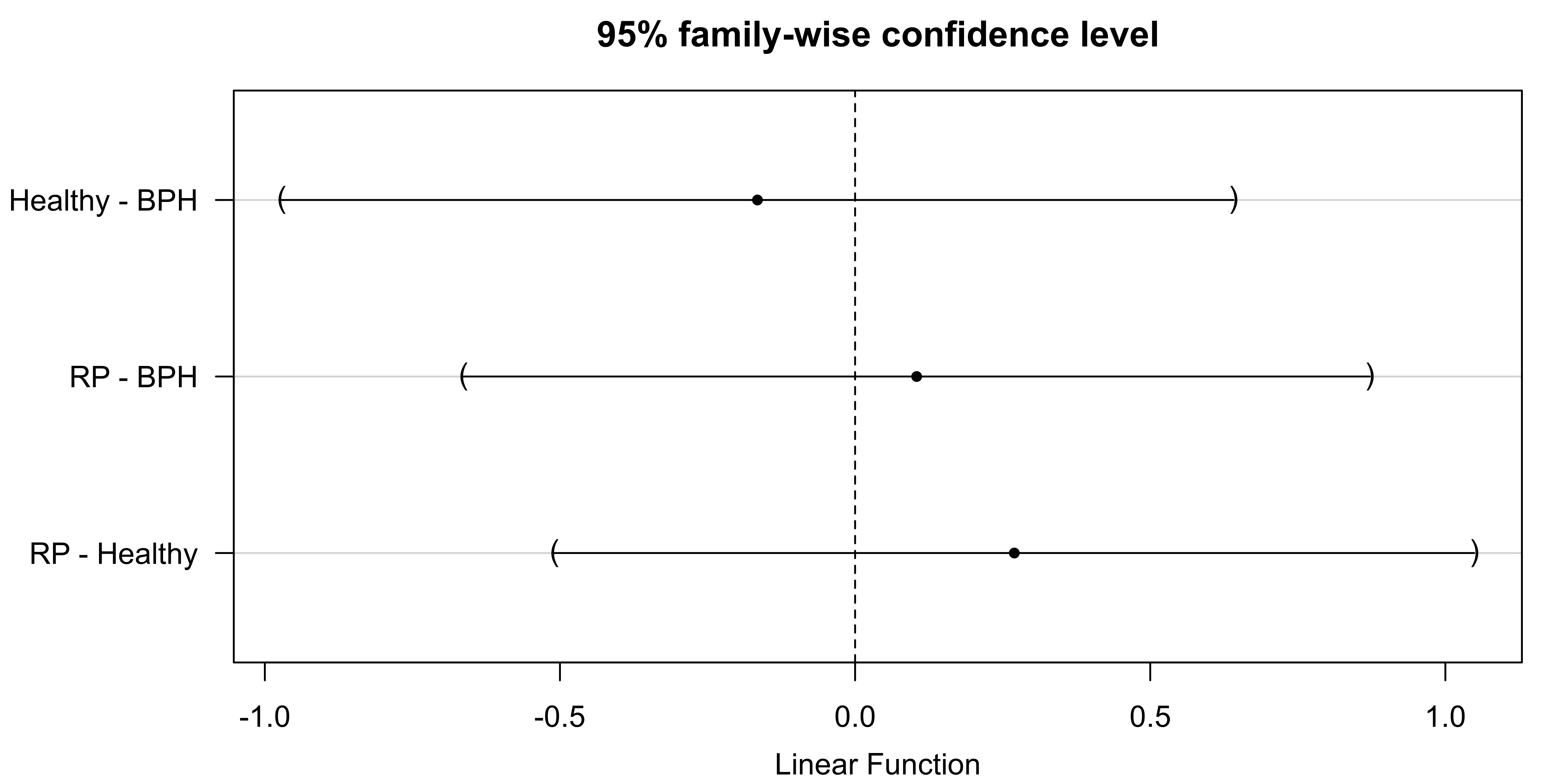
This line indicates that the D2/D1 ratio is not significantly different between the three groups. The results of the ANOVA test show that
ANOVA test results
Linear Hypotheses:
Estimate Std. Error
The aim of this study was to design a low cost, high sensitivity and specificity trial that can help in the early diagnosis of prostate cancer along with PSA and GS indices. Various studies around the world have been carried out to accomplish this. Among the studies designed to use serum proteins and gold nanoparticles to measure the size of gold nanoparticle changes using DLS, are studies by Zheng et al., Huo et al., and Wang [1, 4, 6, 7]. These trials, with a focus on prostate cancer, have yielded positive results. Proteins are one of the most important biological molecules in the body of living things that are associated with a wide range of diseases. But these same disease-related proteins have low levels when the disease is in its early stages, which makes diagnosis very difficult. Therefore, it is necessary to find methods for early and better detection of blood proteins that are inexpensive and accurate.
In 2012, Huo et al. used gold nanoparticles in a study of mouse prostate tissue as well as human prostate tissue using prostate tissue degrading enzyme (DLS) techniques. Given the inverse relationship between the average size of soluble nanoparticles and the GS (Gleason Score) tissue texture, gold nanoparticles potentially have a higher and more accurate diagnostic capability than other available indices [6].
In 2015, a clinical trial was conducted by Zheng et al. on the blood serum of healthy individuals and individuals with BPH and RP (Radical Prostate) in Florida hospitals, using the technique of DLS to serum immunoglobulins and measuring the change of micronutrient resonance at two sizes of 40 nm and 100 nm, indicating that this simple test could be 90% to 95% specificityand show 50% sensitivity between cancer patients and healthy individuals. This difference demonstrates the superiority of such a test over the PSA assay [1].
No similar studies have been conducted on male Iranians so far. Therefore, this study was designed similar to the above studies and was performed on male Iranians over 40 years old in 3 groups, healthy (without prostate cancer), patients with benign prostate enlargement or BPH, and those with prostate cancer, working from the fact that human serum contains high amounts of protein such as Albumin and Apolipoprotein [4].
Studies have shown that specific autoantibodies against tumor antibodies appear months before the clinical diagnosis of cancer, and that autoantibodies are found in various types of cancers. Autoantibodies are excellent biomarkers for early cancer diagnosis and clinical trials. The activity of the host defense system against the tumor occurs from the very early stages of tumor formation and development. At a later stage, the tumor may develop enough to be able to escape the immune system. This is a key point that indicates that there is a golden time to detect increased immune system activity in cancer patients. Immunoglobulin G (IgG) is an autoantibody that is elevated in the blood of people with early stages of prostate cancer. Immunoglobulin G has been found to be highest in healthy individuals or newly infected patients [1].
However, the results of this study were not consistent with any of the previous studies and no significant differences were observed between study groups. As reported in Table 1, the size changes of the nanoparticles were not significant among the 3 groups. This is contrary to what was proven in previous trials by the significant difference between healthy and benign prostate cancer groups [1].
It seems that what caused the size of the nanoparticles to differ significantly between the three groups was related to the ambient temperature changes that occurred during the preparation of gold nanoparticle solution and serum proteins. Previous research has shown that even slight temperature changes can affect the formation of corona proteins around nanoparticles [8]. The temperature variations may occur from preparation to transfer to the laboratory for DLS. Although prepared after sampling, they were frozen at 4
Of course, the use of the method of measuring light scattering from the surface of the nanoparticles is also a major limitation that limits its capacity for the early detection of diseases. The basis of the diagnosis in this method is the variation of the size of gold nanoparticles in the serum collected from each individual, and therefore it is possible that nonspecific substances can also influence this resizing, while having no association with the disease. When nanoparticles come in contact with biological fluids, proteins and other biological molecules are rapidly absorbed to their surface, forming a coating on the surface of the nanoparticles called the corona protein, which can severely affect the size of the nanoparticles as well as the interaction of the nanoparticles with other molecules.
A major disadvantage of DLS-based methods is the possibility of reacting with complex and heterogeneous sets of biomaterials, including undiluted serum containing a large set of proteins, or various raw cellular components present in the blood serum. In this method, a homogeneous set of nanoparticles is exposed to this bio-heterogeneous set.
It is also likely that changes in the structure of corona protein in samples of patients with prostate cancer did not allow a network of corona proteins to be formed, and saw significant changes in the D2 size of the corona protein in prostate cancer patients compared to the other two groups.
Another possible reason for the lack of significant differences between the healthy and benign groups with the serum of cancer patients is related to the timing and formation of soft corona protein and hard corona protein structures. Since the components of the soft corona protein remain attached to the nanoparticles for only a few minutes, and then replace the hard corona protein with high affinity binding proteins [4, 9], the problem may have arisen due to the long duration until the serum’s mixture and the gold nanoparticles (compared to when the DLS test was performed when the samples were taken, something about 24 hours (probable time interval), whereas in previous research work, the time was between 15 and 30 minutes [1].
It is also possible that due to the long duration of the interaction between proteins and nanoparticles, the aggregation of proteins around the nanoparticles and their binding to each other changed in the three groups, making the hard thickness of the corona within 24 hours identical in all three groups. On the other hand, previous research have shown that the interaction between synthetic nanoparticles and blood serum proteins can lead to the reactivation of the nanoparticles, which may have effects on the corona protein. The interaction itself may be weak, for example, and cannot have a significant effect on the proteins, whereas on the contrary, it is so potent that it can denature or deform or even break down proteins and form polypeptide structures that can interact with each other to alter and modify the structure of the corona protein, as well as appearing in the form of coarse particles in solutions.
The negative charges of citrate on gold nanoparticles can also be lost by interaction with the proteins [7]. Therefore, given the long interaction between proteins and citrated gold nanoparticles in the present experiment, negatively charged ions are separated from the surface of the nanoparticles. Assuming that the levels of immunoglobulin proteins in cancerous sera are higher than in healthy and benign individuals [1], the extensive and prolonged interactions in these sera lead to loss of citrate ions resulting in the accumulation and increase in size of corona protein in cancerous individuals within the 2 groups. The others were approximately identical and showed no significant difference.
Past research have shown that levels of immunog- lobins such as IgG increase in various diseases, even with the increase in other immunoglobulins, such as IgM, in rheumatoid arthritis [10] and in people as a group. Only those with common PSA criterion that was higher than one in all of their results, were considered healthy for prostate cancer; however they were not assessed for other diseases such as rheumatoid arthritis, heart disease or simple infectious diseases such as the common cold. This may justify the high probability of corona protein in healthy individuals.
Conclusion
This study is a simple and somewhat inexpensive test, and research has been reported in other parts of the world on people with prostate cancer, but this laboratory method cannot be used to predict or measure prostate cancer in Iran. Although this test seems simple, it does require consideration of various factors such as the quality of the nanoparticles or the nanoparticle’s material, keeping the ambient temperature constant from the time of serum solution preparation with gold nanoparticles until test time, and also quick access to the DLS device, due to the importance of the interval between the preparation of corona protein solution and the DLS test, which makes it difficult and sensitive to perform. More studies should be performed on the number of male Iranians with prostate cancer and benign malignancies and healthy male individuals over 40 years of age considering the mentioned criteria.
Footnotes
Acknowledgments
This work has been supported by the Center for International Scientific Studies & Collaboration (CISSC) under Contract No. 1843 dated 12/10/1996.
Conflict of interest
There is no conflict of interest among the authors of this article.