LINK-A (long intergenic non-coding RNA for kinase activation) is a newly identified long non-coding RNA with oncogenic function, which leads to the hyperactivation of AKT and HIF1. thereby, fosters cell proliferation, mobility and metastasis. VEGF (vascular endothelial growth factor), a well-known cytokine has an important role in angiogenesis. In this study, we quantified RNA expression of LINK-A and VEGF on 45 tumor specimens obtained from Iranian patients diagnosed with Epithelial Ovarian Cancer (EOC). Our goal was to evaluate expression of LINK-A lncRNA and VEGF mRNA in ovarian cancer tissues and find the probable correlation of LINK-A with VEGF as a major transcription target for HIF1. LINK-A and VEGF were remarkably overexpressed in EOC tissues compared to normal tissues ( value: 0.004, 0.0001, respectively), but we did not find correlation between LINK-A and VEGF RNA expressions in this study. LINK-A was significantly overexpressed in higher stages of cancer and tumor grades. VEGF was only significantly elevated in higher stages. This study confirms importance of novel lncRNA of LINK-A in Iranian EOC patients.
Cancer is the most medical concerning issue that causes mortality in the world. Ovarian cancer is the third gynecologic most common cancer after cervical and uterine cancer [1] and ranks fifth in cancer deaths among women. In comparison with breast cancer, it has lower prevalence. However, the mortality rate is three times more and it has the worst prognosis [2]. It is anticipated by GLOBOCAN that the death rate of ovarian cancer will climb significantly by the year 2040 [1].
One of the most important reasons for the poor survival of ovarian cancer patients is the lack of specific signs and symptoms that causes late diagnosis in advanced stages of disease. Finding biomolecular markers could be cooperative to early diagnosis and raise the survival rate of this cancer. Regarding this matter, clinical and experimental studies on identification of biomolecules involved in pathogenesis and metastasis of this cancer are indispensable and may improve the treatment of this disease [3].
Long noncoding RNAs (lncRNAs) are RNA transcripts longer than 200 nucleotides, which has limited coding potential [4]. They are implicated in a variety of biological process by regulating gene expression in cis or trans modes by various transcriptional and post-transcriptional mechanisms through involving in histone modification, chromatin rearrangement, alternative splicing, dosage compensation and probably many other functions awaiting for discovery [5].
The significant role of lncRNAs in cancer has been reported in numerous studies recently, dysregulation of their expression impinge on tumor cell proliferation, metastasis, and invasion [6, 7]. We examined the expression of LINK-A (long intergenic noncoding RNA for kinase activation) a newly discovered lncRNA in this study. LINK-A is an oncogenic cytoplasmic noncoding RNA that interacts with nonreceptor tyrosine kinases to facilitate their activation and leads to hyperactivation of normoxic HIF1 in triple-negative breast cancer [8], in another pathway, LINK-A is correlated with hyperactivation of AKT and makes resistance to inhibitors against AKT [9].
VEGF (vascular endothelial growth factor), the first identified and most effective angiogenic molecule, is a multifunctional cytokine that leads to the progression of angiogenesis by promoting cell permeability, cell division, and mobility of vascular endothelial cells [10]. Elevated expression of VEGF has been observed in various solid tumors such as ovarian cancer and is associated with poor prognosis in most cancers [11, 12, 13]. Several factors stimulate VEGF expression, HIF1 plays a crucial role in cancer and has a fundamental function in the transcription of VEGF. HIF1 and VEGF correlation has been reported in cancer by several studies. It has been shown that HIF1 is the regulatory mediator between tumor hypoxia and VEGF production in pancreatic cancer [14]. Regulation of VEGF expression influenced by HIF1 in EOCr (EOC) was suggested by Wong et al. in 2003 [15]. In cell culture models, Liu et al. indicated elevated expression of VEGF in mRNA and protein levels. and also, showed VEGF mRNA stability by HIF1 [16], similar results was reported by Forsythe et al. in Hep3B cells by overexpression of HIF1 [17]. Zhu and Zhang informed increased expression of VEGF-A and HIF1 in 76 cancerous lung tissues and the decline of VEGF-A expression after silencing of HIF1 by anti-HIF1 siRNA. They proposed that inhibition of the HIF1/VEGF pathway can reverse the outcomes of disease and resistance to radiotherapy [18]. By considering LINK-A role on hyperactivation and stabilization of HIF1 in triple-negative breast cancer tissues either cell lines besides the importance of VEGF as a main factor in angiogenesis regulated by HIF1, we aimed to examine RNA expression of LINK-A and VEGF in EOC tissue specimens obtained from Iranian patients to figure out LINK-A and VEGF probable correlation and examine their association with clinicopathological features.
Methods and material
Patients
This study includes 53 tissue samples obtained from patients diagnosed with epithelial ovarian carcinoma and with benign ovarian cyst. Samples were collected from surgical specimens at imam Khomeini hospitals (Tehran, Iran). The patient’s ages were between 19 to 71 years with a mean age of 47 years. Tissues were snap frozen in liquid nitrogen after surgery and transported to the pathology laboratory and stored at 80 for confirming diagnosis and histological grading by pathologists. Written informed consent was collected from all patients.
RNA extraction and cDNA synthesis
Total RNA extracted from tissue samples by TRIzol reagent (Ambion, Carlsbad, CA, USA) according to the manufacturer’s instruction. Concentrations of extracted RNAs were determined by the NanoDrop™ 2000 Spectrophotometer (Thermo Fisher Scientific, USA). cDNA was synthesized via 2 g total RNA for each sample, using random hexamer and oligo dT primers by RevertAid First Strand cDNA Synthesis Kit (Thermo Fisher Scientific, Inc), then quantities of LINK-A and VEGF expression for each sample were evaluated relative to -actin. BioFACT™ Real-Time PCR Master Mix (for SYBR Green I) was used to preparation of reactions and the Real time PCR was conducted in MIC System thermo cycler.
Statistical analysis
The expression levels of LINK-A and VEGF RNAs compared between EOC and normal specimens by independent-samples t-tests. Correlation between expression levels and clinicopathological features were evaluated by one-way ANOVA. All variables were analyzed using GraphPad Prism 8.0.2 software. The value 0.05 was considered significant.
Sequence of the primers
GENE
Strand
Primer sequence ()
Primer length
Product length
LINK-A
F
GTGGCCATGGGACAGACAAGGAC
23
181 bp
R
GCATCAGTCAGAGGCCCATGTAAGG
25
VEGF
F
AACTTTCTGCTGTCTTGGGTG
21
179 bp
R
ATGTCCACCAGGGTCTCGATT
21
B-actin
F
AGACGCAGGATGGCATGGG
19
161 bp
R
GAGACCTTCAACACCCCAGCC
21
Clinical data of patients
Clinical factors
Number of cases (%)
LINK-A
VEGF
value LINK-A
value VEGF
Age (Mean SD) (range)
46.84 13.287 (19–71)
50
25 (44.4%)
5.86 2.03
0.72 1.85
0.9
0.8
50
20 (55.6%)
5.73 2.07
0.54 2.28
Cancer stages (%)
Stage I
15 (34.1)
5.83 2.12
0.48 2.17
0.007
0.01
Stage II
10 (22.7)
7.36 1.16
2.20 1.97
Stage III
15 (34.1)
4.63 1.97
0.1 1.59
Stage IV
4 (9.1)
6.37 1.32
1.63 1.24
Tumor grades (%)
I
7 (15.9)
4.42 1.63
0.69 1.76
0.02
0.87
II
7 (15.9)
5.72 2.27
0.11 1.63
III
9 (20.5)
4.91 1.74
0.09 1.70
IV
3 (6.8)
8.23 1.59
0.86 2.71
V
15 (34.1)
6.63 1.87
1.12 2.58
GB (borderline malignancy)
3 (6.8)
5.76 2.03
0.67 2.04
Tumor size (%)
15 mm
25 (55.7)
5.52 1.91
0.50 1.81
0.3
0.4
15 mm
20 (42.3)
6.16 2.17
0.81 2.32
Histology types (%)
Serous
21 (47.7)
5.45 1.89
0.47 1.61
0.7
0.3
Mucious
3 (6.8)
5.64 2.65
0.42 3.9
Endometrioid
7 (15.9)
6.16 2.12
2.04 2.02
Clear cell
6 (13.6)
6.58 2.80
0.33 2.04
Small cell carcinoma
2 (4.5)
7.11 3.16
1.32 3.72
Poorly differentiated
2 (4.5)
5.58 0.99
0.73 1.44
LMP2
3 (6.8)
4.72 0.80
1.30 1.44
Results
Sample characteristics
Fifty-three specimens included in this study, 45 tumor samples were obtained from patients with EOC and 8 normal ovarian tissue samples were got from patients, which had been enduring surgery because of cysts and other benign disease. Malignant tissues comprise stages I/II/III/IV with various histological types (21 serous, 7 endometrioid, 6 clear cell, 2 mucinous, 1 adenocarcinoma, 4 LMP2, 2 poorly differentiated carcinoma, 2 small cell carcinoma) and 8 normal tissue samples. Age range of patients at the time of diagnosis were 19–71 years old with the median ( SD) of 46.87 ( 13.28) years. Table 2 shows the clinical information of patients.
Relative RNA expression assay of LINK-A long non coding RNA and VEGF mRNA
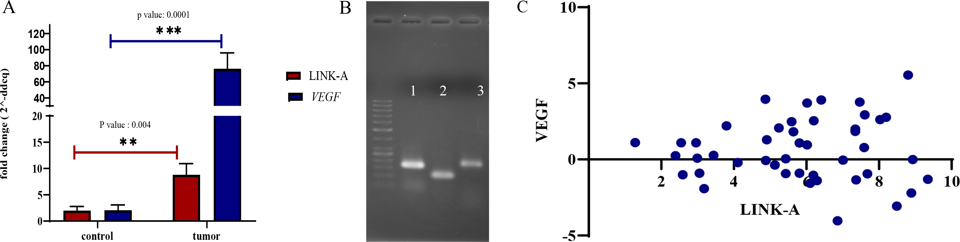
The expression levels of lncRNA of LINK-A and VEGF mRNA in collected specimens were evaluated by Real-Time PCR. Our results indicated remarkable significant upregulation in tumor samples for LINK-A and VEGF RNAs irrespective clinical features of tumors ( value: 0.004, and 0.0001, respectively). LINK-A as a recently introduced oncogenic long non coding RNA and VEGF as a well-known hall mark in cancer showed higher expression in EOC specimens (Fig. 1). For determining significant differences of expression fold changes between tumor and normal groups, we conducted the non-parametric Mann-Whitney test, as the expression fold changes did not have normal distribution.
Plot of relative quantitative expression of LINK-A lncRNA and VEGF mRNA in tumor tissues of EOC patients and normal tissues (A). electrophoresis gel of Real-time PCR products,lane 1,2,3 respectively represents BETA-ACTIN,LINK-A, VEGF PCR products (B). Graph of Pearson correlation test beween expression of LINK-A and VEGF expressions, for each sample used, which had normal distribution (C).
Correlation between LINK-A and VEGF expressions
There was no correlation between expression of LINK-A and VEGF in both normal and tumor tissues. To determine whether there is a correlation between LINK-A and VEGF expressions, we conducted Pearson correlation coefficients analysis between quantitative expression of samples ( value: 0.7, and 0.2, respectively for normal and tumor tissues).
Association between quantitative expression of LINK-A and VEGF RNAs and clinical factors
We performed ANOVA analysis to evaluate association between LINK-A and VEGF RNA expressions and stages of cancer in each tissue specimens. According to the ANOVA test, there is a significant association between LINK-A expression and stages of cancer, which Tukey comparison indicated that this association is between stages II and III ( value: 0.007). ANOVA test revealed a significant association ( value: 0.01) between VEGF expression and stages. This significance was obtained between stages I and II as well as I and IV. Since number of specimens in some groups was small we categorized stages of tumors in two separate groups, as low and high stages, in this condition we compared high and low stages for link-a and VEGF expression by student’s t-test individually. LINK-A expression was significantly elevated in high stages related to low stages ( value: 0.01), and there was no significant differences between VEGF expression and stages of disease ( value: 0.4). We found out significant differences between expression of LINK-A and tumor grades by ANOVA analysis ( value: 0.02). Comparison between high and low grades by student t-test analysis revealed significant difference for expression of LINK-A ( value: 0.001). LINK-A and VEGF expression were independent of tumor size ( value: 0.3, 0.4, respectively), histological types ( value: 0.7, 0.3), and age of patients ( value: 0.9, 0.8, respectively).
Discussion
We observed significant overexpression of LINK-A and VEGF RNAs in EOC tumor tissues grafted from Iranian patients in this study. LINK-A is a recently identified lncRNA in triple-negative breast cancer with oncogenic activity through two imperative molecular pathways. LINK-A is a cytoplasmic lncRNA located near to the cell membrane and provides a scaffold like structure, which protein kinases like BRK and LRRK2 bond to that and place in a conformation, which lead to a more accessible structure by other regulatory proteins and finally facilitates kinase activation of BRK and LRRK2, all these events cause HIF1 phosphorylation and stabilization in normoxic condition [8]. Also, LINK-A directly interacts with AKT and PIP3, facilitates AKT-PIP3 interaction and subsequent enzymatic activation causes to tumorigenesis and resistance to AKT inhibitors, which are in clinical trials [9]. After these first and stunning discoveries about LINK-A by Lin et al., other researchers reported importance of LINK-A in ovarian cancer and glioma cells. We also have obtained some data indicating oncogenic function of LINK-A in a549 cell line (representing non-small cell lung cancer) by RNA interference system (not published yet). Up-regulation of LINK-A has been reported in glioma cells by Wu et al., they proved that silencing of LINK-A inhibit cell proliferation, migration and invasion in U87 and U251 glioma cells and LINK-A influence glycolysis and proliferation by regulating lactate dehydrogenase A (LDHA) [19]. According to the findings of Ma and Xu in ovarian cancer patients, LINK-A levels in plasma is positively correlated with distant tumor metastasis. They found that overexpression of LINK-A led to the increased cell migration and invasion by up-regulation of TGF-1 in ovarian carcinoma cells [20]. In another study performed by Zhang et al. significant higher expression of LINK-A in biopsies and serums of ovarian carcinoma patients has been reported. Their results indicated that overexpression of LINK-A is correlated with promotion of cell migration and invasion, Role of LINK-A in metastasis of ovarian cancer is probably by up-regulation of HIF1 [21]. Our results on Iranian EOC patients confirmed two recent findings about higher LINK-A expression in ovarian cancer patients. By considering molecular function of LINK-A, which finally leads to the stabilization and up-regulation of HIF1 and perceiving the importance of VEGF in multiple cancers besides being under regulated with HIF1 [22] encouraged us to examine LINK-A lncRNA and VEGF expression in ovarian cancer patients. There are some evidences claiming that HIF1 is a major link between tumor hypoxia and VEGF production [14, 17, 18, 23, 24]. Furthermore, as explained before, most types of cancers show elevated expression of HIF1 even in normoxic condition [8, 21, 25]. On the other hand, HIF1 is not the only inducer of HIF1, the AKT/PIP3 pathway, which is usually activated in most types of cancers, mediates activation of VEGF expression through SP1in hypoxia independent of HIF1 [26]. As explained before, LINK-A is involved in hyperactivation of AKT, therefore LINK-A could be effective in induction of VEGF expression through two critical separate pathways. Correlation of VEGF overexpression with advanced tumor stages of ovarian cancer has been reported in some studies [27, 28], although, there are some other reports that did not find significant association between VEGF expression and clinicopathological factors [12, 29, 30]. In this study, we observed significantly higher expression of VEGF in tumor tissues compared to control group. There were significant differences between stages of disease and VEGF expression. However, it was not so strong ( value: 0.01), as we compared differences of expressions between two groups of high and low stages it was not significant anymore. Duncan et al. conducted a study on 339 primary ovarian cancer tissues, which revealed that only a small proportion of patients (7%) had high expression of VEGF, and the same group had significantly poorer survival. They did not find correlation between VEGF expression and clinical factors [30]. Ranjbar et al. found significant increase in expression of VEGF compared with healthy controls in a study on Iranian ovarian cancer patients, but did not recognize correlation between expression of VEGF and clinicopathological factors [31]. Our findings about situation of VEGF expression in ovarian cancer was consistent with recent studies, as aforementioned VEGF was elevated in EOC tumor tissues and except from stages of cancer was independent from other clinicopathological factors. In this study the main aim was to find a probable correlation between LINK-A and VEGF expression in EOC tumor samples. Probable influence of LINK-A on VEGF expression seems to be upstream of transcription system by HIF1 and AKT hyperactivation as explained during this paper. However, our results indicated that LINK-A and VEGF expression was independent from each other. We suggest that correlation between LINK-A and VEGF expression investigated profoundly by larger tumor samples or by precise molecular experiments. Our data revealed significant association between LINK-A expression and stages of cancer and tumor grades. VEGF expression was only related to stages of cancer. This study is the first time that examine LINK-A lncRNA expression in Iranian ovarian cancer patients and its correlation with VEGF expression. Our findings about LINK-A overexpression in EOC tumor tissues were consistent with the last studies. We did not find a correlation between VEGF and LINK-A expression in this study. Multiple factors could be effective in regulation of VEGF expression besides pathways with LINK-A, So deeper studies would be helpful to discover or deny this probable correlation precisely.
Conclusion
Data obtained from this study indicate the elevated expression of LINK-A lncRNA and VEGF in EOC specimens. LINK-A expression in higher stages and tumor grades highlights its importance in ovarian cancer in Iranian patients, which is consistent with two recent studies. Our results do not indicate a correlation between LINK-A and VEGF expression.
Footnotes
Acknowledgments
This work was supported by a research (grant 732) from National Institute of Genetic Engineering and Biotechnology of Iran.
References
1.
BrayF. et al., Global cancer statistics 2018: GLOBOCAN estimates of incidence and mortality worldwide for 36 cancers in 185 countries, CA Cancer J Clin68 (2018), 394–424.
2.
YonedaA.LendorfM.E.CouchmanJ.R. and MulthauptH.A., Breast and ovarian cancers: A survey and possible roles for the cell surface heparan sulfate proteoglycans, J Histochem Cytochem60 (2012), 9–21.
3.
ChienJ. et al., Analysis of gene expression in stage I serous tumors identifies critical pathways altered in ovarian cancer, Gynecol Oncol114 (2009), 3–11.
4.
Durruthy-DurruthyJ. et al., The primate-specific noncoding RNA HPAT5 regulates pluripotency during human preimplantation development and nuclear reprogramming, Nat Genet48 (2016), 44–52.
5.
YanP.LuoS.LuJ.Y. and ShenX., Cis- and trans-acting lncRNAs in pluripotency and reprogramming, Curr Opin Genet Dev46 (2017), 170–178.
6.
GibbE.A.BrownC.J. and LamW.L., The functional role of long non-coding RNA in human carcinomas, Mol Cancer10 (2011), 38.
7.
TsaiM.C.SpitaleR.C. and ChangH.Y., Long intergenic noncoding RNAs: New links in cancer progression, Cancer Res71 (2011), 3–7.
8.
LinA. et al., The LINK-A lncRNA activates normoxic HIF1α signalling in triple-negative breast cancer, Nature Cell Biology18 (2016), 213
9.
LinA. et al., The LINK-A lncRNA interacts with PtdIns (3,4,5) P3 to hyperactivate AKT and confer resistance to AKT inhibitors, Nat Cell Biol19 (2017), 238–251.
10.
PangR.W. and PoonR.T., From molecular biology to targeted therapies for hepatocellular carcinoma: The future is now, Oncology72 (2007), 30–44.
11.
YamamotoS. et al., Expression of vascular endothelial growth factor (VEGF) in epithelial ovarian neoplasms: Correlation with clinicopathology and patient survival, and analysis of serum VEGF levels, British Journal of Cancer76 (1997), 1221–1227.
12.
YuL.DengL.LiJ.ZhangY. and HuL., The prognostic value of vascular endothelial growth factor in ovarian cancer: A systematic review and meta-analysis, Gynecologic Oncology128 (2013), 391–396.
13.
KooP.J.MorgenszternD.BoyerJ.L. and HerbstR.S., Targeting vascular endothelial growth factor in patients with squamous cell lung cancer, Journal of Clinical Oncology30 (2012), 1137–1139.
14.
BüchlerP. et al., Hypoxia-inducible factor 1 regulates vascular endothelial growth factor expression in human pancreatic cancer, Pancreas26 (2003), 56–64.
15.
WongC.WellmanT.L. and LounsburyK.M., VEGF and HIF-1α expression are increased in advanced stages of EOC, Gynecologic Oncology91 (2003), 513–517.
16.
LiuL.X. et al., Stabilization of vascular endothelial growth factor mRNA by hypoxia-inducible factor 1, Biochemical and Biophysical Research Communications291 (2002), 908–914.
17.
ForsytheJ.A. et al., Activation of vascular endothelial growth factor gene transcription by hypoxia-inducible factor 1, Molecular and Cellular Biology16 (1996), 4604–4613.
18.
ZhuH. and ZhangS., Hypoxia inducible factor-1α/vascular endothelial growth factor signaling activation correlates with response to radiotherapy and its inhibition reduces hypoxia-induced angiogenesis in lung cancer, Journal of Cellular Biochemistry119 (2018), 7707–7718.
19.
WuD.ZhaoB.CaoX. and WanJ., Long non-coding RNA LINK-A promotes glioma cell growth and invasion via lactate dehydrogenase A, Oncology Reports38 (2017), 1525–1532.
20.
MaJ. and XueM., LINK-A lncRNA promotes migration and invasion of ovarian carcinoma cells by activating TGF-β pathway, Bioscience Reports38 (2018).
21.
ZhangH.YaoB.TangS. and ChenY., LINK-A long non-coding RNA (lncRNA) participates in metastasis of ovarian carcinoma and upregulates hypoxia-inducible factor 1 (HIF1α), Medical Science Monitor: International Medical Journal of Experimental And Clinical Research25 (2019), 2221.
22.
MayerhoferM.ValentP.SperrW.R.GriffinJ.D. and SillaberC., BCR/ABL induces expression of vascular endothelial growth factor and its transcriptional activator, hypoxia inducible factor-1α, through a pathway involving phosphoinositide 3-kinase and the mammalian target of rapamycin, Blood, The Journal of the American Society of Hematology100 (2002), 3767–3775.
23.
MaxwellP. et al., Hypoxia-inducible factor-1 modulates gene expression in solid tumors and influences both angiogenesisand tumor growth, Proceedings of the National Academy of Sciences94 (1997), 8104–8109.
24.
CarmelietP. et al., Role of HIF-1α in hypoxia-mediated apoptosis, cell proliferation and tumour angiogenesis, Nature394 (1998), 485–490.
25.
KuschelA.SimonP. and TugS., Functional regulation of HIF-1α under normoxia – is there more than post-translational regulation? Journal of Cellular Physiology227 (2012), 514–524.
26.
ChoiS.B.ParkJ.B.SongT.-J. and ChoiS.Y., Molecular mechanism of HIF-1-independent VEGF expression in a hepatocellular carcinoma cell line, International Journal of Molecular Medicine28 (2011), 449–454.
27.
RaspolliniM. et al., Prognostic significance of microvessel density and vascular endothelial growth factor expression in advanced ovarian serous carcinoma, International Journal of Gynecologic Cancer14 (2004), 815–823.
28.
BrustmannH.NaudéS., Vascular endothelial growth factor expression in serous ovarian carcinoma: Relationship with high mitotic activity and high FIGO stage, Gynecologic Oncology84 (2002), 47–52.
29.
SönmezerM.GüngörM.EnsariA. and OrtacF., Prognostic significance of tumor angiogenesis in EOC: In association with transforming growth factor β and vascular endothelial growth factor, International Journal of Gynecologic Cancer14 (2004), 82–88.
30.
DuncanT.J. et al., Vascular endothelial growth factor expression in ovarian cancer: A model for targeted use of novel therapies? Clinical Cancer Research14 (2008), 3030–3035.
31.
RanjbarR.NejatollahiF.AhmadiA.S.N.HafeziH. and SafaieA., Expression of vascular endothelial growth factor (VEGF) and epidermal growth factor receptor (EGFR) in patients with serous ovarian carcinoma and their clinical significance, Iranian Journal of Cancerprevention8 (2015).