
Abstract
BACKGROUND:
Considerable evidence supports that SLE could be related to apoptotic cells and EBV infection.
OBJECTIVE:
The aim of this study was to identify the transcriptional signature of EBV infection in SLE patients for survey of the molecular apoptosis signaling pathways.
METHODS:
The PBMCs gene expression profiles of healthy control and SLE patients were obtained from GEO. Functional annotation and signaling pathway enrichment were carried out using DAVID, KEGG. To validate bioinformatics analysis the changes in genes expression of some of obtained genes, Real time PCR was performed on PBMCs from 28 SLE patients and 18 controls.
RESULTS:
We found that mean viral load was 6013
CONCLUSIONS:
The study indicated that some cellular genes may have an important role in pathogenesis of SLE through apoptosis signaling pathways. Beside, EBV infection as an environmental risk factor for SLE may affect the dysfunction of apoptosis.
Keywords
Introduction
Apoptosis is an important physiological process in development and disease. Apoptotic cells are a major source of self-antigens, but usually evade immune responses [1]. Clearing apoptotic cells inefficiently and accumulating apoptotic cell debris provoke a chronic inflammatory response, which may lead to the breakdown of self-tolerance [2].
Systemic lupus erythematosus is considered as a chronic autoimmune connective tissue disease and one of the most common forms of heterogeneous autoimmunity. Autoimmunity in systemic lupus erythematosus (SLE) is believed to be driven by autoantigens [3]. Considerable evidence supports the notion that SLE autoimmunity could be related to impaired or delayed clearance of apoptotic cells. Although the precise causes of SLE are unknown, increased apoptosis rate, defect in cleaning the products of apoptosis, and high activity of B lymphocytes have been often related to a higher incidence of SLE [4, 5]. Recognizing autoantigens by Toll like receptor (TLR), as well as nucleic acid sensors in the antigen presenting cells (APC) results in creating the constitutive activation of type I interferons and changes in immune cell signaling [6, 7]. Some believe that genetic and environmental factors determine the risk of initiation and progression to SLE. The Epstein-Barr virus (EBV) is regarded among the environmental factors which can contribute to SLE development and progression since seroconversion and viremia are reported in these patients [8, 9].
An increase of 5–40 times in EBV blood load was demonstrated in SLE patients compared with control group which seems to be independent of treatment with immunosuppressive agents [10, 11]. Apoptosis and clearance of apoptotic cells are tightly regulated processes, the dysfunction of which is considered in the etiology of SLE. Lack of control in apoptosis pathways results in clearing exogenous agent like EBV infection defectively [12, 13]. EBV latency gene products are involved in viral persistence and oncogenesis through interfering with multiple cellular processes such as apoptosis pathways [14]. Latent membrane proteins 1 (LMP1) and 2A (LMP2A) are expressed in many EBV-associated malignancies and play essential roles in maintaining latent infection, EBV-induced B-cell transformation and apoptosis signaling pathway [15, 16]. In addition, some genetic factors induce abnormality in apoptosis pathways [17, 18].
The dual-specificity phosphatase 1 (DUSP1) known as MAPK Phosphatase-1 can inhibit MAPK pathway, which plays an important role in the apoptotic pathway, which is mediated through inhibiting the MAPK pathway in the process of growth and cell death. Some studies have been conducted on the role of dual-specificity phosphatase (DUSP) family in cell signaling pathways and SLE pathogenesis [19, 20, 21]. The lysosome-associated membrane glycoprotein 3 (LAMP3), which was reported to be highly expressed in SLE patients, is thought to play role in regulating cell viability and apoptosis [22, 23]. The leukocyte tyrosine kinase (LTK) and neuropilin-1 (NRP1) genes are known for their role in immunological disorders through different immune cell subsets and regulation of various molecular pathways [24, 25].
To the best of our knowledge, few studies have focused on regulating the apoptosis signaling pathways in SLE patients with EBV infection. The present study aimed to use available high-throughput data in SLE patients in order to identify the genes involved in apoptosis signaling pathwaysand the relationship between SLE and EBV infection through their role in regulating the apoptosis signaling pathways.
Materials and methods
In silico study
First, the Peripheral blood mononuclear cells (PBMCs) gene expression profile of control group and SLE patients was obtained from GEO database. The dataset code GSE50772 with the GPL570 platform code performed by Affymetrix Gene Chip Human Genome U133 plus 2.0 [HG-U133_Plus_2] which contains the gene expression data of the PBMCs samples including 61 SLE patients and 20 control samples was conducted at the University of Michiganto confirm the presence of the genes involved in interferon and other genes. Then, the Deferential Expressed Genes (DEGs) were extracted using GEO2R tool. In addition, the STRING and GeneMania biological network databases were used to evaluate the interactions and communications of these genes together with other genes and pathways involved in apoptosis. Further, the interaction network of the gene list was analyzed using Cytoscape software. In the next step, the network was analyzed and clustered to select the genes which are separate clusters. Finally, primer designing tools, Primer-Blast program, and Oligoanalyzer software were implemented for selecting primers in both selected and controlled genes.
Subjects
The samples included the patients diagnosed with SLE based on American Rheumatology Association criteria and referred to the rheumatology clinic and Autoimmune Diseases Research Center of Kashan Shahid Beheshti Hospital for regular follow-up treatment. In addition, the control group was recruited from the same hospital without having any lupus. However, the SLE patients with other simultaneous rheumatologic, active infectious diseases, and other viral infections such as HBV, HCV, and HIV were excluded Considering the results of our previous study indicating that the amount of infection with EBV in SLE patients was high, 18 control and 28 systemic lupus patients who had EBV viral load (VL) were examined in the present study [10]. Since the proportion of the SLE is nine times more common in women than that of men, just the females aged 13–63 years were selected.
PBMCs preparation
PBMCs were immediately isolated using Ficoll-Paque
DNA extraction and EBV viral load quantitation
First, high Pure Viral Nucleic acid kit (Roche, Germany) was used for extracting DNA from PBMCs. Then, the concentration of the extracted DNA was determined by assessing absorbance at 260 nm. In addition, the purified DNA was washed with 50
RNA extraction and quantitative real-time PCR
The RNA from 200
List of primer sequences used for RT-PCR analysis
List of primer sequences used for RT-PCR analysis
F: Forward primer. R: reverse primer.
The 2
Results
Demographic and clinical characteristics of SLE patients with EBV viral load
Table 2 shows demographic and clinical characteristics of 28 SLE patients with EBV viral load who followed up in the rheumatology clinic of Shahid Beheshti Hospital, Kashan. All patients were women with a mean
Clinical characteristics of SLE patients with EBV viral load
Clinical characteristics of SLE patients with EBV viral load
Note: SD Standard Deviation, ANA Anti-Nuclear Antibody.
The PBMCs count was not significantly different between SLE patients and control group, a higher PBMCs count was reported among SLE patients (914500
Comparison of the PBMCs count between SLE patients and control group.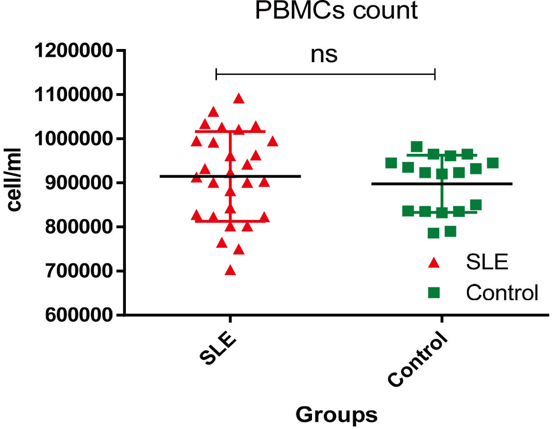
Comparison of the of EBV viral load in SLE patients and control group.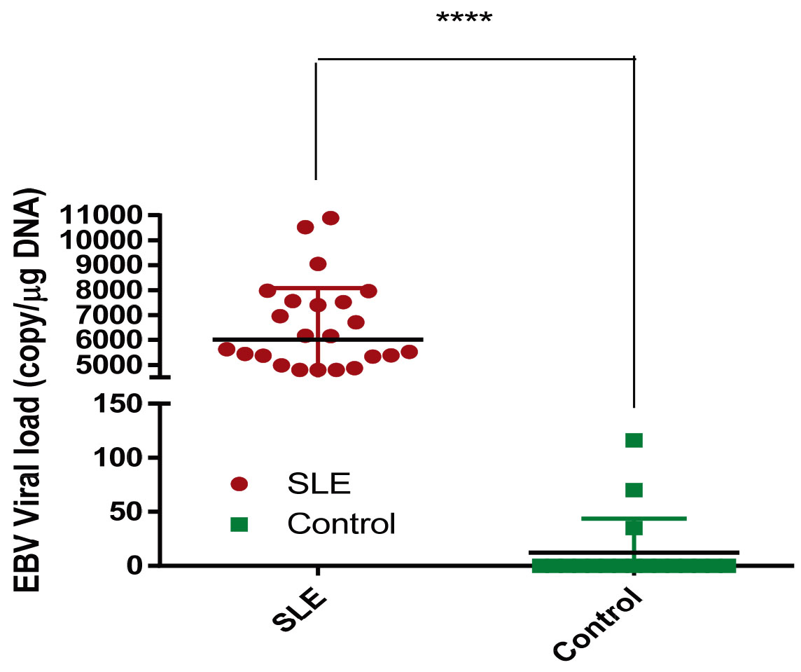
Based on the analysis of interaction network in the selected effective genes in SLE, DUSP1, LAMP3, LTK, and NRP1 were clustered separately. The DUSP1 and LAMP3 were overexpressed while LTK and NRP1 were under-expressed in microarray data analysis. Figure 3 displays the interaction network of effective genes in the SLE.
The parts of interaction network of effective genes in the SLE. Candidate genes are shown in square boxes.
Based on the results, elevated expression of the DUSP1 and LAMP3 genes in PBMCs samples of SLE patients with EBV viral load was higher compared to the control group. While the mRNA levels of LTK was significantly lower in the patients than control group, no significant difference was found in the mRNA transcriptional levels between SLE patients with EBV viral load in expressing the NRP1 gene compared to the control group (Table 3).
In-silico selected biomarker validation in SLE patients with EBV viral load
In-silico selected biomarker validation in SLE patients with EBV viral load
Note: Quantification expressed as mean change fold
The sensitivity and specificity of DUSP1 (75% vs. 83.33), LAMP3 (92.86% vs. 83.33%), LTK (53.57% vs. 94.44%) and LMP2A (96.43% vs. 55.56%) were statistically significant. In contrast, the sensitivity and specificity of NRP1 (82.14% vs. 44.44) was not significant (Fig. 4A–J). Based on the results, DUSP1, LAMP3, LTK genes, and viral LMP2A gene can be considered as candidate genes as biomarkers in SLE patients with EBV viral load
Determining biomarker role using dct and Roc curve analysis in candidate genes. qRT-PCR quantification of candidate genes (dct) between control and SLE patients with EBV viral load for (A) DUSP1, (C) LAMP3, (E) LTK, (G) NRP1 and (I) LMP2A are shown. Student’s 
Continued.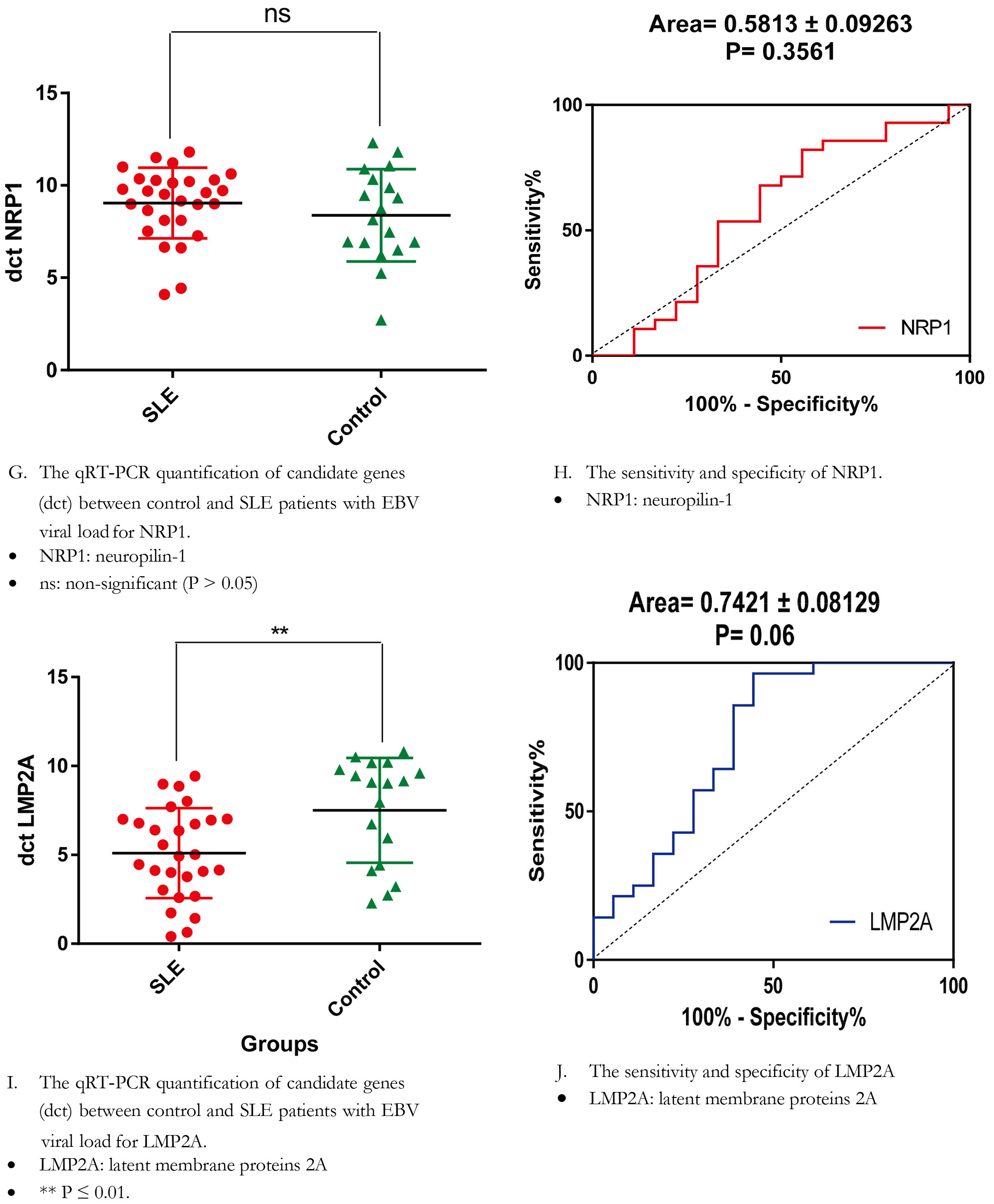
Increased apoptosis, defects in apoptotic clearance, and high activity of B lymphocytes may be considered as the underlying reasons although the exact cause of SLE is unknown [27]. In addition, EBV as an environmental factor is important in the etiology of SLE. In fact, the virus produces some proteins which interfere in various phases of the apoptosis process. Therefore, the number of viral and host factors plays an important role in developing the SLE and associated malignancies [28]. Systems biology approaches depend on high-throughput techniques with data analysis platforms which can assess proteins, metabolites, genes, and network analysis of complex biologic or pathways influenced during specific autoimmune diseases. Better-coordinated multi-omics approaches and standardized translational research, along with the skills related to clinicians, engineers, biologists, and bioinformaticians are required to contribute to the discovery of validated and qualified biomarkers [29, 30]. In the present study, regarding the network and pathway analysis, four differentially-expressed genes including DUSP1 and LAMP3 as two highly-expressed genes and LTK and NRP1 as two low-expressed genes were selected for evaluating the results among Iranian SLE patients with EBV infection. The LMP2A gene was selected to study the EBV latent genes in SLE patients since the persistent expression of LMP2A may be considered as an important risk factor in EBV-associated diseases and apoptosis signaling pathways.
DUSP1is known as MAPK Phosphatase-1 and MAPK pathway inhibitor. Involvement in the apoptotic pathway is considered as one of the important roles of this gene, which is mediated through the inhibitory role of the MAPK pathway in the process of growth and cell death [20]. The present study showed an increase of 14.72-fold DUSP1 gene expression in SLE patients with EBV virus compared to the control group, which is consistent with the previous studies indicating that DUSP family can be a diagnostic biomarker in the SLE [31, 32]. Chuang et al. reported that the reduced expression of DUSP22 in human T cells is an important biomarker in diagnosing lupus erythematosus nephritis [33]. The result of another study showed that DUSP23 as another protein of this family had an increased expression in TCD4
The LAMP3 gene is located in the lysosome, which is involved in the process of antigen presentation in dendritic cells through transferring between lysosomes, endosomes, and plasma membrane via exocytosis. Increased expression of LAMP3 in TCD4
Based on the result, neuropilin-1 (NRP1) is low- expressed gene in selected microarray data of SLE patients. Similarly, the laboratory results of this study confirm low-expressed NRP1 with 2.28 fold in SLE patients with EBV VL (
LTK as another gene has been emphasized in different studies. However, the tyrosine kinase ligand and its physiological role are not well-understood. Some have shown the role of polymorphisms in this protein in signaling pathways and highlighted the important role of this gene product in the process of cell proliferation and death [40, 41]. Some have shown that these polymorphisms result in increasing PI3K pathway and promoting autologous B cell proliferation in the SLE patients. The PI3K as one of the cascading signaling pathways regulates various cellular processes such as cell proliferation and survival, and plays a significant role in autoimmune allergies. On the other hand, some reported that these tyrosine kinases are involved in the phosphorylation of JAK/STAT pathway proteins, which are included in cell survival homeostasis. Therefore, increased expression of LTK with increased cell survival has been considered in many studies [24, 41, 42]. The present study showed a 1.78-fold decrease in the expression of this gene although no significant difference was observed between SLE and control group. The generalization of these genes to SLE and their role in apoptosis, as well as the recent advances in high throughput technology, is regarded as a promising point for identifying important diagnostic biomarkers and identifying the pathway for personalized medicine.
Furthermore, apoptosis is the most extensively investigated programed cell death during viral infection. Apoptosis elicited by virus infection has both negative and positive influence on viral replication. Host cells eliminate virally infected cells via apoptosis, which aborts virus infection [43, 44]. On the other hand, some viruses take advantage of inducing apoptosis as a way to release and disseminate progeny viruses. EBV-infected cells release Fas ligand in exosomal fractions and induce Fas-mediated extrinsic pathway in a number of different cell types including B, T, and epithelial cells. Both EBV latent membrane protein 1 (LMP1) and protein 2A (LMP2A) sensitize the infected B cells to Fas-mediated apoptosis through increasing Fas expression, which are susceptible to elimination by the immune system [45]. EBV is an interesting factor contributing to SLE since seroconversion and viremia are reported in the patients. In addition, the presence of LMP2A gene in this study showed that this latency protein has high expression levels in these patients. Further, apoptosis, as an important phenomenon in EBV infection and SLE, should be considered in the complexity of SLE pathogenesis.
Conclusion
Based on the results, the evaluation of the relationship between SLE and EBV infection through considering their role in regulating the apoptosis signaling pathway will provide new insight into SLE complex pathogenesis.
Footnotes
Acknowledgments
The authors of the study are grateful to Dr. Kaveh Sadeghi and Mrs. Mona Marzban for assisting with sample collection. This research was supported by Kashan University of Medical Sciences (Grant No: 9656).
Conflict of interest
The authors declare no conflict of interest.