
Abstract
Nanomaterials are finding increasingly diverse medical uses as technology advances. Researchers are constantly being introduced to new and improved methods, and these applications see widespread use for both diagnostic and therapeutic purposes. Early disease detection, efficient drug delivery, cosmetics and health care products, biosensors, miniaturisation techniques, surface improvement in implantable biomaterials, improved nanofibers in medical textiles, etc. are all examples of how biomedical nanotechnology has made a difference in the medical field. The nanoparticles are introduced deliberately for therapeutic purposes or accidentally from the environment; they will eventually reach and penetrate the human body. The exposed nanoparticles interact with human blood, which carries them to various tissues. An essential aspect of blood rheology in the microcirculation is its malleability.
As a result, nanomaterial may cause structural abnormalities in erythrocytes. Echinocyte development is a typical example of an induced morphological alteration. The length of time it takes for these side effects to disappear after taking a nano medication also matters. Haemolyses could result from the dangerous concentration. In this experiment, human blood is exposed to varying concentrations of chosen nanomaterial with potential medical applications. The morphological modifications induced were studied by looking at images of erythrocyte cells. That’s a picture of a cell taken using a digital optical microscope, by the way. We used MATLAB, an image-analysis programme, to study the morphometric features. Human lymphocyte cells were used in the cytotoxic analysis.
Introduction
The fields of Nano science and Nanotechnology focus on the creation, manipulation, control, and application of materials in the Nano scale size scale/range. Nanotechnology isn’t a brand-new field of study, but rather has deep roots in traditional scientific inquiry. There have been many advances in nanotechnology in recent years, and it has found use in many fields, including science, technology, and medicine. However, it’s still in its infancy compared to the natural world. Given that nature itself produces complex nanostructured materials, the study of bio inspired ones has received a lot of recent attention [1]. To investigate the unique characteristics of nanomaterials, we need to learn a great deal from nature. Researchers in the field of nanotechnology are making steady progress, expanding the possibilities for using their findings to improve people’s lives. When creating nanostructures, one can either reduce the size of the substance or assemble the atoms. The creation of nanostructures can be done in either a top-down or bottom-up fashion. Nanostructured materials are distinguished by their shape- and size-dependent characteristics. Nanomaterials’ unique combination of electrical, thermal, mechanical, magnetic, and optical capabilities has opened up a wide range of potential uses. Cosmetics, medical fabrics, and dressings are just a few examples of consumer goods that make use of nanoparticles in some capacity. Nanomaterials have several potential uses in the field of medicine, including diagnostic biosensors for genes and metabolites, tracking cancer cells, and biomedical imaging [2]. Nanoparticles can take the form of either spherical or capsule-shaped nanoscale materials. Scientists and researchers have recently shifted their focus toward creating and constructing synthetic nanoscale biomaterials, which can operate at the low molecular level under the integrated rules of physics, chemistry, and biology, due to their unique nano size-dependent capabilities. Over the past few decades, scientists have developed and used a wide variety of inorganic nanoparticles in a variety of biomedical applications, including those based on carbon (carbon nanotubes), magnetic materials (iron oxides), plasmonic materials (Gold nanoparticles), semiconducting materials (quantum dots), and ceramics (silica nanoparticles).
Size has a significant impact on the physical, chemical, mechanical, and optical properties of nano-sized materials. Researchers have a difficult time understanding the affinity and interaction of nanoparticles with biomolecules. Nanomaterials have varying degrees of interaction with both small and large molecules in the human body. The size, concentration, and interaction time of nanomaterials with biomolecules have a significant role in determining their toxicity and biocompatibility. More than 98% of the components of blood are erythrocytes. Disc-shaped, nucleus-free, flexible erythrocytes transport oxygen and carbon dioxide in the blood. To fit through the narrow passages of micro vessels, red blood cells reform into a disc shape. Deformation of erythrocytes is a known phenomenon. Drugs and foreign substances can alter plasma composition and hence affect the deformability of erythrocytes [3].
Nanotubes are a type of carbon allotrope that consists of a single rolled-up layer of the graphite structure. This framework is a series of nanometre-scale cylinders with lengths ranging from a few nanometres to a millimetre. This carbon molecule has sp2 hybridization between the atoms. Carbons nanotubes can be broken down into two distinct types depending on the amount of layers they have. Bio-persistence, solubility, aggregation propensity, and hydrophobicity are only a few of the issues that prevent CNTs from being used in a physiological setting [4]. CNTs can stay in the body for a long period since they are poorly biodegradable. The use of CNTs in biomedical applications faces a number of challenges; however, these challenges can be overcome through a variety of methods. Due to advances in surface modification and enzymatic techniques, CNTs are increasingly being used in therapeutic applications like drug delivery, gene therapy, cancer therapy, and tissue engineering.
Human blood cells
Connective tissue that has been fluidized to form blood. Human blood is made up of cells floating in the plasma. Plasma proteins, macromolecules, nutrients, hormones, vitamins, minerals, metabolites, and other absorbed components make up the plasma of the blood. Erythrocytes, leukocytes, and platelets are all produced components of human blood. The erythrocyte is the most common blood cell, constituting more than 98% of all produced elements in the blood [5]. Red blood cells’ primary function is to carry off gas from the lungs to the rest of the body. The platelets and leukocytes that help the blood clot and stop bleeding. The red blood cells float in the plasma as biconcave disc-shaped cells. Red blood cells typically range in size from 7 to 8 microns. Hemoglobin, an oxygen-carrying protein, accounts for 95 percent of red blood cells. In the microcirculation, erythrocytes are able to alter their shape because they are non-nucleated and malleable. Erythrocyte deformability refers to their ability to change shape in response to fluid dynamics. Red blood cells’ shape and deformability play a significant influence in the blood’s microcirculation [6].
Toxicity of the nanoparticles
Despite widespread use in medicine, little is known about the safety of these substances. Many parameters, including dose, composition, size, surface characteristics, and other physiochemical properties, influence a nanomaterial’s toxicity. Size matters when it comes to a material’s physiochemical qualities [7]. Toxic levels can vary, and bulk compounds can generate nano-specific toxicity.
Toxic effects and physiochemical qualities are key to understanding nano toxicology. Toxicological effects are being evaluated in a variety of ways. The cell culture method is used in some investigations, whereas simulation and modelling of biological models are used in many others. Many in vitro experiments have also been documented. However, the internal physiological environment cannot be replicated in an exterior experiment. Research in vivo is not always practical, but it is essential for evaluating the interaction between the biological system and nanoparticles [8]. There needs to be a greater focus on physicochemical qualities and nanotoxicology in toxicology investigations.
Simulator used
Matrix Laboratory is where the Matrix Laboratory was developed, hence the name MATLAB. Developed and supported by MathWorks Inc., MATLAB is a popular computer programme. Matrix computations are what inspired the creation of MATLAB. It’s a technical language designed for use in a high-level computer environment, and can be used for calculations, visualisations, and other tasks. To perform scientific computing in academic and research settings, MATLAB is the best option. The extensive range of possible computations is made possible by the robust in-built algorithms. Toolboxes are collections of various programmes that can be used for various purposes [9]. There are toolboxes for various areas of applied science and engineering, including image processing, computational biology, signal processing, symbolic computation, control theory, simulation, and optimization. Diagnostic utility may be found in recent research on digital analysis of morphometric features of photographs. The field of cell metrology and analysis is rapidly expanding as a result of the widespread adoption of computer image processing technology. According to multiple source will be helpful for comparing results and pinpointing precise deviations from normal that can be attributed to a variety of factors. Therefore, the influence of nanoparticles on blood cells has been accurately studied thanks to the software-based investigation.
Existing work done
Due to their characteristics, iron oxide nanoparticles are finding usage in biological settings. Iron oxide nanoparticles have been particularly beneficial due to their magnetic properties, where they have found applications as a contrast agent in magnetic resonance imaging (MRI), a target medication delivery system, and thermos ablation techniques [10]. Drugs used to treat cancer may also be damaging to healthy tissue. Incorporating iron oxide nanoparticles into an anti-cancer medicine makes it possible to concentrate the drug in a specific area by using an external magnetic field. As an added benefit, an external magnetic field might regulate both the dosage and timing of drug delivery. The stability, biocompatibility, and surface characteristics of iron oxide nanoparticles are excellent. It’s biodegradationally slow and has a long half-life in the bloodstream [11]. As a low-toxicity, surface-modified iron oxide nanoparticle, it has many potential medical uses. Colloidal silica, fumed silica, silica aerogel, and silica gel are all examples of the many forms in which silicon dioxide nanoparticles can be found.
Silica nanoparticles have been found to have greater potential in biomedical applications than other inorganic nanoparticles due to their unique optical properties, high surface area to volume ratio, adsorption capacity, low density, and capacity for encapsulation. Silica nanoparticles, thanks to their large specific surface area, rapid fabrication time, and inexpensive cost, offer great promise as a tool for the targeted adsorption and separation of proteins. In a visual diagnostic assay, Ester-functionalized polypyrrole-Silica nanoparticles have been proposed due to their ability to instantly get flocculate through the formation of a covalent link with human serum albumin [12, 13, 14]. Preparing and employing silica-based nanoparticles for attaching precisely to histidine-tagged proteins, isolating or purifying them, and labelling the protein with several fluorophore species site selectively. Over the past 20 years, silica nanoparticles have been successfully utilised as a medication delivery mechanism. Incorporating small-molecule medicines, DNAs, RNAs, peptides, and proteins into silica nanoparticles has been used as a targeted drug delivery mechanism for several disorders. Researchers have created and employed hollow silicachitosan hybrid nanoparticles for the administration of tumour necrosis factor alpha (TNF-). Antitumor medications like docetaxel and doxorubicin were delivered using silica nano rattles produced by the Tang group and featuring a novel ball-in-ball organisation [15, 16, 17]. As a result, silica nanoparticles coupled with fluorescent dyes were widely used as an optical imaging probe in a wide variety of biomedical applications. Nanoparticles of 20–30 nm in size coated with fluorophores and manufactured to be photo stable and 20 times brighter than the fluorophores used alone. Doped with iron oxide to create a dual imaging probe, scientists use silica nanoparticles for magnetic resonance imaging (MRI) cell labelling and grafting [18].
The nanomaterial has a wide variety of interactions with biomolecular components. It can communicate with the cell membrane, subcellular components, and a wide range of biomolecules including proteins, lipids, and sugar moieties [19]. Cell signalling, hormone function, and metabolic pathways are just a few of the physiological processes that nanomaterials have the potential to disrupt, either directly or indirectly. Therefore, the greatest worry when using nanomaterial preparations for biological applications is the potential for harmful consequences [20, 21, 22].
Concerns about the toxicological effects of nanoparticles on environmental health centre mostly on how well they mix with blood. Hemocompatibility is a complex problem that arises while designing medical devices and supplies. There is no discernible etymological distinction between the phrases “hemotoxicity” and “hemocompatibility,” therefore they can be used interchangeably [23, 24, 25]. Hemolysis, fibrinolysis, thrombus formation, change in coagulation parameters, immunological alterations, and complement activation are just some of the negative effects caused by nanomaterials interacting with blood. There is currently no defined approach for researchers to use when determining a material’s hemocompatibility. Blood is a fluid connective tissue made up of many different chemical components [26]. The blood plays a crucial part in homeostasis maintenance, thanks to the produced elements (Red blood cells [RBC], White blood cells [WBC], and platelets) that are suspended in the plasma and continuously flowing in the blood vessels [27, 28, 29].
Besides proteins, minerals, hormones, urea, and glucose can all be found in blood plasma. Red blood cells, also called erythrocytes, make up more than 98% of the total cell volume in a person’s blood. Red blood cells (erythrocytes) are biconcave disc-shaped cells that are both flexible and non-nucleate. The ability of the erythrocyte to deform means that it can undergo structural changes in response to microcirculatory stress without undergoing hemolysis. Rheological features of blood cells in the microcirculation affect the deformability of erythrocytes [30].
The proposed algorithm
We used MATLAB’s image processing tool to study the shape of the erythrocytes. The RGB image of an erythrocyte cell. In the end, an image processing programme was used to change the colour picture into a binary picture. The median filter gets rid of the extreme values (the noise in the image) that aren’t needed. The sobel edge detecting technique was used to determine where the cells ended. In images with irregular edges, the sobel edge operator receives reliable data. Information on the size and dimensions of the image sensors’ pixels. From the pixel data, we determined the area, perimeter, and form factor of the erythrocyte cells. The form of normal blood cells is that of a biconcave disc. After being fixed in the slide, these cells will take on a spherical form. Form factor can be used to determine the extent to which an object is not round. When analysing dimensionless values in terms of measurable ones, the form factor (also known as the shape factor) is applied. Here, we examine how far red blood cells deviate from a perfect sphere using a measure called the form factor. The precise circle has a form factor of 1.
A deviation from the norm of one is seen in the form factor of damaged or diseased blood cells. In order to determine the extent of the damage, the shape information contained inside the cell image provided by the form factor is crucial. Pixel dimension data can be used to infer cellular morphometry. The application was tested using a MATLAB phantom image.
Flow chart for cell parameters evaluation algorithm.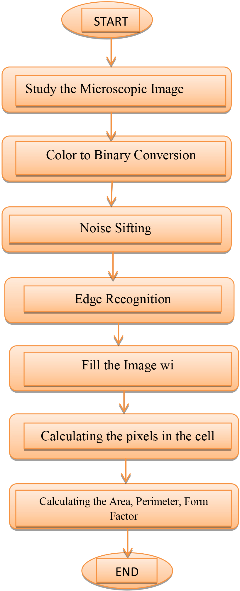
In an aberrant or physiologically variable environment, red blood cells have a tendency to aggregate. Aggregated cells reveal their affinity for one another by their density and composition. Analysing the effects of nanoparticle interaction on erythrocyte aggregation and contrasting the results with those from untreated controls Analysis of the aggregation’s pixel density revealed that it was quite compact [31]. With the help of MATLAB, we were able to transform the colour aggregation image into a black and white one. The original image had its background eliminated. Using the image processing programme, we evaluated the pixel density distribution. The National Institutes of Health (NIH) provides a free, open-source programme called ImageJ that operates on the Java platform (NIH). The image’s grey scale versus distance in pixels (Fig. 2). The pixel distribution is dependent on the compactness of the cells aggregation.
Erythrocytes aggregation investigation algorithm.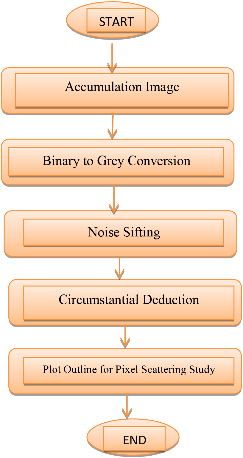
Treated standard erythrocyte cell carbon copy.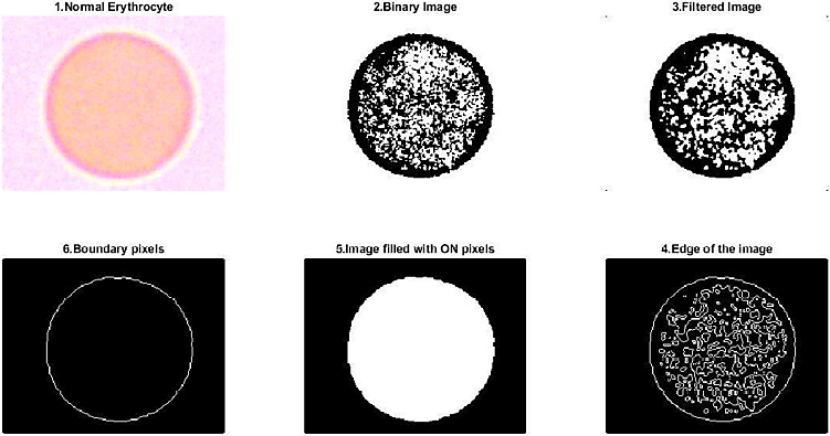
The form factor is defined by following formula:
Where A is area and P is perimeter of the circle.
The pixel area was calculated by following formula:
Attained outcomes of regular erythrocyte is Area
Multi-walled (-OH functionalized) carbon nanotubes interacted with erythrocyte cells in plasma free of plasma to determine the hemolytic concentration. Supernatant was collected and light absorption evaluated at 540 nm after incubation at various concentrations of MWCNT (-OH functionalized).
Hemolysis (%) persuaded by multi-walled carbon nanotubes
Echinocyte recording in MWCNT preserved blood
Hemolysis (%) persuaded by multi-walled carbon nanotubes.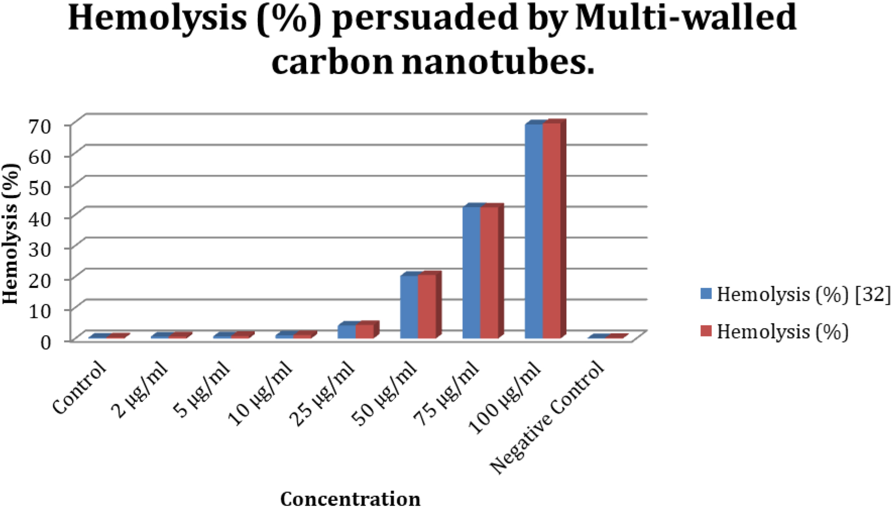
We have compared the hemolysis percentage value of proposed method with existing approach [32]. The blue colour bars are representing existing research work performance and red colour bars are representing the proposed work performance and from Fig. 4, we can conclude that proposed work performed better than existing method by giving higher percentage value of hemolysis.
Multiwalled carbon nanotubes (-OH functionalized) were exposed to human blood samples from a variety of healthy individuals. Echinocytes, the cells impacted, were counted and scored. A blood smear made using the thin-film, wedge-slide method. Echinocytes (crenated cells) were counted as a percentage of total cells in duplicate blood smears made from one thousand cells. The following table displays the typical amount of damaged cells.
Increasing the nanoparticle concentration led to a greater rise in the number of Echinocyte as shown in Fig. 5. The slow but steady rise in Echinocyte count is indicative of the length of time and nanoparticle concentration in the blood that may cause hemotoxicity.
Plasma-free erythrocyte cells were exposed to multi-walled carbon nanotubes [33, 34] to determine the hemolytic concentration. The supernatant was collected after incubation with several concentrations of MWCNT (pure), and light absorption was measured at 540 nm.
Hemolysis (%) tempted by MWCNT (pristine)
Hemolysis (%) tempted by MWCNT (pristine)
Number of Echinocyte plotted against concentration.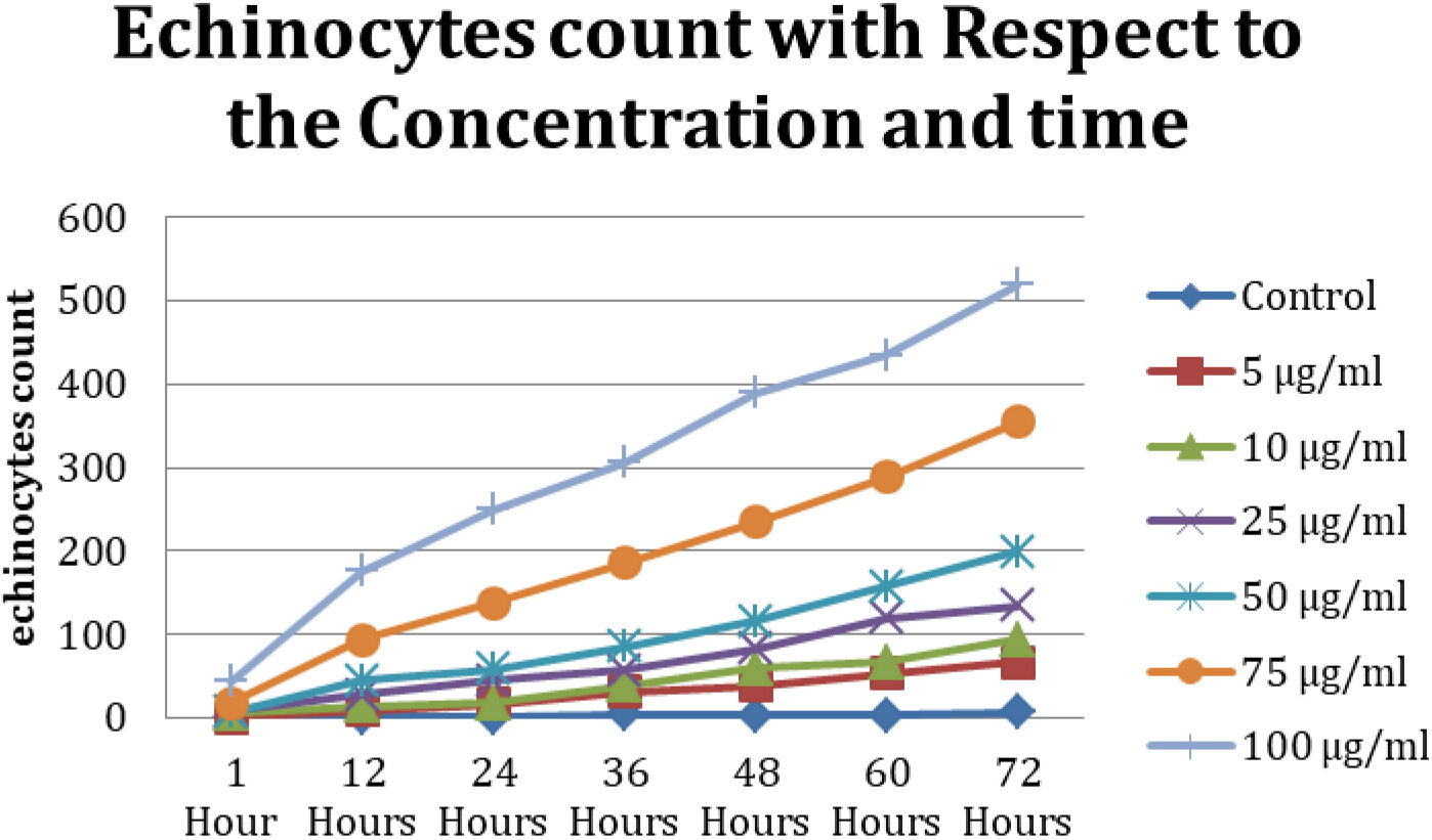
We have compared the hemolysis percentage value of proposed method with existing approach [32]. The blue colour bars are representing existing research work performance and red colour bars are representing the proposed work performance and from Fig. 6, we can conclude that proposed work performed better than existing method by giving higher percentage value of hemolysis.
Multiwalled carbon nanotube (pristine) reacted with human blood obtained from healthy individuals at varying concentrations. Echinocytes were counted and rated for severity. An example of a blood smear made using the thin-film, wedge-slide method. Blood smears were made in duplicate, and the percentage of echinocytes (crenated cells) per thousand total cells on each slide was recorded. The following table displays the typical amount of damaged cells [35, 36].
Echinocytes recording in MWCNT (pristine) preserved blood
Hemolysis (%) tempted by MWCNT (pristine).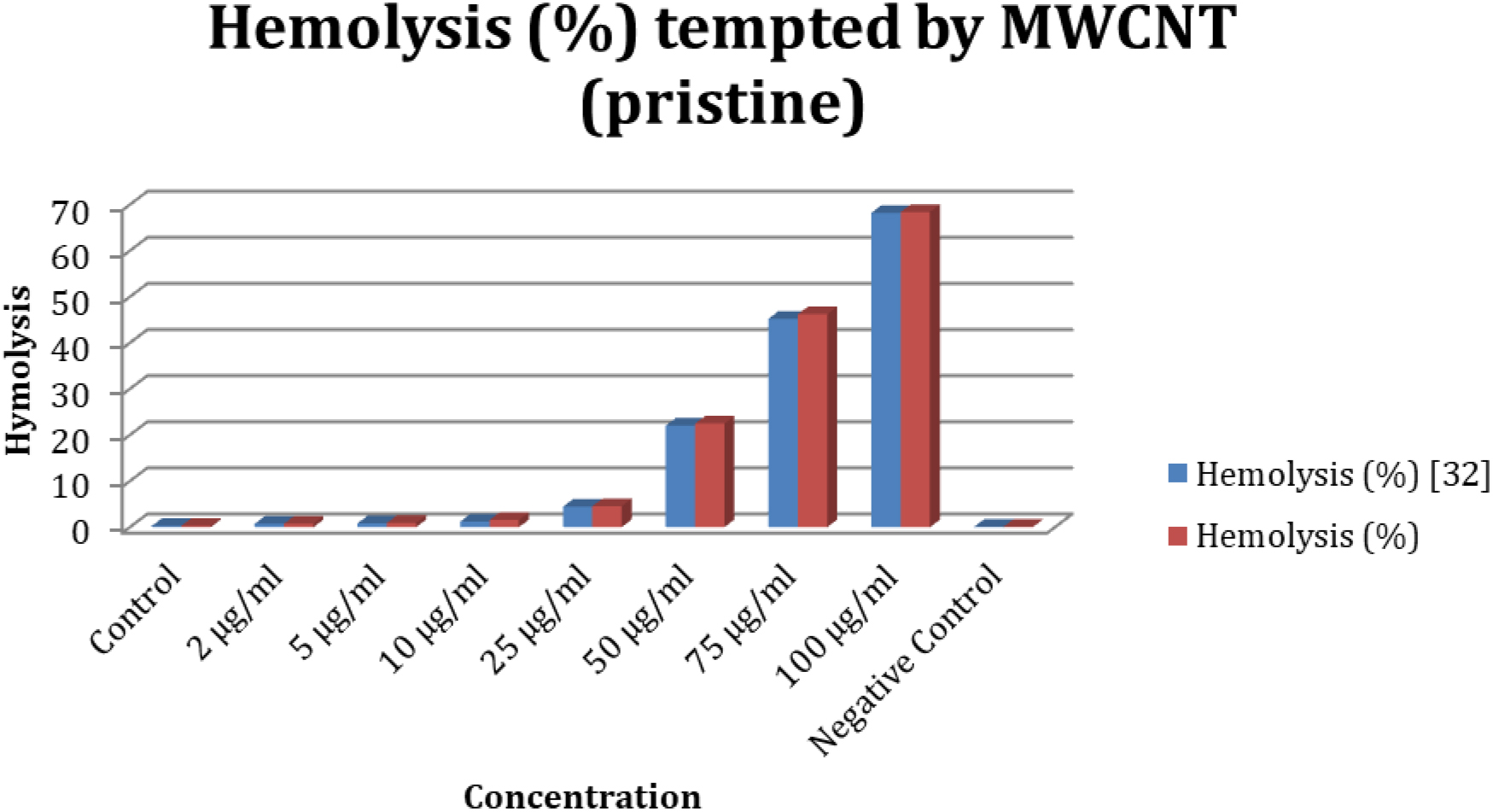
Echinocyte count plotted against concentration.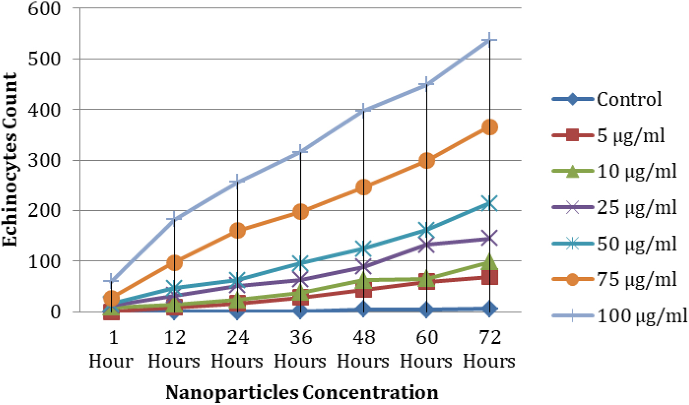
Echinocyte counts rise as nanoparticle concentrations rise, as predicted. This slow but steady increase in Echinocyte count suggests that prolonged nanoparticle exposure at high concentrations may be hemotoxic.
Human erythrocyte cells reacted with nanoparticles of iron (II) oxide, iron (III) oxide, silicon dioxide, and hydroxyl-functionalized and pristine multiwalled carbon nanotubes. Nanoparticle-induced morphological alterations vary with particle concentration and exposure duration. Metal oxide nanoparticles showed greater toxicity than carbon nanotubes but this was not statistically significant, and MWCNT and pristine SWCNT induced more changes in the erythrocyte morphology than the -OH functionalized nanoparticles. Nanoparticles in their unmodified state are more cytotoxic than their modified counterparts.
Our findings imply that nanoparticles circulating in the bloodstream have significant clinical implications. Reduced toxicity from nanoparticles is possible by functionalization, but more research is needed into immune response, concentration, and membrane penetration before they may be used in medicine.
Nanoparticle-induced echinocyte formation is a function of both interaction time and nanoparticle concentration. Cells in the plasma-free blood sample reacted with a variety of nanoparticles. Toxic levels were higher in plasma-free cells. Nanoparticles interacting with plasma proteins could be to blame. Different nanomaterial concentrations and incubation times were tested, and their effects on erythrocyte morphometric parameters were compared. The development of echinocytes increased with both concentration and duration.
The virgin forms of multi-walled carbon nanotubes were shown to be more hazardous than the functionalized versions. More echinocytes were stimulated by single-walled carbon nanotubes than by either multi- or single-walled varieties.
Footnotes
Acknowledgments
Princess Nourah bint Abdulrahman University Researchers Supporting Project number (PNURSP2023R 155), Princess Nourah bint Abdulrahman University, Riyadh, Saudi Arabia.
Conflict of interest
The authors of this manuscript declared that they do not have any conflict of interest.
Data availability statement
The corresponding author may provide data to back up the conclusions of this study upon request.