
Abstract
Background:
High-density lipoprotein (HDL) containing apolipoprotein A-I is associated with the pathogenesis of Alzheimer’s disease (AD). HDL particle size is modified in the presence of pathological conditions, while the significance of the HDL particle size remains controversial.
Objective:
The aim of this study was to investigate the HDL lipoprotein subclasses in mild cognitive impairment (MCI) and AD.
Methods:
This cross-sectional study included 20 AD patients, 17 MCI patients, and 17 age-matched controls without cognitive impairment, selected from the database of the Study of Outcome and aPolipoproteins in Dementia (STOP-Dementia) registry. The diagnoses of AD and MCI were performed by expert neurologists according to the Diagnostic and Statistical Manual of Mental Disorders-Fifth Edition criteria. Serum HDL subclasses were measured by electrophoretic separation of lipoproteins using the Lipoprint System. The neutrophil-lymphocyte ratio (NLR), a marker of inflammation, was calculated by dividing the neutrophil count by the lymphocyte count.
Results:
Small-sized HDL particle levels in the MCI group were significantly higher than in the control group, although there was no difference in serum HDL-cholesterol levels between MCI and control groups. NLR in the MCI group was higher than in the control group, but this difference was non-significant (p = 0.09). There was no difference in HDL subclasses or NLR between the AD and control groups.
Conclusion:
These findings suggest that HDL subclasses might be associated with the development of MCI.
Keywords
INTRODUCTION
Alzheimer’s disease (AD) is an age-related neurodegenerative disorder, and it is estimated that the worldwide prevalence of AD will be greater than 131,000,000 people by 2050 [1]. The cause and mechanism of AD has been partially elucidated, with the progressive deposition of amyloid-β (Aβ) and tau protein being considered to be neuropathological hallmarks of AD [2, 3].
High-density lipoprotein (HDL) and apolipoprotein A-I (apoA-I) promote the efflux of excess cholesterol via cholesterol transporters such as the ATP-binding cassette transporter A1 (ABCA1) [4, 5]. Brain Aβ elimination across the blood-brain barrier (BBB) is modulated by the natural chaperone apoA-I. ABCA1 is involved in the pathogenesis of AD [4, 5]. Disturbances in HDL metabolism may influence a number of functions, including cognition, and neuronal growth and repair, thereby leading to AD [6].
Chronic inflammation is also a hallmark of AD [7–9]. While we do not know whether it is directly involved in the pathogenesis, or is simply a downstream consequence of neuronal death, the blood neutrophil-lymphocyte ratio (NLR) is reported to be an inflammatory biomarker for AD [7–9]. It is also known that the metabolism of HDL and apoA-1 is modified in the presence of inflammation [10, 11].
A recent study reported that patients with mild cognitive impairment (MCI) and AD had a smaller low-density lipoprotein (LDL) particle size than control subjects [12]. Although an inverse relationship between plasma HDL-cholesterol (HDL-C) levels and cardiovascular disease is well known [13], recent clinical trials aimed at reducing cardiovascular disease by raising HDL-C levels showed disappointing results [14]. HDL shows a highly heterogeneous particle size [15]. Under some pathological conditions, including inflammation and cardiovascular disease, the size of HDL particles changes [16–18]. The change is often thought to be a more useful indicator of disease than HDL-C levels alone, although the significance of HDL particle size remains controversial [19–21]. Currently, only a few studies have examined the link between small-sized HDL particles and the risks of MCI and AD. The aim of this study was therefore to investigate the associations of small-sized HDL particles and inflammation with MCI and AD.
MATERIALS AND METHODS
Participants and study design
This cross-sectional study included 20 AD patients, 17 MCI patients, and 17 age-matched controls without cognitive impairment, selected from the database of the Study of Outcome and aPolipoproteins in Dementia (STOP-Dementia) registry. Only adults aged ≥65 years with AD and MCI who visited the Department of Neurology at the National Hospital Organization Kyoto Medical Center were included in the study. Subjects with diffuse Lewy body disease, frontotemporal dementia, other neurodegenerative disease (i.e., progressive supranuclear palsy and corticobasal degeneration), immune-mediated disease in need of steroid therapy (i.e., atypical encephalitis), and reversible dementia (i.e., hypothyroidism, chronic subdural hematoma, Wernicke’s encephalopathy, and chronic alcoholism), and subjects judged as unsuitable for the study by the investigator or medical doctor for other reasons were excluded from the study. The study protocol conformed to the ethical guidelines of the 2013 Declaration of Helsinki and was approved by the Ethics Committee of Kyoto Medical Center (approval number: 15-090). This trial was registered with UMIN as 000019992.
Neurological findings, cognitive function tests, and diagnosis
The diagnoses of AD and MCI were performed by expert neurologists according to the Diagnostic and Statistical Manual of Mental Disorders-Fifth Edition (DSM-V) criteria [22], the International Classification of Diseases-10 (ICD-10), and brain single-photon emission computed tomography (SPECT) [23] and magnetic resonance imaging (MRI) findings [24, 25]. Cognitive functions were assessed using the Mini-Mental State Examination (MMSE) [26], Hasegawa dementia scale revised (HDS-R) [27], Frontal Assessment Battery (FAB) [28, 29], and Japanese version of the clinical dementia rating-sum of boxes (CDR-SB) scale to rate the severity of each participant’s dementia symptoms according to information on six areas including memory, orientation, judgment and problem-solving, community affairs, home and hobbies, and personal care [30, 31].
Data collection and blood tests
Information on demographic characteristics (i.e., body mass index [BMI], weight, and height) and medications was extracted from electronic medical records. The total cholesterol, LDL-C, HDL-C, serum triglyceride, and fasting plasma glucose levels were measured using an automatic biochemical analyzer (AU580; Beckman Coulter, Inc., Tokyo, Japan). The NLR, which is a marker of peripheral inflammation, was calculated by dividing the neutrophil count by the lymphocyte count [32].
APOE genotyping
Genomic DNA was extracted from the venous blood (2.0μl) of each participant using a DNA Extract All Reagents kit (Applied Biosystems, Yokohama, Japan), according to the manufacturer’s instructions. After extraction, the genomic DNA was immediately stored at –30°C. APOE genotyping was determined using predesigned TaqMan SNP genotyping assays (Applied Biosystems, Foster City, CA, USA) for rs429358 (ABI#C_3084793_20) and rs7412 (ABI#C_904973_10), with analysis on an ABI prism 7300 (Applied Biosystems, Yokohama, Japan) [33, 34]. In brief, 5μl GTXpress Master Mix (Applied Biosystems, Yokohama, Japan), 0.5μl SNP-specific TaqMan genotyping assay mix (Applied Biosystems, Yokohama, Japan), 2.5μl nuclease-free H2O, and a 2.0μl DNA solution were added per well. The denaturation began at 95°C for 20 s, with 40 cycles of incubation at 95°C for 15 s, then annealing and extension at 60°C for 1 min. The allele frequencies of SNP genotypes were tested for the Hardy-Weinberg equilibrium.
HDL subclass analysis
HDL subclasses were analyzed electrophoretically using the Lipoprint System (Lipoprint HDL System; Quantimetrix Corporation, Redondo Beach, CA, USA) [35, 36]. This method uses electrophoresis with a liquid loading gel and lipophilic dye in a precast linear polyacrylamide gel. For the HDL particles, the system used very-LDL (VLDL)/LDL as the starting reference point (Rf = 0) and albumin as the leading reference point (Rf = 1). Ten HDL subclasses were observed between the two points. Subclasses 1–3 represented large-sized HDL particles, subclasses 4–7 represented intermediate-sized HDL particles, and subclasses 8–10 represented small-sized HDL particles.
Brain SPECT
SPECT data were analyzed using the Specific Volume of Interest Analysis of the easy Z score Imaging System (e-ZIS; 99mTc-ECD Fujifilm RI Pharmacy, Tokyo, Japan) [23]. Brain region of interest (ROI) was set up in posterior cingulate gyrus and parietal lobe (chiefly precuneus). Three indicators (severity, extent, and ratio) were calculated automatically and compared between the AD and MCI groups.
Brain MRI
MRI was performed using a 1.5T MR system (Achieva and Ingenia; Philips Healthcare, Best, The Netherlands). White matter hyperintensities were evaluated using the Fazekas scale [24], a well-validated and established qualitative visual rating method, which separately categorizes the severity of deep and periventricular lesions, on a scale from 0 to 3 (0: none or a single punctate WHM lesion, 1: multiple punctate lesions, 2: beginning confluency of lesions (bridging), and 3: large confluent lesions). T1-weighted images were viewed in the coronal plane, and medial temporal lobe atrophy scores for the left and right hemispheres were given. The scale rates atrophy on a 5-point scale (0 point, absent; 1 point, minimal; 2 points, mild; 3 points, moderate; and 4 points, severe) based on the height of the hippocampal formation and the width of the choroid fissure and temporal horn [25].
Statistical analysis
Data are expressed as the mean±standard deviation (SD) or median (25–75% tile). The NLR was log-transformed. Differences among the three groups (AD, MCI, and control groups) were analyzed using one-way analysis of variance (ANOVA) with Scheffe’s post hoc test (in parametric analysis) or Mann-Whitney U test with Kruskal-Wallis test (in nonparametric analysis). Categorical variables were compared between the three groups using the chi-square test. Correlation analysis was performed using Pearson’s correlation.
p-values of <0.05 were considered significant. All statistical analyses were performed using SPSS Statistics for Windows version 20.0 (IBM Corp., Armonk, NY, USA). Cases with missing data were deleted in each analysis.
RESULTS
Compared with the control group, the AD and MCI groups had significantly higher total cholesterol levels. However, there were no differences in plasma HDL-C levels among the three groups. There was also no difference in blood pressure and medication rates among the three groups. Additionally, there were no differences in the prevalence of current smoking, drinking, and antidiabetic drugs among the three groups. The uptake of radioisotope decreased in the brain ROI of the AD group compared to the MCI group. The mean severity arbitrary unit (a.u.) and extent (%) of the brain SPECT data in the AD group were significantly higher than those in the MCI group. The allele frequency was higher in the MCI group, but there was no statistically significant difference in allele frequency among the three groups (Table 1).
Demographic characteristics of the AD, MCI, and control groups
HDL, high-density lipoprotein; LDL, low-density lipoprotein; HbA1c, hemoglobin A1c; MMSE, Mini-Mental State Examination; FAB, Frontal Assessment Battery at Bedside; RCPM, Raven’s colored progressive matrices; CDR-SB, Clinical Dementia Rating Sum of Boxes; SPECT, single-photon emission computed tomography; a.u., arbitrary unit; MRI, magnetic resonance imaging; MTA, medial temporal lobe atrophy; PVWM, periventricular white matter; DWM, deep white matter. Data are presented as means±standard deviation. *p < 0.05 versus AD; †p < 0.05 versus Control.
The MCI group had significantly higher levels of small-sized HDL particles than the control group (Fig. 1). The small-sized HDL particle did not correlate with HDL-C levels (r = –0.09, p = 0.51). The small-sized HDL/total HDL ratio in the MCI group tended to be higher than that in the control group (13.4±7.6 versus 8.1±2.7% p = 0.09). The log NLR was highest in the MCI group [0.43 (0.32–0.51)], but was not significantly different from that in the control group. There was not a significant correlation between small-sized HDL levels and CDR-SB scores in patients with MCI. Also, there was not a significant correlation between small-sized HDL levels and CDR-SB scores in patients with MCI or AD.
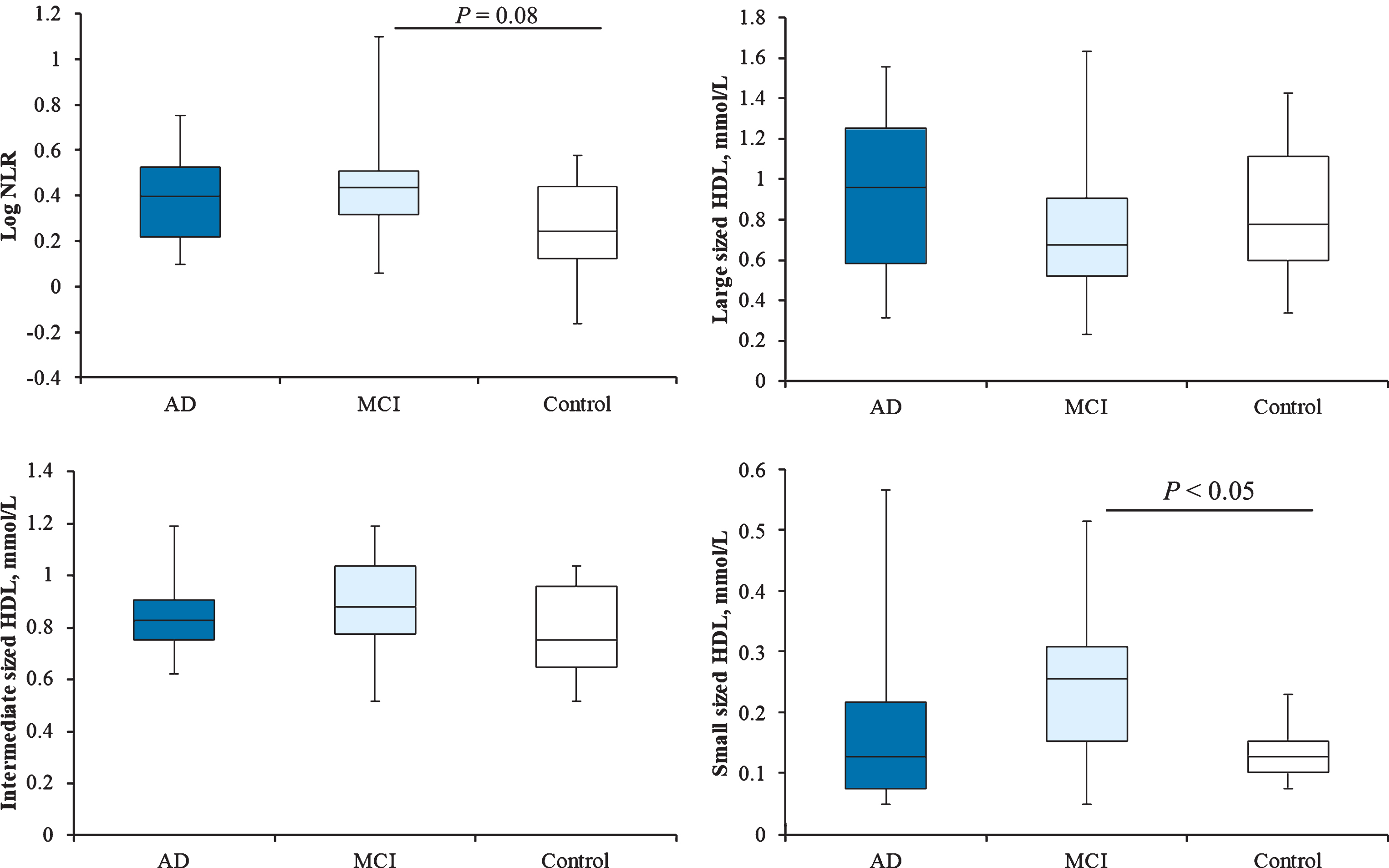
NLR and HDL subclasses in the AD, MCI, and control groups. NLR, neutrophil to lymphocyte ratio; HDL, high-density lipoprotein.
DISCUSSION
This study showed a relationship between small-sized HDL particle levels and MCI, although there were no significant differences in serum HDL-C levels among the groups. Higher small-sized HDL particle levels in MCI than in AD may suggest that small-sized HDL particles have a neuroprotective effect. It was reported that the HDL particle size correlated with the cholesterol esterification rate in 141 healthy male volunteers [37]. In our study, the small-sized HDL/total HDL ratio in the MCI group tended to be higher than those in the control group (p = 0.09). We noticed that comparison of small sized HDL/total HDL ratio might reduce the variance. The distribution of HDL subclasses provides relevant information on the anti-oxidant potential of HDL [38]. Small-sized HDL particles typically contain paraoxonase 1 (PON1) as an anti-oxidant molecule [39], and PON1 activity is decreased in AD [40]. However, the significance of the HDL particle size remains controversial [19–21], and further mechanistical investigations are required.
Lipoproteins in the central nervous system have been studied mainly in the cerebrospinal fluid (CSF). LDL, VLDL, and chylomicrons do not cross the BBB [41]. Lipoproteins in CSF called HDL-like particles, including apoE and apoA-I, are similar in size to plasma HDL particles [42]. As apoE in the peripheral circulation cannot cross the BBB, apoE is produced in the brain by astrocytes and microglia. However, the apoA-I in the brain comes from the peripheral circulation, because the brain does not produce apoA-I, and apoA-I-HDL levels in the circulation can affect apoA-I-HDL levels in the brain, as CSF and circulating apoA-I-HDL levels are similar [43]. Moreover, some oxidized cholesterol metabolites can diffuse through the BBB in both directions [44]. Degenerative disorders such as AD are associated with low apoA-I-HDL levels in the circulation and/or CSF [45]. Aβ is produced by the brain and binds to HDL, and thereby maintains its solubility in CSF and plasma. This HDL-Aβ interaction could prevent the deposition of Aβ into the brain. Therefore, HDL subclass distributions may have an important implication in the modulation of Aβ clearance, and consequently in AD pathogenesis.
There are several limitations to the present study. First, the cross-sectional design was a limitation, and therefore the results should be evaluated with a degree of caution. Second, this was a single-center study with a small sample size especially control subjects, because of the difficulty in recruiting age-matched control subjects without cognitive impairment in the same department, and the data should be confirmed by large scale studies.
NLR is a major marker of systemic chronic inflammation. However, the statistical power was not sufficient. Additionally, the number of controls should have been increased to raise the importance of this study. The sample sizes of the MCI and control groups, 32 and 96 participants, respectively, were required to increase the statistical power.
Cumulative evidence suggests that apolipoproteins, complement system, and transthyretin are involved in AD pathogenesis through sequestration of Aβ. A combination of apolipoprotein A1, complement C3, and transthyretin achieved an area under the curve of 0.89 (sensitivity 91% and specificity 80%) in MCI versus healthy controls and also discriminated individuals with mild cognitive decline from healthy controls [46]. A set of sequester proteins for the assessment of early stages of cognitive decline could be blood-based biomarkers. Small-sized HDL particle levels might also be blood-based biomarkers for MCI. Further examinations including prospective studies are required to clarify these issues.
Third, we did not measure HDL levels in the CSF. Fourth, the lipoprotein subclassification was performed using only the Lipoprint System. However, nuclear magnetic resonance and other methodologies will be necessary in future studies.
Conclusion
High small-sized HDL particle levels may be associated with the development of MCI. Further research, including large multicenter patient samples, is needed to clarify the association between small-sized HDL particles and MCI.
Footnotes
ACKNOWLEDGMENTS
This work was supported by JSPS KAKENHI Grant Number 17K19871 and Innovation, SIP (Project ID 14533567), Technologies for creating next-generation agriculture, forestry and fisheries (Bio-oriented Technology Research Advancement Institution, NARO). The superb technical assistance of Kokoro Tsuzaki is gratefully acknowledged. We thank Seiko Miyata and Ayaka Aizawa for assessing cognitive function tests, and Kana Kuroda for collecting and managing data.