
Abstract
Alzheimer’s disease (AD), an aging-related neurodegenerative disease, is a major cause of dementia in the elderly. Although the early-onset (familial) AD is attributed to mutations in the genes coding for amyloid-β protein precursor (AβPP) and presenilin1/presenilin 2 (PS1/PS2), the cause for the late-onset AD (LOAD), which accounts for more than 95% of AD cases, remains unclear. Aging is the greatest risk factor for LOAD, whereas the apolipo protein E4 allele (APOEɛ4) is believed to be a major genetic risk factor in acquiring LOAD, with female APOE ɛ4 carriers at highest risk. Nonetheless, not all the elderly, even older female APOE ɛ4 carriers, develop LOAD, suggesting that other factors, including environmental exposure, must play a role. This review summarizes recent studies that show a potential role of environmental exposure, especially ozone and particulate matter exposure, in the development of AD. Interactions between environmental exposure, genetic risk factor (APOE ɛ4), and sex in AD pathophysiology are also discussed briefly. Identification of environmental risk factor(s) and elucidation of the complex interactions between genetic and environmental risk factors plus aging and female sex in the onset of AD will be a key to our understanding of the etiology and pathogenesis of AD and the development of the strategies for its prevention and treatment.
INTRODUCTION
Alzheimer’s disease (AD), a neurodegenerative disease, is a major cause of dementia in the elderly with healthcare costs tripling the general population age 65 or older. According to the Alzheimer’s Association, 5.8 million Americans and 26.6 million people worldwide suffer from AD currently. With the increase in the lifespan, the prevalence of AD will be increased significantly. Despite extensive studies, there is still no effective treatment for this devastating disease due to an incomplete understanding of its etiology and pathogenesis. Early-onset (familial) AD, which accounts for less than 5% of AD cases, is attributed to mutations in the genes coding for amyloid-β protein precursor (AβPP) or presenilin1 (PS1) or presenilin2 (PS2), leading to increased production and deposition of amyloid-β peptide (Aβ) in the brain. The causes for the late-onset Alzheimer’s disease (LOAD, also called sporadic AD), which accounts for >95% of all AD cases, however, remain unclear.
Several risk factors for AD have been identified, including old age, carrying the apolipoprotein E4 (APOE ɛ4) allele, and being postmenopausal women (Fig. 1). Of 5.8 million Americans who have AD, 81% are age 75 or older [1]. It is estimated that the incidence of AD doubles every 5 years after age 65 and that 32% of the population at the age of 85 or older suffer from AD. Therefore, aging is considered to be the greatest risk factor for the development of AD [1–3]. Nonetheless, not all elderly suffer from AD, indicating that aging is a critical risk factor but alone is not sufficient to cause the disease. Human apolipoprotein E (apoE), existing in three isoforms (apoE2, apoE3, and apoE4), encoded by three distinct alleles ɛ2, ɛ3, and ɛ4, is a major carrier of lipids and cholesterol. Both epidemiology and animal studies suggest that the APOE ɛ4 allele, which is carried by approximately 15% of the population worldwide [4–7], is a major genetic risk factor for AD [7, 8]. It has also been well documented that two-thirds of AD patients are women and postmenopausal women are at increased risk [9–11]. Importantly, APOE ɛ4 has been shown to have more effect on the susceptibility of women to AD than it has on men [4, 11–16]. Nonetheless, not all of the APOE ɛ4 carriers, even older female APOE ɛ4 carriers, develop AD, suggesting that other factors, including environmental exposure, must play a role (Fig. 1).
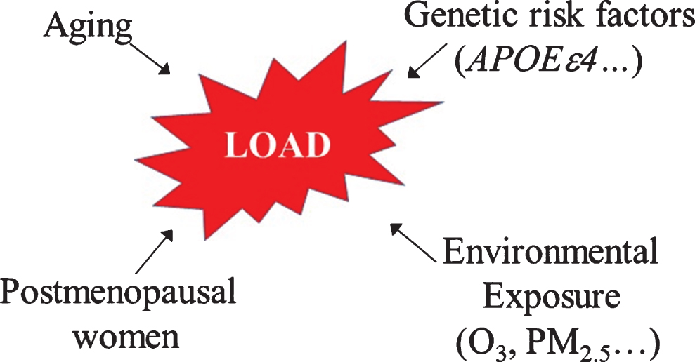
Schematic flow chart of potential risk factors for the development of late-onset Alzheimer’s disease (LOAD).
Ozone (O3) and particulate matters (PMs) are two of most abundant air pollutants in suburban and urban settings. Accumulated evidence from both epidemiology and animal studies suggests that exposure to unhealthy levels of O3 and/or PMs, especially particulate matter with an aerodynamic diameter of 2.5 micrometers (PM2.5) or smaller, may contribute to the onset of AD (Fig. 1). Nonetheless, which environmental pollutant(s) is (are) responsible and how environmental pollution contributes to the development of AD remains unclear. In this review, we summarized the recent findings that support a potential link between environmental exposure, especially O3 and PM2.5 exposure, and cognitive impairment. Interactions between environmental exposure, genetic risk factor APOE ɛ4, and sex in the development of AD are also discussed briefly.
AMBIENT OZONE EXPOSURE AND AD
O3 is a highly reactive oxidant and one of the most abundant urban pollutants. Over 30% of the population in the United States live in areas with unhealthful levels of O3 (American Lung Association State of the Air, 2012). Besides, some workers (e.g., pulp mills and outdoor construction) are intermittently exposed to relatively high levels of O3 (0.3 ppm–1 ppm) through their working environment [17–24]. Although the lung is the primary target, emerging evidence from epidemiology and animal studies indicates that O3 inhalation causes pathological changes in other tissues/organs beyond the respiratory system and that exposure to high levels of O3 may be a risk factor for AD [25–37].
Evidence from human studies
Accumulation of Aβ42 in the hippocampus and frontal cortex, the key brain regions required for learning and memory, is a pathological feature of AD. Several studies have shown that children and young adults who lived in the areas heavily polluted with O3 and PMs, like Mexico City, had an increased level of Aβ42 in their brains, compared with same-aged children and young adults who lived in low polluted areas [38–42]. Although the mechanism underlying the increased Aβ load in the brain of the children/young adults living in heavily polluted areas is unclear, it has been reported that these children and young adults also have augmented neuroinflammation, disrupted blood-brain barriers, and damaged epithelial and endothelial cells in their brains compared to children and young adults who lived in clean air areas [38, 40–42]. These data suggest that dysfunction of endothelial and epithelial cells as well as blood-brain barriers may contribute to brain Aβ accumulation in these children/young adults exposed to polluted air. It should be pointed out, although O3 and PM are the major air pollutants in Mexico City, other types of pollutants also exist. None of these human studies have examined the correlation between neuropathological changes with the concentration of any of these specific air pollutants. Therefore, the results from these studies can only suggest a potential link between air pollution and neuropathology in general and cannot distinguish the effects of O3 and PM or other air pollutants.
To determine whether exposure to O3 and/or PMs contributes to the onset of AD, several epidemiology studies have been conducted recently [25–30]. By analyzing the Neurobehavioural Evaluation System-2 (NES2) data from 1,764 adults participating in the Third National Health and Nutrition Examination Survey between 1988 and 1991 as well as the ambient PM10 (PM with aerodynamic diameter <10 μm) and O3 data from EPA database, Chen et al. reported that there was no association between PM10 exposure and cognitive function measured by the NES-2 after adjustment for sociodemographic factors [25]. However, each 10 ppb increase in annual O3 level was associated with 3.5–5.3 years of aging-related decline in cognitive performance [25]. Using a case-control comparison strategy, Wu et al. reported that long-term exposure to high concentrations of PM10 (≥49.23 μg/m3) or O3 (≥21.56 ppb) was significantly associated with an increased risk of AD in Taiwan [26]. Gatto et al. examined cross-sectional associations between various ambient air pollutants, including O3, PM2.5, and nitrogen dioxide (NO2), and six measures of cognitive function and global cognition among healthy, cognitively intact individuals residing in the Los Angeles basin. They found that exposure to high levels of PM2.5 was associated with lower verbal learning, exposure to NO2≥20 ppb was associated with a lower logical memory, and exposure to 49 ppb of ambient O3 was associated with a lower executive function [28]. By conducting a cohort study with 95,690 participants age ≥65, another group of scientists from Taiwan reported a weak association between ambient O3 concentration and AD risk at baseline but a 211% increase in risk with every increase of 10.91 ppb of ambient O3 concentration over the follow-up period from the year 2000 to the year 2010 [27]. Their studies suggest that long-term exposure to O3 above the current US Environment Protection Agency (EPA) standards is associated with an increased risk of AD. In a huge population study aimed to evaluate the association between long-term exposure to air pollution and first hospitalization for dementia, Cerza et al. reported that there was a negative association between exposure to NO2 and dementia hospitalization, whereas exposure to O3 was positively associated with dementia hospitalization, especially senile dementia [30]. By assessing the cognitive performance of individuals from National Alzheimer’s Coordinating Center, the average age of 76.8 years, and ambient O3 and PM2.5 concentrations established using a space-time Hierarchical Bayesian Model, Cleary et al. reported that the increased levels of O3, but not PM2.5, correlated with an increased rate of cognitive decline after adjustment for key individual and community-level risk factors [29]. Most interestingly, their data showed that individuals harboring one or more APOE ɛ4 alleles had a faster rate of cognitive decline and that the deleterious association of O3 was confined to individuals with normal cognition when entering the study [29]. Their findings suggest that APOE ɛ4 may accelerate O3 exposure-associated cognitive decline in the elderly and that healthy individuals are more sensitive to O3 effect than memory-impaired individuals. Potential interaction between APOE ɛ4 and environmental exposure in the onset of AD will be further discussed later in this review. Together, these epidemiologic studies suggest that exposure to high levels of O3 is a risk factor for AD in old population.
It should be pointed out that controversial results have also been reported [43, 44]. Using retrospective cohort from primary care data in London, Carey et al. reported that there was no association between exposure to higher levels of O3 and dementia, although they found that exposure to high concentrations of NO2 or PM2.5 was associated with dementia and that this association was more consistent for AD than vascular dementia [43]. In another large population study (approximate 2.1 million individuals) conducted in Ontario, Canada, Chen et al. reported that no association was found between O3 exposure and the incidence of dementia between 2001 and 2013, although a positive association was found between exposure to PM2.5 and dementia incidence [44]. The reason for such a discrepancy between different population studies remains to be determined.
Evidence from animal studies
Epidemiology studies, although important, can only establish a correlation between an exposure and a disease, not a cause-effect relationship. Moreover, it is difficult to elucidate the underlying molecular mechanism from epidemiology studies. In contrast, animal studies can help establish a cause-effect relationship and dissect the molecular mechanisms underlying the pathophysiology. In this regard, several studies have been conducted to explore the potential link between O3 exposure and AD-like neuropathophysiology, using experimental animal models [31, 45–49]. Rivas-Arancibia et al. showed that exposure to 0.2 ppm–1 ppm of O3 for a short period (4 hours) impaired long-term memory, reduced dendritic spines, and induced brain lipid peroxidation in young rats [34, 50]. This group also reported that O3 exposure stimulated Aβ42 production, impaired mitochondrial function, and reduced brain repair capacity, whereas treatment with antioxidant vitamin E attenuated O3-induced hippocampal lipid peroxidation and memory deficits in rats [36, 46–48], suggesting that increased oxidative stress is responsible for O3-induced memory deficit. Moreover, they have shown that O3 exposure induced endoplasmic reticulum stress and apoptosis in rat hippocampus [49], suggesting that O3 inhalation impairs memory through multiple mechanisms.
O3 in the atmosphere (troposphere) is formed from chemical reactions between different air pollutants such as nitrogen oxides and volatile organic compounds (methane, organic solvents, etc.) under sunlight. Therefore, O3 concentration in the atmosphere is high during the day and low in the evening. Moreover, several days of elevated O3 are usually followed by a long period of “clean” air in most urban settings, although air pollution can last much longer time in heavily polluted areas such as Mexico City than in other areas. To mimic what occurs in the urban settings, we investigated, in a previous study, whether exposure to a cyclic O3 exposure protocol, which consists of 5 days of O3 exposure (0.8 ppm, 7 hours/day) followed by 9 days of filtered air exposure, may contribute to AD pathology, using AβPP/PS1 double transgenic mice, a well-established murine model of familial AD [31]. We found that exposure of 6-week-old non-transgenic littermates (wild type mice) to O3 through a cyclic exposure protocol for 8 cycles had no significant effects on memory of either male and female wild type mice [31]. However, exposure of 6-week-old AβPP/PS1 mice to such a cyclic O3 exposure protocol accelerated learning/memory function loss in male AβPP/PS1 mice, although it had no significant effect on female AβPP/PS1 mice [31]. We also found that male AβPP/PS1 mice had lower levels of antioxidants (glutathione and ascorbate) and augmented oxidative stress response (increased NADPH oxidase expression and lipid peroxidation) as well as increased neuronal apoptosis upon O3 exposure, compared to female AβPP/PS1 mice [31]. On the other hand, female AβPP/PS1 mice had higher basal levels of Aβ42 and Aβ40 compared to male AβPP/PS1 mice; O3 exposure had no significant effect on brain Aβ load in either sex. Together, our data suggest that exposure to O3 under the conditions that mimic human exposure scenarios per se may not cause AD but can accelerate the pathophysiology of AD in genetically predisposed populations [31]. Our data also suggest that exposure to O3 under the conditions mimic to human exposure scenarios impairs memory probably through inducing oxidative stress and neuronal cell death, rather than increasing brain Aβ accumulation.
Potential signals mediating lung-brain effects of O3
The most challenging question that remains to be answered is how inhaled O3 affects brain structure and function. In other words, what is (are) the signaling molecule(s) that mediate(s) the lung-brain effects of inhaled O3? As a highly reactive oxidant, O3 reacts quickly with multiple substrates in airway surface lining fluid and produces secondary reactive species such as ozonide radicals, singlet oxygen, antioxidant radical intermediates, and lipid peroxidation products [51, 52]. The extrapulmonary effects are most likely induced by these secondary reactive species rather than by O3 itself. Several hypotheses have been proposed to explain the extrapulmonary effects of O3, including inflammatory mediators [53], nitric oxide [54], and reactive lipid peroxidation product(s) [55] (Fig. 2). Erickson et al. reported that exposure of female BALB/c mice to 2 ppm O3 for 2 hours increased the level of acute-phase serum amyloid A (A-SAA) protein in the liver, serum, and brain, although it had no significant effect on the blood levels of 23 cytokines/chemokines, except keratinocyte chemoattractant (KC/CXCL1), suggesting that A-SAA may be an important systemic signal of O3 exposure to the CNS [56]. Mumaw et al. further showed that exposure to 1 ppm O3 for 4 hours caused a persistent activation of brain microglia but had no significant effect on the levels of circulating cytokines such as CCL2, CCL11, TNFα, IL-6, and IL-1β in 8-weeks old Sprague-Dawley rats [57]. Interestingly, they showed that the serum from O3 exposed mice induced an augmented immune response to LPS in hippocampal mixed glia cultures from old rats, compared to hippocampal glia cultures from young rats, and that monoclonal macrophage 1 antigen (MAC1) blocking antibodies eliminated O3-induced priming effect [57]. These data suggest that O3 exposure leads to increases in non-cytokine-related signals in the circulation, which promote brain inflammatory responses [57].
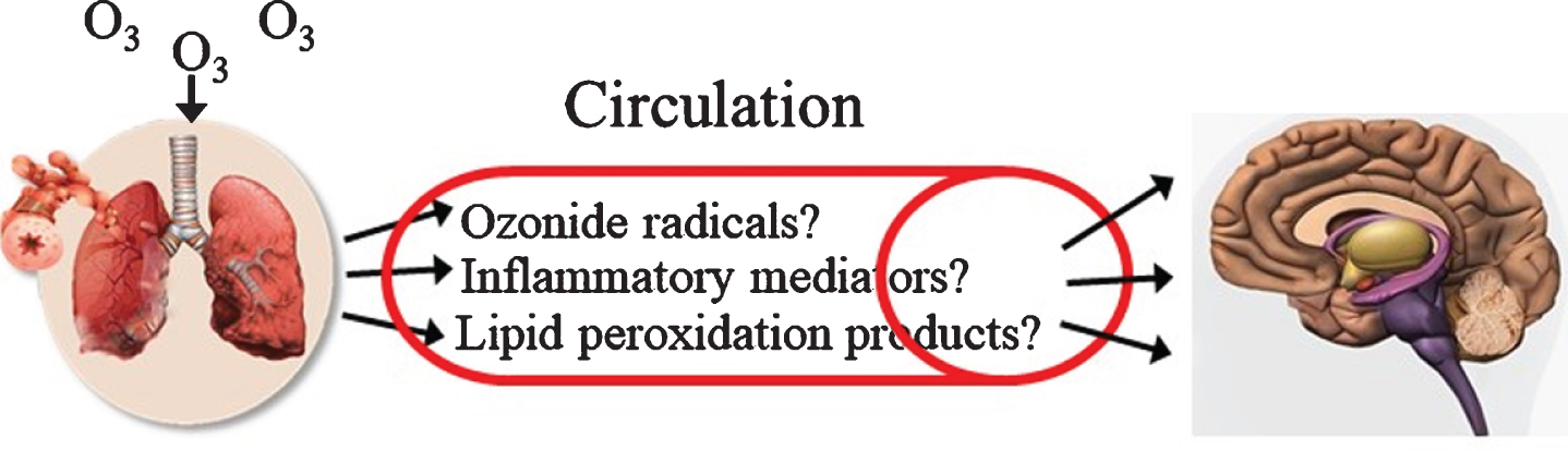
Potential signal molecules involved in O3 inhalation-induced brain pathophysiology.
Malondialdehyde (MDA) is a lipid peroxidation end product. Cretu et al. reported that exposure of rats to 0.5 ppm O3 for 10 minutes daily for 2 or 4 weeks increased MDA level in the plasma and brain as well as pathological changes in the brain [58]. In a previous study, we showed that male AβPP/PS1 mice had lower levels of antioxidants (glutathione and ascorbate) and augmented induction of NADPH oxidase as well as increased neuronal apoptosis upon O3 exposure, compared to female AβPP/PS1 mice [31]. This was associated with accelerated learning/memory impairment in male AβPP/PS1 mice [31]. We also found that O3 inhalation increased the amounts of 4-hydroxynonenal (4HNE), an end product of lipid peroxidation, in the plasma and hippocampus/cortex of male, but not female, AβPP/PS1 mice [31]. Moreover, treatment of neuroblastoma SHSY5Y cells with 4HNE significantly increased apoptotic cell number, the activity of Caspase-3/7, and expression of p53 and Bax, three apoptosis markers [31]. Together, these data suggest that circulating lipid peroxidation products, such as MDA and 4HNE, which were released potentially from the oxidatively damaged lung, may contribute to brain pathological changes under O3 inhalation condition [31, 58].
PARTICULATE MATTER EXPOSURE AND AD
Particulate matters (PMs) are liquid or solid matters suspended in the atmosphere.The major components of airborne particulates are sulfates, nitrates, ammonium, chloride, elementals, organic carbons, biological materials, and minerals, although the exact composition of PMs varies considerably depending on the location, weather, the season, time of day, emission sources, and many other factors [59]. Besides natural sources, such as volcanic activities and wildfires, human activities, including the combustion of fossil fuels, mining, and agricultural activities contribute importantly to PM pollution. PMs are arbitrarily grouped into ultrafine PMs with aerodynamic diameter <100 nm (PM0.1), fine PMs with aerodynamic diameter 2.5 micrometers (PM2.5), and coarse PMs with aerodynamic diameters between 2.5 and 10 microns (PM10). As PM2.5 and PM0.1 can penetrate and deposit into the deep lung tissue and enter the circulation, these forms of PMs have greater potential to cause extrapulmonary diseases than PM10. Accumulated evidence from human and animal studies suggests that exposure to particulate matters contributes importantly to cognitive decline and neuropathological changes [43, 60–74].
Human studies
Emerging evidence from epidemiology studies suggests that exposure to PMs is an important environmental risk factor for the development of AD [26, 62–67], although negative findings have also been reported [25, 29]. A longitudinal study conducted with 645 pairs of cognitively impaired subjects and their caregivers showed that aggravated neuropsychiatric symptoms were associated with exposure to high levels of PM2.5 in South Korea [63]. A similar result was reported in another study conducted with 2,896 adult Korea aged 70 to 84 years [74]. The authors concluded that air pollutions, especially PM2.5, were associated with cognitive impairment, including global cognition, attention, memory, and executive function in Korean old adults aged ≥70 years [74]. Through analysis of 130,978 adults aged from 50 to 79 years residing in London as well as the average annual concentrations of nitrogen dioxide (NO2), PM2.5, and O3 at 20×20 m resolution from dispersion models, Carey et al. reported that there was a positive exposure-response relationship between dementia and NO2 or PM2.5, but not O3, exposure and that the association was more consistent for AD than vascular dementia [43]. By following approximately 9.8 million subjects in 50 cities of the northeastern US, Kioumourtzouglou et al. reported that exposure to PM2.5 is associated with significant increases in the hazard ratios for dementia, AD, and Parkinson’s disease [61]. AD is associated with reduced hippocampal volume. Using brain-imaging data from UK Biobank, a large community-based dataset, and air pollution data, Hedges et al. showed that exposure to PM2.5 was associated with a smaller left hippocampal volume, whereas none of the other air pollutions, including PM2.5–10, PM10, nitrogen oxide, and nitrogen dioxide, was associated with hippocampal volume change [67]. By analyzing the data from four cohort studies conducted in Canada, Taiwan, the UK, and the US involving 12 million elderly aged ≥50 years, Tsai et al. found that exposure to a 10 μg/m3 increase in PM2.5 from 2015 to 2018 was significantly and positively associated with increases in dementia and AD cases [64]. An early decline of episodic memory is reported in preclinical AD patients. To determine whether PM2.5 exposure is associated with episodic memory decline and underlying neuroanatomic changes, Younan et al. conducted a longitudinal study using the data from Women’s Health Initiative Study of Cognitive Aging and the Women’s Health Initiative Memory Study of Magnetic Resonance Imaging [65]. Episodic memory was assessed by the California Vernal Learning Test, including measuring immediate free recall/new learning and delayed free recall, and by two brain MRI scans. A spatiotemporal model was used to estimate 3-year average PM2.5 exposure before 1st MRI image analysis. Using multilevel structural equation models, they found that PM2.5 was associated with greater declines in immediate recall and new learning but not in delayed-recall or composite scores [65]. Their findings suggest that PM2.5 exposure is associated with memory decline at the preclinical stage [65].
Animal studies
Numerous studies in experimental animals have also shown that exposure to particulate matters induces neuroinflammation and AD-like pathology and impairs learning and memory [59, 75–79]. Kim et al. reported that exposure of wild type mice to nickel nano particles increased brain levels of both Aβ40 and Aβ42 as well as the ratio of Aβ42 to Aβ40, a more disease-related change [71]. Bhatt et al. further showed that exposure of wild type mice (C57BL/6) to PM2.5 for 9 months increased brain beta-site AβPP cleaving enzyme (BACE) protein level, AβPP processing, and Aβ40 as well as cyclooxygenase 1 and 2 protein levels in mouse brain, although exposure to PM2.5 for 2 months had no significant effects [75]. No significant effect of PM2.5 exposure on the oxidative stress markers or synaptic marker was observed in the brain of these PM2.5 exposed wild type mice [75]. Some studies have been conducted to determine whether PM exposure accelerates neuropathological changes in AD model mice [73, 77]. Jew et al. reported that exposure of middle-aged (12.5 months old) mice to human exposure relevant concentrations of particulate matters (29–132 μg/m3 of Harvard ultrafine concentrated ambient particles (HUCAPS) for 2 weeks significantly impaired spatial learning of both 3×TgAD mice, a murine model of familial AD, and wild type mice [73]. Jang et al. showed, on the other hand, that exposure Neuro2A cells in the ex vivo hippocampus tissue from 3×Tg-AD transgenic mice to fine PMs dose-dependently activated poly(ADP-ribose) polymerase (PARP-1), decreased NAD+, increased Aβ levels, and activates glial cells, mostly in CA1 region [77]. Inhibition of PAR-1 activity reversed PM-induced Aβ accumulation and glial activation, suggesting that increased PARP-1 activity is responsible for PM-induced AD pathological changes [77]. Human beings are usually exposed to multiple environmental agents simultaneously. Some of these agents exhibit synergistic, whereas others show additive or antagonist effects when exposed to them simultaneously. To test the potential synergistic effects between two air pollutants, Liu et al. exposed wild type C57BL/6 mice to formaldehyde (0.155 mg/kg/day) and PM2.5 (0.193 mg/kg/day) alone or together for one week [72]. They found that exposure to either compound alone had little or no adverse effects on the mouse brain. However, when mice were exposed to PM2.5 and formaldehyde simultaneously, AD-like pathological changes were observed, suggesting a synergistic effect between these two compounds [72]. Using GC-MS, LC-MS, western blotting, and immunohistochemistry techniques, Park et al. found that exposure to PMs induces alterations in metabolic pathways involved in redox homeostasis, neuroinflammation, and Aβ metabolism in the hippocampus, olfactory bulb, cortex, and cerebellum, with the changes in the hippocampus most significant [79]. Together, these studies suggest that PM exposure can impair memory through multiple mechanisms, including inducing oxidative stress, neuroinflammation, and brain Aβ accumulation.
TRAFFIC-RELATED EXPOSURE AND AD
Living near traffic roads is associated with exposure to many biohazardous chemicals as well as noise. Although exposure to particulate matters is one of the major concerns associated with traffic-related exposure, people living near roads are usually exposed to multiple hazardous chemicals besides particulate matters, including nitrogen oxides, O3, heavy metals, volatile organic compounds, and polycyclic aromatic hydrocarbons [80]. Accumulated evidence suggests that living near major traffic roads is associated with increased brain structure and function changes that may lead to neurodegenerative diseases such as AD and Parkinson’s disease. Several large population studies and animal studies have been conducted to assess the effects of living near major traffic roads on the incidence of neurodegenerative diseases including AD [80–89]. In this section, we will specifically address the potential risk associated with traffic-related exposure in the development of AD as the effects from traffic-related exposure represent complex interactions between multiple air pollutants and noise and have been studied as an independent entity by many investigators.
Human population studies
Ranft et al. studied 399 women aged 68–79 years who lived for more than 20 years at the same residential address to address the relation between long-term exposure to traffic-related PMs and incidence of mild cognitive impairment [81]. After adjusted for potential confounders using regression analysis, they found distance (dose)-dependent effects of traffic-related particulate matters on neuropsychological performance [81]. Using a 15-year longitudinal human study data and annual mean nitrogen oxide data from the residential address of the participants as markers for traffic-related air pollution exposure, Oudin et al. demonstrated that there were dose-dependent increases in AD and vascular dementia in Northern Sweden [83]. By assessing two population-based cohorts with approximate 2.2 million people who resided in Ontario, Canada on April 1, 2001, Chen et al. further showed that the closer people lived to major traffic roads the higher the incidence of dementia, but not Parkinson’s disease or multiple sclerosis, in Ontario, Canada [80]. These studies demonstrate a strong correlation between exposure to traffic-related pollutants and cognitive decline, although the exact disease-causing factors remain to be identified.
Animal studies
Aβ accumulation and tau protein phosphorylation in the hippocampus and frontal cortex are the pathological features of AD. Levesque et al. found that exposure of male Fisher 344 rats to diesel exhaust (DE) at concentrations of 992, 311, 100, 35, or 0 μg PM/m3 for 6 months (subchronically) led to increased levels of inflammatory cytokine TNFα at high DE concentrations in all of the brain regions except the cerebellum [85]. No increase in IL-6 in any concentration of DE tested was observed, although IL-1β was increased in the groups treated with high concentrations of DE. They also showed that the levels of Aβ42 and phosphorylated tau (pS199) were significantly elevated at the higher concentrations of DE (992 and 311 μg PM/m3) exposed groups in both temporal and frontal lobes, suggesting that DE exposure induces AD-like pathological changes [85]. Increased brain Aβ load has also been reported in mice after exposure to diesel engine exhaust or traffic-related air pollution [86, 89]. Woodward et al. reported that exposure of young (3 months) and middle (18 months) aged mice to nanoscale PM (nPM) induced neurite atrophy, decreased myelin basic protein, and increased the amounts of Iba1, a microglial marker, and TNFα mRNA in the hippocampal CA1 region of young mice [87]. The dentate gyrus region, however, was unaffected [87]. Old control mice had similar changes as nPM exposed young mice and nPM exposure had no further effect on old mice, probably due to an age-ceiling effect [87]. In a recent study, the same group further showed that nPM exposure increased the levels of 4HNE, a lipid peroxidation product, and Aβ in lipid raft [89]. Treatment of mouse neuroblastoma N2a cells with nPM also caused dose-dependent increases in the levels of nitrogen oxide, 4HNE, and Aβ. Treatment with antioxidant N-acetyl-cysteine, on the other hand, attenuated nPM-induced oxidative stress responses and alterations of AβPP processing in lipid raft [89]. Their results suggest that traffic-related air pollutants promote neuronal pathological changes through inducing oxidative damage to lipid rafts [89].
INTERACTION BETWEEN GENE AND ENVIRONMENTAL EXPOSURE IN THE ONSET OF AD
It has been well documented that responses to environmental toxicants vary depending on the races, animal species, and even different individuals within the same race or species, suggesting that genetic background may affect the responses to environmental agents [90]. Besides aging, accumulating evidence indicates that genetic variations other than mutations in AβPP and PS1 or PS2 genes, which lead to the development of early-onset (also called familial) AD, affect the susceptibility to the late-onset AD. As of 2018, 21 of AD-related susceptibility genes have been identified, which are involved in various biological processes, including lipid homeostasis, inflammation and immunity, endocytosis, apoptosis, and Aβ processing and clearance [90–96]. Of these identified susceptibility genes, the APOE ɛ4 allele is believed to have the strongest link with AD [7]. Human apoE, existing in three isoforms (apoE2, apoE3, and apoE4), encoded by three distinct alleles ɛ2, ɛ3, and ɛ4 is a major carrier of lipids and cholesterol. Both epidemiology and animal studies have shown that the APOE ɛ4 allele, which is carried by approximately 15% of the population worldwide [4–7], is a major genetic risk factor for AD [4, 11–16]. Nonetheless, not all of the APOE ɛ4 carriers, even in old ages, develop AD, suggesting that other risk factors, including environmental exposure, must play a role. It is believed that AD may develop as a consequence of complex interactions between aging, genetic risk factors, and environmental risk factors.In this section, we will further discuss such interactions in the development of AD, focusing on the APOE ɛ4 allele.
Human studies
Mexico City is heavily polluted with PM2.5 and O3. Several studies have shown that Mexico City residents, including infants, children, and young adults, exhibit increased brain oxidative stress, inflammation, and AD-like pathological changes as well as cognitive deficits [70, 97–101]. Importantly, it has been reported that APOE ɛ4 carriers have higher levels of these pathophysiological changes compared to non-APOE ɛ4 carriers [97–101]. Calderon-Garciduenas et al. examined the brain metabolic rations, short-term memory, and IQ for 50 Mexico City residents with an average age of 13.4 years who carry either APOE ɛ3 and APOE ɛ4 allele [98]. They found that, compared to APOE ɛ3 children, APOE ɛ4 children had a decreased N-acetylaspartate/creatine ratios in the right frontal white matter and decrements on attention, short-term memory, and below-average scores in Verbal and Full-Scale IQ [98]. The same group of investigators also showed that infants, children, and young adults living in metropolitan Mexico City have increased cortical tau pretangles, neurofibrillary tangles, and amyloid in substantia nigra, auditory, oculomotor, trigeminal, and autonomic systems [101]. However, APOE ɛ4 carriers have 23.6 times higher odds of neurofibrillary tangle stage V compared to non-APOE ɛ4 carriers who have similar cumulative PM2.5 exposure and age [101]. This group of investigators also showed that APOE ɛ4 heterozygous children (APOE ɛ4/ɛ3) have decreased attention and short-tern memory subscale and below-average scores in verbal, performance, and full-scale IQ compared to APOE ɛ3/ɛ3 carriers [99]. Moreover, they showed that female heterozygous APOE ɛ4 carriers with 75–94% of normal BMI are at the highest risk of severe cognitive deficits [99].
Several large population studies have also indicated that APOE ɛ4 carriers are more sensitive than non-APOE ɛ4 carriers to environmental exposure-induced cognitive decline [29, 102–104]. By assessing 5,419 participants with an average age of 76.8 years in the US, Cleary et al. reported that exposure to low-level of O3 was associated with cognitive decline in the elderly and that individuals harboring one or more APOE ɛ4 alleles exhibited a faster rate of cognitive decline compared to non-APOE ɛ4 carriers [29]. They also found that the deleterious association of O3 was confined to individuals with normal cognition who eventually became cognitively impaired [29]. Cacciottolo et al. reported that older women from the Women’s Health Initiative Memory Study (WHIMS) who resided in places with fine PM exceeding EPA standards had increased risk of developing global cognitive decline and all-cause dementia with stronger adverse effects in APOE ɛ4 carriers [102]. They further showed that exposure of female EFAD transgenic mice (5×FAD+/–/human APOE ɛ3 or ɛ4+/+) to urban nanosized PM (nPM) for 15 weeks significantly increased cerebral Aβ load, which was worsened by APOE ɛ4.Their data suggest strongly that nPM exposure promotes AD pathological changes in the aging brain in women and that APOE ɛ4 further enhances such sensitivity [102]. Alemany et al. reported that the associations between behavior/memory performance (behavior problem scores, caudate volume, and cognitive performance trajectories) and outdoor concentrations of polycyclic aromatic hydrocarbons and nitrogen dioxide were stronger in/limited to APOE ɛ4 carriers [103]. A 6-year follow up study with 4,821 participants showed that high ambient concentrations of PM2.5, NO2, or PM10 were associated with a rapid decline of cognitive function and that the cognitive decline was faster in APOE ɛ4 carriers than in non-APOE ɛ4 carriers in responding to all of these pollutants [104]. Together, the evidence presented in these studies supports a synergistic effect between APOE ɛ4 and environmental exposure in promoting memory decline or AD.
It should be mentioned that not all of the studies support such a synergistic effect. Oudin et al. reported that, although air pollution was associated with dementia incidence in a longitudinal study conducted in Northern Sweden, APOE ɛ4 has no significant effect on the outcome [105]. The discrepancies between these studies suggest that other factors, genetic and/or environmental, are involved as well. It should also be pointed out that none of these epidemiology studies has addressed sex-dependent interactions between APOE genotype and environmental exposure in AD pathophysiology. It is unclear whether APOE ɛ4 enhances the sensitivity to the environmental pollutants-induced memory impairment primarily in female APOE ɛ4 carriers or in both male and female APOE ɛ4 carriers. More studies are required to address this important issue.
Animal studies
Humanized apoE4 and apoE3 mice, also known as apoE3 or apoE4 targeted replacement (TR) mice, in which the endogenous murine APOE gene is replaced with the human APOE ɛ4 or APOE ɛ3 (common human form) gene, respectively [106], have been widely used to study the mechanisms by which APOE ɛ4 modulates AD pathophysiology. These mice have also been used to study the interactions between genetic risk factor APOE ɛ4 and dietary or environmental exposure in the development of AD [107–119], including particulate matters and O3 exposure [32, 102]. Engstrom et al. reported that exposure to lead through drinking water for 12 weeks impaired memory in both apoE4 and apoE3 female mice but memory loss occurred earlier in apoE4 than in apoE3 mice [110]. ApoE4 mice, both male and female, are also more sensitive to cadmium-induced memory loss than apoE3 mice [119]. As in humans, the APOE ɛ4 allele mainly affects the memory of female mice under unchallenged conditions [8, 120–122]. Consequently, many studies have been conducted using female apoE4 TR mice only. Cacciottolo et al. reported that exposure of female EFAD (5XFAD+/–/human APOE ɛ3 or ɛ4+/+) transgenic mice to nanosized PM induced more dramatic AD-like pathological changes in the brain in apoE4 mice than it did in apoE3 mice [102].
Although some studies have shown that apoE ɛ4 TR mice are more sensitive to environmental factors-induced memory loss than apoE ɛ3 TR mice, opposite observations have also been reported [32, 111]. Peris-Sampedro et al. found that exposure to an organophosphate pesticide chlorpyrifos impaired spatial memory in male apoE3, not male apoE4, TR mice [109]. Using different exposure strategy, the same group further showed that postnatal chlorpyrifos exposure impaired spatial memory of apoE3 but not apoE4 female mice [111]. In a recent study, we tested the hypothesis that O3 exposure synergizes with the genetic risk factor APOE ɛ4 and aging leading to AD, using young and old male apoE4 and apoE3TR mice as men are more likely exposed to high levels of O3 via working environments and few studies have addressed APOE ɛ4 effects on males [32]. We found, surprisingly, that O3 exposure impaired memory only in old apoE3 male mice, while old apoE4 or young apoE3 and apoE4 male mice were spared [32]. Associated with memory loss, old apoE3 male mice exhibited increased protein oxidative modifications and neuroinflammation upon O3 exposure compared to other groups [32]. In contrast to apoE3 male mice, old apoE4 male mice have significantly increased expression/activities of several enzymes involved in antioxidant defense, diminished protein oxidative modifications, and neuroinflammation upon O3 exposure [32]. Together, these findings highlight the complexity of the interactions between APOE genotype, age, and environmental exposure in AD pathophysiology. These findings also suggest that the interactions between gene-environment in AD pathophysiology may be sex-dependent.
SEX, ENVIRONMENTAL EXPOSURE, APOE ɛ4, AND AD
Of the 5.6 million people age 65 or older who suffer from AD in the US, 3.5 million are women and 2.1 are men, indicating that the prevalence of AD is higher in women than in men [2, 123–133]. The mechanism underlying increased susceptibility of old women to AD is unknown, although a loss of estrogen in menopause [126, 130] and increased oxidative stress [134–140] is believed to contribute. Studies have shown that males and females respond differently to environmental pollutant-induced neurodevelopment and cognitive impairment [74, 141–146]. Moreover, it has been reported that APOE ɛ4 differentially affects the susceptibility of males and females to environmental pollutant-induced memory decline [90, 147]. It should be pointed out that controversial data have been reported. Some studies showed that APOE ɛ4 enhanced the sensitivity of females to environmental challenges, whereas other studies showed an opposite trend that APOE ɛ4 males were more sensitive than APOE ɛ4 females [110, 119]. Nonetheless, how sex affects the susceptibility to AD and how sex interacts with the APOE ɛ4 gene and environmental risk factors leading to AD remains largely unknown. In this section, we summarize a few published studies conducted in humans and animals, which address sex-related responses to environmental exposure in terms of neurodevelopment and cognitive function.
Although it has been long recognized that males and females respond differently to many challenges, very few studies have been conducted to dissect sex-dependent differences in response to environmental pollutant-induced neuropathophysiology. Prenatal exposure has been increasingly recognized to affect later-life susceptibility to some diseases. To explore the potential linkage between prenatal exposure to environmental pollutants and later life memory decline, Cowell et al. examined 258 mother-child dyads enrolled in a Boston, Massachusetts pregnancy cohort [142]. They found that, after stratification, the associations between prenatal exposure to traffic-related black carbon and memory scores were stronger in boys than in girls, although no significant effect was detected on learning and memory with non-stratified data [142]. Chiu et al. further showed that the responses to prenatal particulate exposure-induced neurodevelopment differ between boys and girls, depending on the time windows of prenatal exposure [146]. After adjusting for maternal age, education, race, and smoking status, they found that exposure to higher levels of PM2.5 at 31–38 weeks was associated with lower IQ, at 20–26 weeks with increased omission errors, at 32–36 weeks with slower hit reaction time, and at 22–40 weeks with increased hit reaction time standard error among boys [146]. On the other hand, higher PM2.5 exposure at 18–26 weeks was associated with reduced visual memory, and at 12–20 weeks with reduced general memory in girls [146]. Lertxundi et al. also found that prenatal exposure to PM2.5 and NO2 was negatively correlated with the neurological development of 4–6 years old children, especially boys [145]. In a recent study, we examined whether exposure to ambient fine particles (PM2.5) at residential locations affects intelligence quotient (IQ) during pre-/early- adolescence (ages 9–11) and emerging adulthood (ages 18–20) in a demographically-diverse population residing in Southern California [148]. We found that increased ambient PM2.5 levels were associated with decreased IQ scores, especially the Performance IQ (PIQ) and that the average PIQ score decreased by 3.08 points for each inter-quartile (7.73 μg/m3) increase in one-year PM2.5 preceding each assessment [148]. Interestingly, we found that the adverse effect was 89% stronger in males, compared to their counterparts [148]. Animal studies also showed that prenatal exposure to concentrated ambient ultrafine particles impaired the fixed interval schedule-controlled performance of male mice, while adult exposure impaired fixed interval of female mice, although concentrated ambient ultrafine particle exposure impaired short-term memory of both sexes [141]. Interestingly, it has been reported that the treatment of rats with estradiol prevents O3-induced brain oxidative stress and social recognition impairment [35, 149], supporting the notion that a decrease in estrogen level may underling increased susceptibility of old females to O3-induced AD-like pathophysiology.
Although it has been well documented that female APOE ɛ4 carriers have the highest risk of acquiring AD, how APOE genotype interacts with female sex leading to increased susceptibility to AD remains largely unknown. A few published studies also suggest that APOE ɛ4 may differentially affect the susceptibility of males and females to environmental pollutant-induced memory decline, although the underlying mechanism is unknown [90, 147]. Some studies indicate that APOE ɛ4 enhances the sensitivity of females to environmental challenges [110], whereas other studies show an opposite trend [119]. Lead (Pb) is an important traffic-related environmental pollutant. It has been reported that Pb exposure impairs spatial working memory, assessed by novel object recognition test, in both male and female human APOE ɛ3 or APOE ɛ4 knock-in (ApoE ɛ4 and ApoE ɛ3 KI) mice [110]. ApoE ɛ4 KI mice, however, show memory impairment earlier than ApoE ɛ3 KI mice, with female ApoE ɛ4 KI mice manifesting memory deficit the earliest [110]. Cadmium (Cd) is another important environmental pollutant and a component of particulate matter. Studies have shown that exposure of ApoE ɛ4 KI and ApoE ɛ3 KI mice, both male and female, to Cd through drinking water impairs their spatial learning and memory function [119]. The memory deficits manifested earlier in ApoE ɛ4 KI mice than in ApoE ɛ3 KI mice within the same sex [119]. Importantly, they showed that male ApoE ɛ3 and ApoE ɛ4 KI mice display memory deficits earlier than their corresponding female KI mice, suggesting that APOE ɛ4 affects the sensitivity of male mice to Cd-induced memory more than it does to female mice [119]. Nonetheless, more studies are warranted to dissect the complex interactions between APOE genotype, sex, and environmental exposure in the development of AD.
CONCLUSION
The etiology of LOAD is unknown. Accumulating evidence suggests that LOAD is caused most likely through complex interactions between genetic risk factors, environmental risk factors, and female sex plus aging. Both epidemiology and animal studies have shown that exposure to O3 and PM is associated with cognitive decline; whether these air pollutants arethe culprit for AD, however, remains to be determined. Moreover, how female sex, APOE ɛ4, aging, and environmental risk factors interact leading to AD remains largely unknown and warrens further investigation. Understanding the complex interactions between these risk factors is a foundation for the development of strategies for the prevention and treatment of this devastating disease.
DISCLOSURE STATEMENT
Authors’ disclosures available online (https://www.j-alz.com/manuscript-disclosures/20-0435r1).