
Abstract
Background:
Masitinib is a selective tyrosine kinase inhibitor that modulates mast cells activity. A previous phase II study reported a cognitive effect of masitinib in patients with Alzheimer’s disease.
Objective:
We aimed to shed light on the mode of action of masitinib in Alzheimer’s disease.
Methods/Results:
We demonstrated here that chronic oral treatment of APPswe/PSEN1dE9 transgenic mice modeling Alzheimer’s disease restored normal spatial learning performance while having no impacts on amyloid-β loads nor on neuroinflammation. However, masitinib promoted a recovery of synaptic markers. Complete genetic depletion of mast cells in APPswe/PSEN1dE9 mice similarly rescued synaptic impairments.
Conclusion:
These results underline that masitinib therapeutic efficacy might primarily be associated with a synapto-protective action in relation with mast cells inhibition.
INTRODUCTION
Alzheimer’s disease (AD) is a neurodegenerative pathology that affects 40 million people worldwide. There are currently no available therapies to stop or even slow down AD neurodegeneration [1].
In recent years, a number of preclinical and clinical observations have drawn attention to the role of mast cells (MCs) in the physiopathology of AD. A randomized, placebo-controlled, phase II study was performed with masitinib (AB1010) [2], a selective tyrosine kinase inhibitor with a known inhibitory action on the survival, differentiation, and degranulation of MCs through targeting the c-Kit receptor [3]. Masitinib given orally as an adjunct therapy was found to slow down cognitive decline in AD patients [2] and a multicenter phase III study was subsequently initiated in patients with mild to moderate AD (NCT01872598).
Besides playing a key role in innate immunity, MCs have been involved in different neurological conditions (for review, see [4, 5]) and the beneficial effect of masitinib in brain pathologies (e.g., multiple sclerosis, amyotrophic lateral sclerosis) has been exemplified [6–8]. As described above, masitinib treatment mitigates cognitive symptoms in AD patients but its exact mode of action remains to be determined. Inhibition of MCs at the blood-brain barrier vicinity may reduce permeability and penetrance of proinflammatory mediators. Alternatively, chymotrypsin-like protease released by MC can catabolize the amyloid precursor protein to generate amyloid-β (Aβ)-containing fragments [9] and inhibition of MCs may therefore lower Aβ pathology.
The aim of the present study was to better clarify the effects of masitinib in AD using transgenic mice (APPswe/PSEN1dE9 model) modeling the disease and displaying brain amyloid pathology [10], neuroinflammation [11], and synaptic loss [12]. To evaluate the hypothesis that the masitinib effects observed after treatment are related to MCs inhibition we generated a new model of APPswe/PSEN1dE9 mice expressing a Wsh mutation leading to a specific depletion of MC populations.
MATERIAL AND METHODS
Animals and treatments
Transgenic APPswe/PSEN1dE9 mice co-express human APP and PS1 genes with AD familial mutations on a C57BL/6J background [10]. APPswe/PSEN1dE9 (TgAD) mice as well as wild-type (WT) littermates were purchased from Jackson Laboratory and bred in-house.
For the present study, 12-month-old TgAD and WT male mice were treated with masitinib (AB1010, AB Science) at 75 mg/kg/day or vehicle (distilled water) in a single dose by oral gavage for 2.5 months, 6 days/week (WT-Vehicle, n = 10; WT-masitinib, n = 10; TgAD-Vehicle, n = 8; TgAD-masitinib, n = 9).
Mast cell-deficient APPswe/PSEN1dE9 mice were produced by mating APPswe/PSEN1dE9 males with female mice harboring a c-Kit receptor mutation (homozygous Wsh line 13) [13] at the Centre de Recherche en Cancérologie (Marseille, France). Ten-month-old male (M) and female (F) mice were used (WT, n = 6 M + 4 F; Wsh, n = 5 M + 4 F; TgAD, n = 5 M + 5 F; TgAD/Wsh, n = 7 M + 8 F).
Animals were held in accordance with the French animal welfare act and the EU legislation (council directive 2010/63/EU). The present research project was positively evaluated and authorized by the regional ethics committee (Project N°04311.03) and all protocols were carried out according to ARRIVE guidelines.
Cognitive evaluation
Learning and memory of mice were evaluated using the Morris water maze task as described in the Supplementary Methods.
Histology
All mice were sacrificed at 10 or 14 months of age. They were transcardially perfused with phosphate buffered saline following deep anesthesia with sodium pentobarbital. Their brains were collected and cut by half. The right hemisphere was immediately frozen in liquid nitrogen and then transferred at 80°C before subsequent biochemical analysis. The left hemisphere was fixed by immersion in 4% phosphate buffered paraformaldehyde at 4°C for 48 h and then cryoprotected in 2% DMSO –20% glycerol PBS at 4°C, and finally sliced on a freezing microtome (serial 40μm thick sections).
Amyloid load assessment was performed following Congo red staining according to Puchtler’s method [14]. For each APPswe/PSEN1dE9 mouse a whole series of coronal sections spanning the entire rostro-caudal extent of the brain were stained and subsequently analyzed.
Immunohistochemistry was performed on other batches of serial sections using the standard ABC (Avidin-Biotin Complex) detection method with diaminobenzidine as chromogen. Used antibodies were a polyclonal anti-Iba1 antibody (Wako, 1 : 3000), a polyclonal anti-GFAP antibody (Dako,1 : 5000) or a monoclonal anti-synaptophysin antibody SY38 (Millipore, 1 : 1000) that were incubated overnight at 4°C.
All histological sections were scanned with a NanoZoomer 2.0-RS slide scanner (Hamamatsu Photonics, pixel size 0.25 and image analysis was performed using different methods (Supplementary Material).
Biochemical analysis
Aβ peptides, IL-1β, and chemokines dosages were performed according to protocols described in the Supplementary Methods.
Statistical analyses
All data were expressed as mean±SEM and analyzed with student t-tests or ANOVA using GraphPad Prism 6 (GraphPad Software, La Jolla, CA, USA) or Statistica 10–13 (StatSoft, Inc., Tulsa, OK, USA) software packages. Statistical significance was defined as p < 0.05. In ANOVA designs, multiple comparisons relied on post-hoc Bonferroni tests. All neuropathological and biochemical measures were normalized to reference groups. This allowed to take into account variability between discrepant factors (e.g., brain region analyzed) to better assess the effect of main factors of interest (i.e., treatment and genotype).
RESULTS
Cognitive function
We first addressed the effects of masitinib treatment on the cognitive performance of TgAD mice trained in the Morris water maze task.
The analysis of navigation strategies (Fig. 1A) underlined that all but the TgAD-Vehicle group succeeded to develop spatial navigation strategies with continuous training. As a noticeable observation, TgAD mice treated with masitinib reached levels of performance very close to those obtained by WT mice. ANOVA confirmed these observations underlining a significant effect of the group factor (F(3,32) = 4.3; p < 0.025). Post-hoc comparisons indicated that TgAD-Vehicle mice performed a reduced number of spatial trials in comparison to WT-Vehicle mice (p < 0.025) while WT and TgAD mice treated with masitinib performed equally (p > 0.99). Besides, TgAD-masitinib mice performed better than TgAD-Vehicle mice (p < 0.05).

Masitinib mitigates spatial learning deficits in TgAD mice. Analyzed data of the Morris water maze task during the spatial learning phase and during the memory probe test. A) Number of spatial trials performed during the training phase. B) Total distance travelled during training. C) Percent time spent in the target quadrant during the memory probe test. All values are expressed as mean±SEM. *p < 0.05, **p < 0.01.
The learning accuracy, assessed by the distance travelled in the maze, paralleled navigation strategy data (Fig. 1B). ANOVA results indicated an effect of the group factor (F(3,32) = 7.023; p < 0.001). Post-hoc tests showed that TgAD mice were strongly impaired in comparison to WT mice in the vehicle condition (p < 0.001) but not following treatment with masitinib (p > 0.99). TgAD-masitinib mice performed indeed better than TgAD-Vehicle mice as they displayed lower travelled distance (p < 0.05).
During the probe test (Fig. 1C) WT mice showed a clear preference for the target quadrant in both vehicle and masitinib drug conditions indicating memory for the platform location (t-tests to compare percentage of time in the target quadrant to 25% chance level, p < 0.05 for both WT groups). On the contrary, transgenic mice did not perform above the chance level (all p-values >0.7). A two-way ANOVA confirmed a genotype effect on memory retention performance with TgAD mice being globally impaired as compared to WT mice (F(1, 31) = 6.30, p < 0.025) with no significant effect observed for the treatment factor or treatment×genotype interaction (Fs < 1).
Brain amyloidosis and neuroinflammation
Next, we explored if the beneficial effect of masitinib on the cognitive deficits of TgAD mice were associated with a decrease of Aβ accumulation and brain inflammation. In 14-month-old TgAD mice, that display numerous aggregated amyloid deposits, no differences between vehicle- and masitinib-treated mice were observed in terms of Aβ brain concentrations, regardless of peptide isoform (A, A) or conformation (soluble, insoluble) (all ts(15)<0.61; ps > 0.55; see Fig. 2A). Also amyloid loads assessed from brain sections stained with Congo red did not show any differences between TgAD-Vehicle and TgAD-masitinib mice in frontal and hippocampal areas (all ts(15)<0.25; ps > 0.81; Fig. 2B). As anticipated, both transgenic groups showed highly increased IL-1β concentrations in comparison with WT mice confirming a neuroinflammatory background in TgAD mice (WT-Vehicle versus TgAD-Vehicle: t(16) = 5.20; p < 0.0001; WT-Vehicle versus TgAD-masitinib: t(17) = 5.00; p < 0.0001; Fig.2C). Analysis did not however reveal any difference between the two TgAD groups (t(15) = 0.70; ns), indicating no effect of masitinib treatment on the proinflammatory IL-1β cytokine levels. In addition, brain levels of different chemokines (CCL-2, CCL-3, CCL-4) were found to be comparable in the two transgenic groups (All ts(15)<2.117; ps > 0.051; Supplementary Figure 1B).
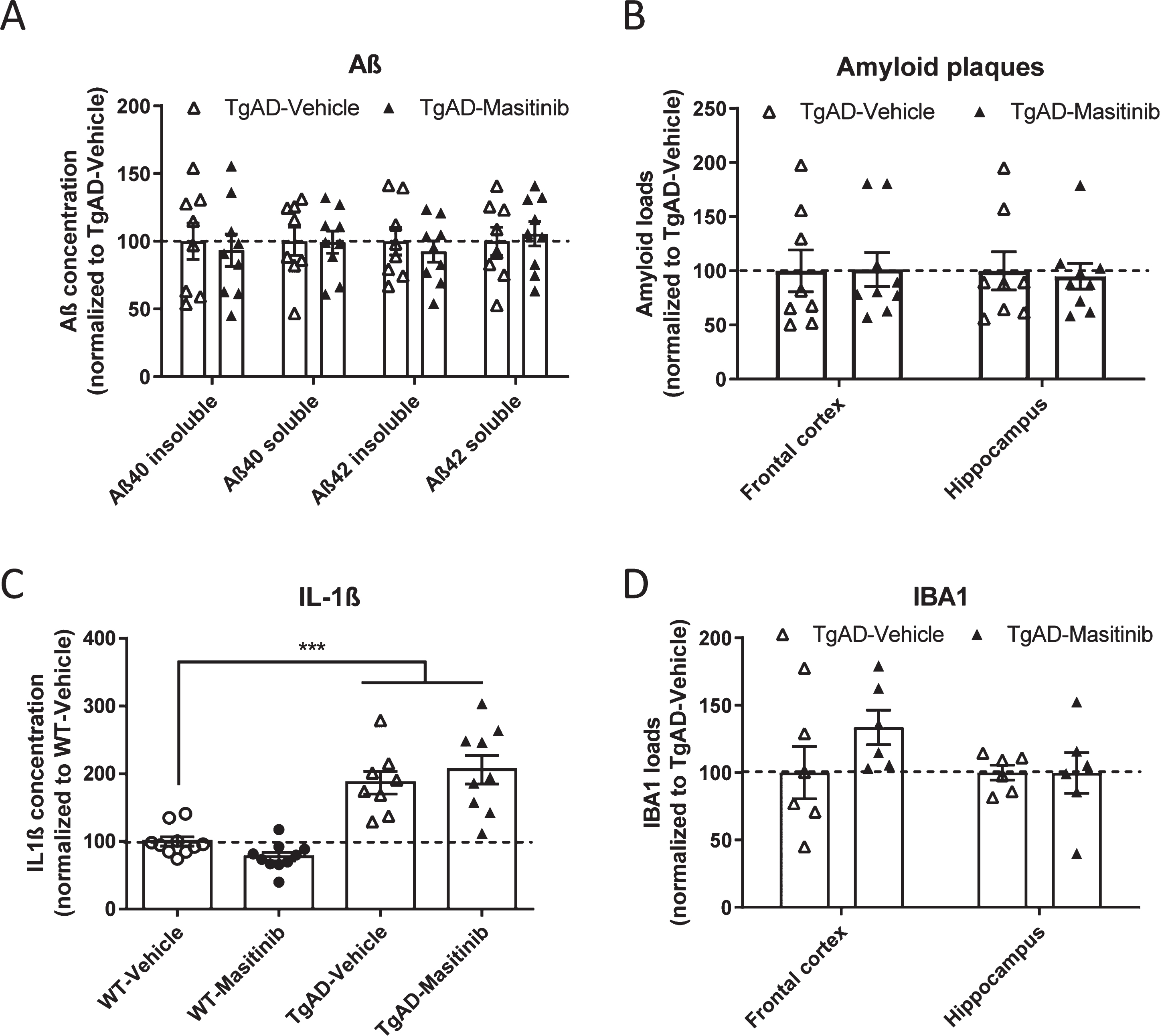
Masitinib does not impact brain amyloidosis and inflammation in TgAD mice. A) Biochemically-assessed brain Aβ (soluble and insoluble) concentrations in TgAD mice. B) Histologically-assessed amyloid loads of TgAD mice in the frontal cortex and hippocampus. C) Brain (whole hemisphere) dosage of IL-1β in WT and TgAD mice. D) IBA1 loads of TgAD mice in the frontal cortex and hippocampus. Data expressed as mean±SEM. ***p < 0.0001.
The two TgAD groups under vehicle or masitinib treatment showed comparable IBA1 loads in brain tissue underlining an overall constant number of microglial cells (comparison of IBA1 loads in TgAD-Vehicle versus TgAD-masitinib groups, in frontal and hippocampal areas: all ts(10)<1.44; ps > 0.18; Fig.2D). Similarly, no difference in GFAP loads were observed in the two transgenic groups (All ts(10)<0.54; ps > 0.84) indicating equivalent levels of astrocytocis (Supplementary Figure 1A).
Synaptic integrity
To evaluate possible correlates of pro-cognitive effects of masitinib treatment in TgAD mice, we assessed the drug’s effect on synaptic markers (Fig.3A). As expected, a strong decrease in synaptophysin immunoreactivity was detected in TgAD-Vehicle mice when compared with WT-Vehicle animals (t(15) = 4.4; p < 0.0005), confirming synaptic anomalies in this transgenic line. Chronic treatment with masitinib significantly increased by 20% synaptophysin immunoreactivity in TgAD mice (comparison Tg-Vehicle versus TgAD-masitinib: t(15) = 2.92; p < 0.01). No difference were observed between WT-Vehicle versus TgAD-masitinib mice (t(16) = 2.11; ns) indicating recovery of synaptic integrity in treated Tg mice.
To assess the hypothesis that masitinib protective effect on synapse is related to MCs inhibition, we generated a new model of APPswe/PSEN1dE9 mice in which c-Kit expression was knocked-down leading to a complete depletion of MC populations. This allowed us to investigate the consequences of mast cell deficiency in TgAD mice. Paralleling observations in 14-month-old mice (see above), a lower but still significant decrease in synaptophysin immunoreactivity was evidenced in younger (10-month-old) TgAD mice as compared with WT littermates (t(17) = 2.111; p < 0.05; see Fig. 3B). Mast cell deficiency in TgAD mice promoted an increase of synaptophysin immunoreactivity (comparison TgAD versus TgAD/Wsh: t(22) = 2.80; p < 0.025), mimicking the effects of pharmacological treatment with masitinib and allowing a full recovery of synaptic integrity in this genotype (comparison TgAD/Wsh versus WT: t(23) = 0.56; ns).
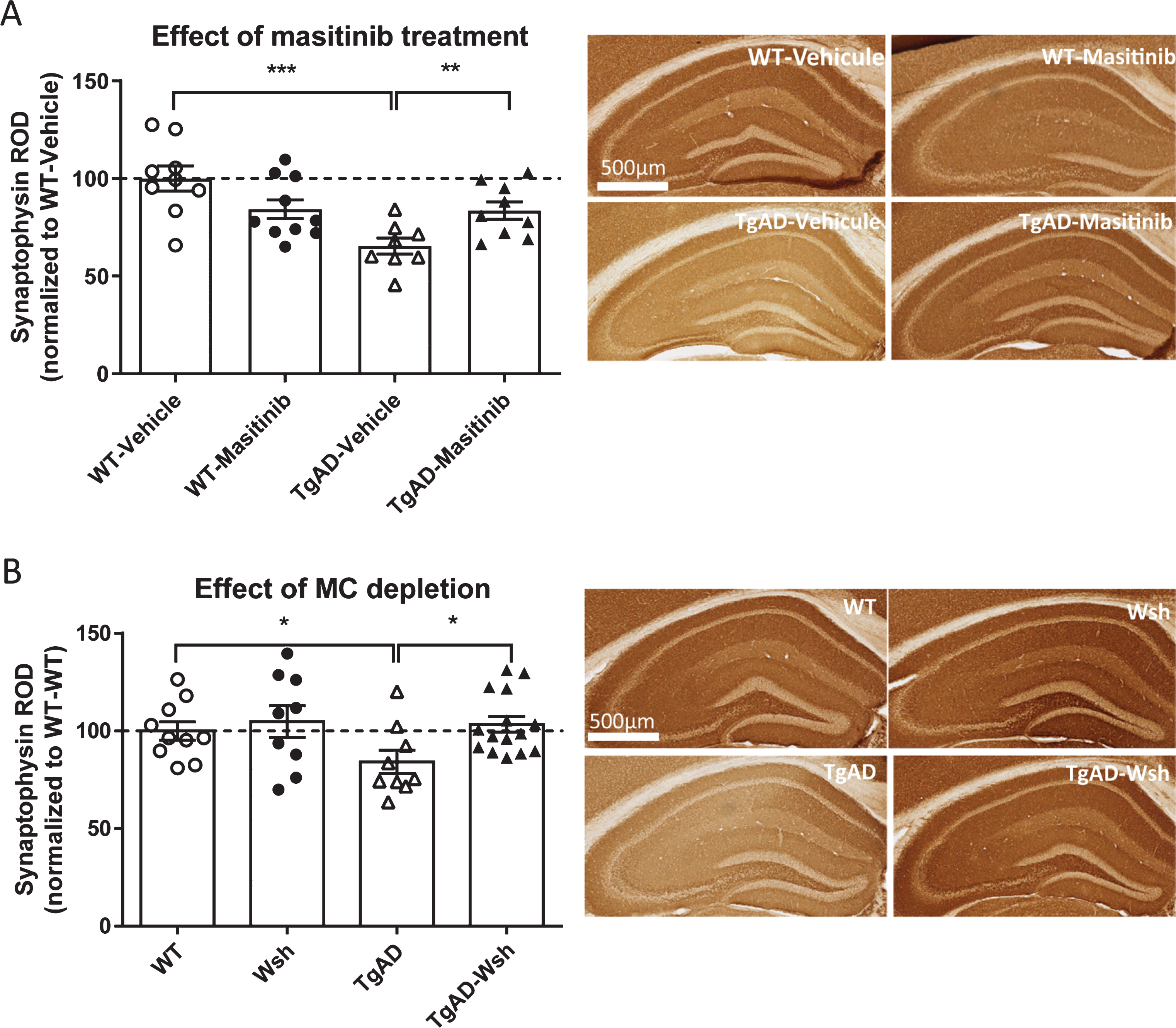
Masitinib and mast cell-deficiency induce a recovery of synaptic markers in TgAD mice. A) Relative optical density of synaptophysin immunoreactivity in the hippocampus of WT and TgAD mice treated with Vehicle or masitinib (left part) and representative microphotographs illustrating synaptophysin immunoreactivity levels in the four studied groups (right part). B) Relative optical density of synaptophysin immunoreactivity in the hippocampus of WT and TgAD mice with or without mast cells depletion induced by the Wsh mutation (left part) and representative microphotographs illustrating synaptophysin immunoreactivity levels in the four studied groups (right part). Data expressed as mean±SEM.***p < 0.0001, **p < 0.01, *p < 0.05.
DISCUSSION
The functions of MCs, initially focused to the immune system and in particular to allergic responses, have been broadened in recent years. MCs are now viewed as key actors in different pathologies including neurological conditions. The mechanism of action of masitinib leading to cognitive improvement in AD [2] remains, however, unidentified and several pathways could be involved relying on blockage of MC activity, and/or inhibition of different kinases [15].
In the present study we investigated the effects of oral masitinib treatment in APPswe/PSEN1dE9 transgenic mice that develop amyloid plaques and are cognitively impaired. We showed that a 2-month chronic treatment mitigates spatial learning impairment in 14-month-old animals, therefore replicating the clinical amelioration described by Piette and collaborators in AD patients [2]. In addition to an amelioration of cognitive phenotypes, APPswe/PSEN1dE9 mice treated with masitinib also demonstrated synaptic protection, as previously demonstrated in a rat model of amyotrophic lateral sclerosis [16]. These effects were not however paralleled by a reduction of Aβ brain levels or of histologically-assessed amyloid plaques loads. In addition, we did not find evidence that chronic treatment with masitinib modified brain dosage of IL-1β cytokine, a key pro-inflammatory mediator expressed in the APPswe/PSEN1dE9 mice used in the present study [11] and pro-inflammatory chemokines remained similarly unaffected after drug treatment. Also, microglial and astrocytic densities were not modified by masitinib treatment. These observations argue against the hypothesis of an effect of masitinib based on a strong modulation of brain amyloidosis or of neuroinflammation.
Importantly, a synapto-protective action of masitinib was evidenced in APPswe/PSEN1dE9 mice. This effect was mimicked in mice depleted in MCs (APPswe/PSEN1dE9/Wsh model) strongly suggesting that targeting MCs was critical in the mode of action of masitinib. To our knowledge, this is the first indication of a deleterious effect of MC on synapses in an AD background.
Therapeutic effect of masitinib may be directly related to lower secretion of specific mediators that would be toxic for the synapses. Indeed, MCs are at the source of a vast array of cytokines and other molecules known to impact on synaptic structure and function [17, 18]. MCs can behave as an early sensor of Aβ accumulation in the brain [19] and trigger the production of neuro- and synapto-toxic factors. Reducing the release of such mediators is expected to be beneficial. Further investigations are needed to unravel the detailed mechanisms involved.
In summary, our study indicates that masitinib mitigates synaptic pathology and cognitive anomalies in APPswe/PSEN1dE9 mice, through an MC-dependent mechanism. These results provide new experimental support and compelling biological rationale for the use of masitinib in the treatment of AD.
Footnotes
ACKNOWLEDGMENTS
The behavioral studies and the histological studies of this work were carried out respectively on the PHENOPARC platform and the HISTOMICS platform of the ICM and we thank all technical staff involved.
This work was supported by BPI France, INSERM, CNRS, Université Sorbonne, and program “Investissementsd’avenir” ANR-10-IAIHU-06 (IHU-A-ICM). The funders had no role in the study design, data collection and analysis, decision to publish, or manuscript preparation. The content is solely the responsibility of the authors.