
Abstract
BACKGROUND:
Murta, a native berry from southern Chile, has been used in Chilean folk medicine to treat inflammatory and infectious diseases among other ailments.
OBJECTIVE:
This work assessed the influence of different drying methods: freeze drying (FD), convective drying (CD), vacuum drying (VD), sun drying (SD), and infrared drying (IRD) on the antimicrobial activity of murta berries against four microbial species.
METHODS:
Murta berries were subjected to five drying methods. Measurement of bioactive compounds that include: phenolic compounds by HPLC, total flavonoid content by a spectrophotometric method; and anthocyanins by HPLC–MS. Determination of antioxidant capacity by DPPH and ORAC methods and antimicrobial activity by means of agar well diffusion assay.
RESULTS:
Murta extracts obtained by FD and CD showed the highest antimicrobial activity, with Staphylococcus aureus the most susceptible species. Drying induced a significant loss of bioactive compounds and antioxidant activity although minimal losses were observed in FD, CD and VD extracts. In these extracts, the abundance of bioactive compounds correlated with the antimicrobial activity. Eight phenolic compounds were identified in murta extracts where pyrogallol’s abundance increased in all dried samples.
CONCLUSIONS:
Our results suggest that murta dried with FD, CD and VD have the highest potential to be used as a functional ingredient in the food industry.
Introduction
Across history, plants have been regarded as a rich source of antimicrobial compounds. However, not until recent, special attention has been paid to edible plants, especially those rich in secondary metabolites (essential oils, polyphenols, etc.). Today, there is a growing interest in the antimicrobial activity of the phytochemicals consumed in the human diet [1]. Among plant phytochemicals, phenolic compounds have received a great deal of attention due to their diverse biological functions, mainly to their antimicrobial activity [2]. Among polyphenols, flavonoids are organic heterocyclic compounds which have been used in many cultures over the centuries as traditional medicine to treat and prevent various infectious diseases and intoxications. These include wound, urinary tract and respiratory infections, ulcers, acne and gastrointestinal diseases. Not in vain, the flavonoid family is the subject of much antibacterial research [3].
Consumption of berry fruits has become important in the promotion of human health, mainly due to the presence of phenolic compounds, which have been associated with protection against various pathologies and due to their anti-inflammatory, gastroprotective, antimicrobial and other biological activities [4]. Consequently, there has been a growing interest to identify natural antioxidants and antimicrobials from berry plants. Several edible Myrtaceae fruits including the Chilean berry murta (Ugni molinae Turcz) are a good source of polyphenolic antioxidants. Chilean native cultures as the Mapuche, Puelche and Pechuenche have long consumed this berry for their wholesome and anti-inflammatory effects [4]. According to previous studies [5], the content of phenolic acids found in murta berry extracts could be a source of potential antimicrobial substances.
Drying is the most common food processing method employed to increase the shelf life of food and food products. The reduction of moisture content during drying inhibits microbial growth and delays deteriorative biochemical reactions [6]. Several studies have evaluated the effects of different drying methods on the abundance and composition of bioactive compounds. Farag et al. [7] reported that freeze drying retained more bioactive compounds in garlic. The authors also showed that freeze dried garlic extracts had higher inhibitory responses towards Bacillus subtilis compared to microwave and air-drying. The aim of this study was to determine how different drying methods (freeze drying, convective drying, vacuum drying, sun drying, and infrared drying) impact the antimicrobial activity of murta berry extracts and to relate these changes in antimicrobial activity with phenolic profile, antioxidant capacity and anthocyanin content.
Materials and methods
Raw materials and drying methods
Murta (Ugni molinae Turcz) berries were purchased from local markets in Valdivia (38°48′30′′S and 73°14′30′′W). The berries were hand selected by color and size to provide a homogeneous group and stored at 5°C before the drying process.
Five different methods were used to study the effect of drying. Freeze-drying (FD) was carried out in a freeze-dryer (Virtis Benchtop model, 3 L, Gardiner, NY, USA) for about 48 h with the condenser temperature and chamber vacuum at – 50° C and 12.5 Pa, respectively. Fresh murta berries were previously frozen at – 80°C before the freeze-drying process. Convective drying (CD) was performed at 60°C and constant air flow of 2.0±0.2 m/s with a load density of 4.6±0.4 kg/m2 during 12 h until samples reached constant weight in a convective dryer designed and built at the Food Engineering Department of the Universidad de La Serena [8]. Vacuum drying (VD) was performed at 60°C and 15 kPa for near 4 h in a vacuum oven (Memmert, model VO 400, Frankfurt, Germany) until samples reached constant weight. Sun drying (SD) was conducted for two weeks during the months from March to May in Elqui Valley, Chile, in an open glass container (90×60×40 cm) with approximately 8 hours sunlight and a maximum temperature of 50°C measured in the middle of the container. Fresh murta (900 g) was spread onto a glass receptacle with a charge density of 2,5 kg/m2. The drying was variable during day light and recorded with a data logger (lascar EL-USB-2). The temperature ranged from 40.5° to 50.5°C and air humidity ranged from 45.5 to 60.5%. Samples were dried until reaching a constant weight. Infrared drying (IRD) was carried out in an electric infrared radiation oven (Teka, model HT490, Germany) at 60°C for 10 to 12 h at a load density of 4.6±0.4 kg/m2 until samples reached constant weight.
The AOAC method (AOAC n° 934.06, 1990) were used to determine moisture content (AOAC). The initial moisture content of murta berry was 84.25±0.28 g/100 g sample. After drying, the moisture content decreased significantly (P < 0.05) and the final value obtained varied depending on the applied drying method. The initial moisture, expressed in g/100 g dry matter (d.m.), were found to be 12.00±0.20 in FD samples, 15.24±0.22 in CD samples, 14.06±0.30 in VD samples, 9.44±0.12 in SD samples and 17.77±0.14 IRD samples.
Preparation of berry extracts
Berry extracts were prepared according to Shene et al. [9] with some modifications. Briefly, fifteen grams of fresh murta or 5 g of dried murta powder (size <0.5 mm) were mixed with 50 mL ethanol (50%, v/v). The mixtures were incubated with agitation in an orbital shaker (OS-100, Shanghai, China) at 200 rpm for 24 h in the dark at room temperature. Subsequently, the extract was filtered through Whatman N° 1 filter paper and centrifuged at 2,500×g at 5°C for 10 min (Universal 320R centrifuge, Hettich, Tuttlingen, Germany). The supernatant containing the soluble extract was evaporated to dryness in a rotary evaporator at 40°C (Büchi, RE 210, Flawil, Switzerland). Dried extracts were resuspended in methanol (80%, v/v) or in sterile distilled water according to the type of measurement.
Antimicrobial activity
Microorganisms and growth conditions
Berry extracts were tested for antimicrobial activity against four microorganisms, Staphylococcus aureus (ATCC 25923) (Gram-positive), Escherichia coli (ATCC 25922) (Gram-negative), Penicillium sp. (mold) and Saccharomyces cerevisiae (yeast). Microbial strains were maintained as 20% glycerol stocks at – 80°C in nutrient broth (Difco). Prior to the experiments, cultures were transferred to solid or liquid media. The bacterial and fungal strains were grown aerobically for 48 h in 5 mL of Mueller Hinton broth (MHB, Merck), continuously shaken at 120 rpm at 37°C. Cultures were then transferred to tryptone soya broth (TSB, Difco) incubated for 12 to 24 h and used as inoculum source for each experiment.
Antimicrobial activity (in vitro) of extracts
Well diffusion method Initial assessment of the antimicrobial potential of berry extracts was evaluated by the well diffusion method according to Kaymak et al. [10], with some modifications. Six mm discs were impregnated with 10 μL berry extract dissolved in sterile distilled water and placed on agar (15 mL) inoculated with 0.5 mL bacteria (0.5 McFarland standard turbidity) or fungi homogeneously distributed in Petri dishes. Amoxicillin (30 μg/mL) was used as the reference antibacterial agent, while fluconazole (25 μg/mL disc) was used as the reference antifungal agent. Negative controls were prepared using the same solvent employed to dissolve the extracts. Plates inoculated with bacteria were incubated on tryptone soya agar (TSA, Difco) at 37°C for 24 h and plates with fungi on Potato Dextrose Agar (PDA, Difco) at 30°C for 48 h. The diameter of the inhibition halo was measured with a digital caliper in millimeters.
2.3.2.2. Minimum inhibitory concentration (MIC) and minimum bactericidal concentrations (MBC). To quantify the antimicrobial activity of berry extracts, the MIC and MBC were determined. The minimal inhibitory concentration (MIC) of all extracts was determined by the tube broth dilution assay [11]. The stock solution of each berry extract (100 mg/mL) was diluted to concentrations ranging from 0.02–50 mg/mL in tryptone soya broth (TSB, Difco). After careful mixing, a 10 μL inoculum containing 1×10⁸ CFU/mL of each microorganism was added to media containing berry extract dilutions. Samples were incubated at 37°C for 24 h (bacteria) or 30°C for 48 h (fungi). The MIC was determined as the lowest extract concentration that prevented the appearance of visible growth. The minimum bactericidal concentration (MBC) was determined from MIC samples in which there was no visible turbidity. Samples were plated on TSA (Difco) and incubated as described above. Plates showing no growth indicated a bactericidal effect of berry extracts.
Bioactive components analysis
Determination of total phenolic and flavonoid content
The total phenolic and flavonoid content from berry extracts was determined spectrophotmetrically. Total Phenolic Content (TPC) was determined colorimetrically by using the Folin–Ciocalteu reagent according to Uribe et al. [12]. The TPC content was calculated from a gallic acid (GA) calibration curve (y = 0.003x + 0.036, R2 = 0.994) and results were expressed as mg GA/100 g dry matter (d.m.). Determination of the total flavonoid content (TFC) was performed according to the aluminum chloride colorimetric method described by Dini et al. [13]. Quercetin (QE) was used for establishing the standard curve (y = 0.0025x + 0.0088, R2 = 0.992). Results were expressed as mg QE/100 g d.m.
Identification and quantification of phenolic compounds by HPLC
Phenolic compounds were identified and quantified according to Rodríguez et al. [14] in a High-Performance Liquid Chromatography (HPLC) system (Agilent 1200, Santa Clara, CA, USA), equipped with a high-pressure pump, an automatic injector, an UV-visible-diode array detector (DAD). Quantification of the identified compounds was performed by comparing their retention times, spectra and the peak area of maximum absorption wavelength with the calibration curves of the corresponding standards. The content of phenolic compounds was expressed in mg per 100 g dry matter (mg/100 g d.m.). All reagents were of analytical HPLC grade Merck KGaA, Darmstadt, Germany) and standards were from Sigma Chemical Co. (St Louis, MO, USA).
Determination of in vitro antioxidant capacity (DPPH and ORAC assays)
The antioxidant capacity of berry extracts was evaluated in vitro by two methods.
Extraction of anthocyanins
Anthocyanins were extracted in an acidified methanol solution according to Brauch et al. [16]. Approximately 8 g of ground fresh murta or 4 g of dried murta powder (size <0.5 mm) were mixed with 10 mL acidified 100% methanol (0.1% HCL, v/v) for 30 s using a probe sonicator (Branson, 2510 E-MTCT, Danbrury, USA). After centrifugation for 3 min at 1048×g (Eppendorf 5804R, Hamburg, Germany), the methanolic phase was separated, and solid residues were re-extracted 3–4 times until the berries or berry powder became colorless. Supernatants were combined, concentrated to dryness at 40°C using a rotary evaporator (Büchi, RE 210, Flawil, Switzerland) and resuspended in sterile filtered deionized water (0.45 μm), and the resulting solution was freeze-dried.
Identification of anthocyanins by HPLC–MS
Anthocyanins were identified by HPLC in an Agilent 1100 system (Agilent Technologies Inc., CA-USA) connected to an Ion Electrospray Trap Mass Spectrometer Esquire 4000 ESI-IT (Bruker Daltonik GmbH, Germany). Anthocyanins were separated using an end-capped Kromasil 100-5C18 analytical column (Eka Chemicals, AB, Sweden). ChemStation for LC 3D software (Agilent Technologies Inc., CA, USA) was used for control of the HPLC and EsquireControl 5.2 software (Bruker Daltonik GmbH, Germany) of the mass spectrometer. The analysis was performed at room temperature by the injection of 20 μL of blank (water/acetonitrile/formic acid; 87/3/10, v/v/v) and samples (30 mg/mL), using a gradient system composed by two phases, (A) water/acetonitrile/formic acid (87/3/10, v/v/v) and (B) water/acetonitrile/formic acid (40/50/10, v/v/v). Gradient elution was: 0–15 min 6.0% B, 15–30 min 30% B, 30–35 50% B, 35–41 min 60% B and 41–45 min 6.0% B. Flow rate was 0.8 mL/min and UV detection at 520 nm. The ionization process (nebulization) by electrospray was performed at 3,000 V using nitrogen as nebulizer gas at 365°C, a pressure of 50 psi and a flow rate of 10 L/min. Mass spectra were obtained in a positive polarity.
Statistical analysis
All analyses were performed in triplicate and results were expressed as mean± standard deviation (SD). The experimental design used consisted of one factor (k = 1) to study at six levels (n = 6, five drying methods and the fresh sample as control). The software Statgraphics® Centurion XV.I (Statistical Graphics Corp., Herndon, VA, USA) was used for data analysis (ANOVA). Differences among the media were analyzed using the least significant difference (LSD) test at significance level of α= 0.05 and a confidence interval of 95% (P < 0.05). A multiple range test (MRT) was also performed to demonstrate existence of homogeneous groups within each of the parameters.
Results and discussion
Antimicrobial activity
The antimicrobial activity of ethanolic murta extracts was evaluated on four microbial species, a Gram-negative (Escherichia coli), a Gram-positive (Staphylococcus aureus), a yeast (Saccharomyces cerevisiae) and a mold (Penicillium sp.). Microorganisms were tested for their sensitivity to murta extracts from fruits dehydrated by the five different drying methods, freeze drying (FD), convective drying (CD), vacuum drying (VD), sun drying (SD) and infrared drying (IRD). Antimicrobial activity was initially determined by the well diffusion method as a preliminary screening. All murta extracts (200 mg/mL) produced a clear inhibition halo toward S. aureus which varied from 24.33 mm in VD to 28.33 mm in FD (Table 1). The control (30 μg/mL amoxicillin) produced an inhibition halo of 39 mm. None of the other microbial species showed sensitivity towards murta extracts at the tested concentration. Since the agar-diffusion test is not quantitative and is a simple method to detect microbial growth, the minimum inhibitory concentration (MIC) and minimum bactericidal concentration (MBC) were quantified for all microorganisms studied.
Antimicrobial activity for the berry extracts against some strains tested
Antimicrobial activity for the berry extracts against some strains tested
aDiameters of inhibition zone for the extracts by the agar diffusion method, the values (average of triplicate) in mm at 200 mg/mL. bResults of Minimum inhibitory (MIC) and bactericidal concentration (MBC) by the broth dilution method, mean vale n = 3, the values expressed in mg/mL. (-) No inhibition; *Amoxicillin (30 μg/mL); γ Fluconazole (25 μg/mL).
MIC values ranged from 1.56 to 100 mg/mL for all microorganisms tested and at all drying conditions (Table 1). Taken together, our results show that extracts obtained after FD and CD presented the highest inhibitory activity against all bacterial and fungal species. In agreement with the well diffusion assay, the highest inhibition was against S. aureus (MIC of 1.56 mg/mL) with respect to the rest of the drying methods (Table 1), indicating that S. aureus was the most sensitive microorganism to the extracts examined in this study. The sequence of microorganisms according to their sensitivity expressed as average MICs (in mg/mL) was: S. aureus > Penicillium sp. > S. cerevisiae > E. coli. It should be noted that these results highlight the importance of this novel antimicrobial agent, considering that S. aureus is one of the most common bacteria implicated in food poisoning, producing several types of enterotoxins that cause gastroenteritis, a major food-borne disease in most countries [17]. Drying influenced differently the antimicrobial activity of murta extracts toward each microorganism (Table 1). Among extracts, FD and CD extracts presented the highest antimicrobial activity against all four microbial species. The lower MIC observed after FD and CD correlates with the higher concentration of phenolic compounds quantified in the FD and CD extracts (see Section 3.2.2) implying that phenolics may be responsible for the antimicrobial activity. This statement is supported by additional studies which also suggest that the antimicrobial effect of plant and fruit extracts can be attributed to the presence of phenolic compounds [18]. Mphahlele et al. [19] reported that pomegranate peel extracts freeze- and oven-dried at various temperature ranges exhibited a significant antimicrobial activity against Escherichia coli, Klebsiella pneumoniae, Staphylococcus aureus and Bacillus subtilis. Their results indicated that peel extracts were more effective against the tested bacteria irrespective of the drying methods employed, possibly due to the higher retention of antioxidant activity after drying. Tian et al. [20] characterized the phenolic compounds extracted with acidified aqueous ethanol from several species and cultivars of berries and leaves and investigated and compared the antioxidative and antimicrobial activities of these extracts. Their results indicated higher sensitivity of Gram-positive than Gram-negative bacteria to the phenolic extracts. The total content of phenolic compounds and the content of non-flavonoid phenolics showed stronger association with the inhibitory effects Staphylococcus aureus and Bacillus cereus than the total content of flavonoids.
Puupponen-Pimiä et al. [21] reported that phenolics extracted from berries affected the growth of several bacterial species by different mechanisms, yet the mechanisms were not well understood. These may include complex interactions between the growth media’s pH and the antimicrobials extracted from berries.
According to Michielin et al. [22], natural products can be classified as antimicrobial agents based on their exerted MIC values. Strong inhibitors are considered those with a MIC lower than 0.5 mg/mL, moderate inhibitors with a MIC between 0.6 and 1.5 mg/mL and weak inhibitors with a MIC above 1.6 mg/mL [18]. The MIC values obtained in this study classify murta extracts as weak inhibitors of E. coli and Penicillium sp. and as moderate inhibitors of S. aureus and S. cerevisiae, when murta was subjected to dehydration by FD and CD. Among all four microorganisms, E. coli (Gram-negative) showed the highest resistance against murta extracts, whose growth was not inhibited by 25, 50 or 100 mg/mL (Table 1). This higher resistance might be related to the cell wall complexity of Gram-negative bacteria whose outer membrane contains highly hydrophilic surfaces rendering it less susceptible to the action of many antimicrobial compounds [23]. Ali et al. [24] indicated that Gram-negative organisms are less susceptible to the effect of antibacterial agents, due to the fact that they possess an outer membrane surrounding the cell wall. Concluded that Gram-negative bacteria having more hydrophobicity surface can be substitute by having its porin proteins in the outer membrane. The extracts were not able to penetrate through the outer membrane, which was consisting of a lipopolysaccharide monolayer surrounding the cell wall that limited diffusion of hydrophobic compounds.
Although the antimicrobial activity of murta berries has been scarcely studied, previous studies have shown a higher activity against E. coli, in the range of 1.2 to 8.3 mg/mL for MIC and 1.7 to 10 mg/mL for MBC [23]; these differences may lay on the extraction conditions. On the other hand, murta extracts showed a higher activity toward S. aureus, than that reported for blueberry extracts where MIC and MBC values exceeded 50 mg/mL [25]. To the best of our knowledge, neither MIC nor MBC have been reported against S. cerevisiae and Penicillium for murta berry extracts. However, López de Dicastillo et al. [23] reported antifungal activity of murta extracts against Penicillium expansum where hydroalcoholic extracts produced an inhibition growth of 3.79±0.22%.
Total phenolic and total flavonoid content
As many antimicrobial compounds are detected in the phenolic and flavonoid fractions from ethanolic extracts [4], bioactive compounds were extracted with 50% ethanol and the total phenolic content (TPC) and total flavonoid content (TFC) quantified before and after application of the different drying methods (Fig. 1). Fresh murta contained a total of 1068.59 mg GAE/100 g d.m. phenols and 527.02 mg QE/100 g d.m. flavonoids. These values are similar to those reported by Ramirez et al. [26] in myrtle berries with 924 mg GAE/100 g d.m. for TPC and 554 mg QE/100 g d.m. for TFC. The TPC observed in this work was lower compared to the values reported in murta berries by Augusto et al. [27], and Reyes et al. [28], which were 1935 and 1460 mg GAE/100 g d.m., respectively. Likewise, the TFC value from fresh murta was lower than the 924 mg QE/100 g d.m. reported by Brito et al. [29].
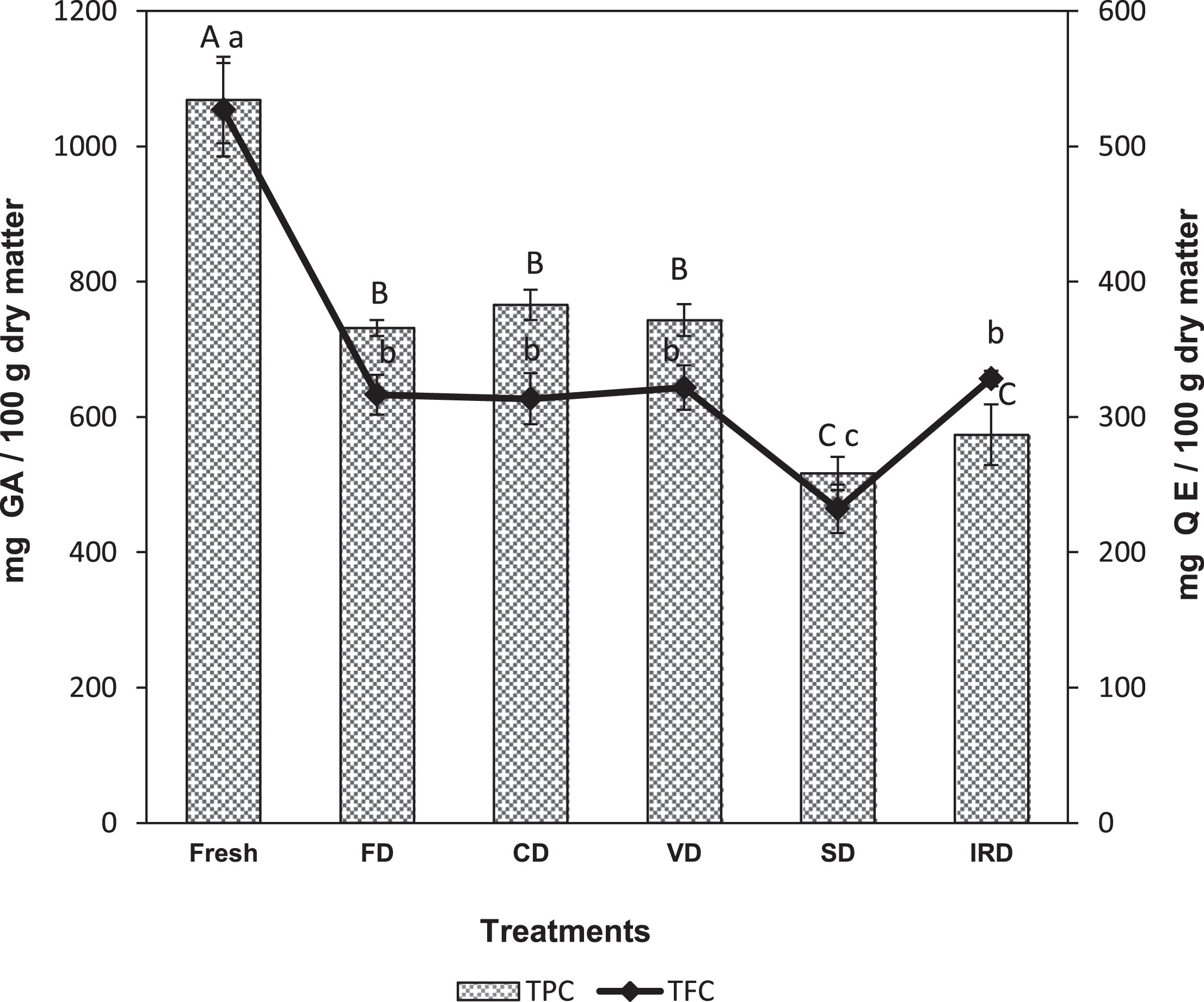
Total phenolic content and total flavonoid content of fresh and dried murta berries. Different letters A, B, C, indicate significant differences (P < 0.05) in TPC between drying treatments and a, b, c (P < 0.05) in TFC between drying treatments.
The TPC in murta dehydrated by the five different methods varied from 516.61 mg GAE/100 g d.m after SD to 765.62 mg GAE/100 g d.m after CD. These values correspond to a significant (P < 0.05) decrease in 28–52% TPC content compared to the fresh murta berries. Among drying methods, FD, CD and vacuum VD retained the highest amount of phenolics, without significant differences between these methods (P > 0.05). Likewise, there were no significant differences (P > 0.05) between SD and IRD murta samples. This behavior was also reported by Henríquez et al. [30] which observed a decrease in TPC from apple peels, indicating that these large losses of phenolic compounds in the dehydration processes are caused by oxidation, degradation and/or enzymatic inactivation and volatilization.
As with TPC, all drying methods caused a 30–56% decrease in TFC compared to the fresh samples. Sun drying induced the highest decrease (56%) whereas the TFC from FD, CD, VD and IRD murta was approximately 30% lower than the fresh samples, without significant differences between these methods (P > 0.05). Similar losses of flavonoids were previously reported in murta dried by VD, CD, FD and SD [31]. The decrease in TPC and TFC after drying may be due to the binding of these compounds to other molecules or macromolecules (such as proteins). Alternatively, alterations in the chemical structure of phenols and flavonoids may result in chemical conversions to other compounds that cannot be quantified by traditional methods [31].
The knowledge of the profile and abundance of phenolic compounds in foods is of paramount importance to assess the health benefits conferred by the food [32]. Eight phenolic compounds were identified in fresh and dried murta berries by HPLC-RP-DAD (Table 2). These compounds were characterised by their retention times and UV absorption spectra. Five phenolic compounds were detected in fresh murta, from which catechin appeared in the highest abundance (171.94 mg/100 g d.m.) followed by tryrosol, pyrogallol, 3-hydroxytyrosol and gallic acid (83.66, 75.52, 24.84 and 4.70 mg/100 g d.m. respectively). These values are higher than those reported in methanolic extracts from fresh murta, which contained a catechin content of 35.1 mg/100 g d.m. and 0.51 mg/100 g d.m. of gallic acid.
Phenolic acid profile of fresh and dried murta berries
Phenolic acid profile of fresh and dried murta berries
*Sample preparation and HPLC determination were performed in triplicate. n.d.: not detectable. Different letters indicate significant statistical difference (P < 0.05).
All drying methods applied caused a greater extractability (3–6.4-fold increase; P < 0.05) of pyrogallol, which was the main compound found in dehydrated murta berries. The highest pyrogallol values were measured in CD murta (482.23 mg/100 g d.m.). Likewise, CD, VD, SD and IRD murta resulted in a 2–2.9-fold increase in gallic acid content compared to the fresh fruit (4.70 mg/100 g d.m.). In contrast, all drying methods caused degradation (P < 0.05) of catechin, tyrosol and 3-hydroxytyrosol with losses in the range of 39–74%, 55–69% and 16–44%, respectively, compared to fresh murta berries.
It is known that the high temperatures and long times employed during drying processes cause thermal degradation of phenolic compounds in fruits and vegetables [6]. This would explain the low diversity and abundance of phenolic compounds detected in SD murta. Sun drying conditions are generally not stable as they depend on climatic factors and require longer times than controlled processes to achieve moisture equilibrium. This leads to an uneven loss of phenolic compounds. In contrast, at all other drying methods (FD, CD, VD, IRD), murta berries were exposed to short drying times, thus reducing the loss of phenolics. This was particularly evident after CD, whose samples presented the highest abundance of the eight phenolic compounds detected. Furthermore, protocatechuic acid, vanillic acid and quercetin were only found in the dehydrated samples. A plausible explanation is this that at the cellular level, phenolic compounds are stored in vacuoles [33]; thermal processing may cause the breakdown of cellular constituents releasing more bound phenolic acids [34] thus explaining the higher content of phenolic compounds in dried fruits compared to the fresh material.
The importance of phenolic compounds goes beyond their correlation with the antioxidant activity that they exercise since in addition, these compounds also exert antimicrobial effects [32]. The mechanisms of antimicrobial activity exerted by polyphenols are still mostly unknown. Thus, understanding the antimicrobial mechanism of action of these natural bioactive molecules can lead to new technologies for the development of food products with particular nutritional functionalities or for food preservation purposes. Berry-derived antimicrobial compounds, especially flavonoids, may have important applications in the future as natural antimicrobial agents such as the food industry and medicine [35]. In general, the mechanisms thought to be responsible for the toxicity of phenolic compounds toward microorganisms include the destabilization of the cytoplasmic membrane, the permeabilization of the cell membrane, the inhibition of extracellular microbial enzymes, direct actions on microbial metabolism, or the deprivation of the substrates required for microbial growth [5].
The main compound found in dehydrated murta berries was pyrogallol, a hydroxylated phenol with proven antimicrobial action [36]. There are few studies on the possible mechanisms of antimicrobial action of pyrogallol. However, Lima et al. [36] showed pyrogallol activity against multi-resistant bacterial and fungal strains and reported a synergistic effect with two of the antibiotics tested (norfloxacin and gentamicin), but only against Staphylococcus aureus. The authors concluded that the mechanism of action of pyrogallol is through enzymatic inhibition by oxidized compounds.
In murta extracts, compounds such as flavan-3-ols and flavonol glycosides among others were attributed to the high antimicrobial activity found [9]. Catechins belonging to the group of flavan-3-ols are flavonoids with higher antimicrobial activity against Gram-positive bacteria than to Gram-negative bacteria [37]. The antimicrobial activity from fresh and dehydrated murta berries might be related to these compounds as catechins were detected in high abundance in fresh and dehydrated murta berries.
The antioxidant capacity of fresh murta measured by DDPH and ORAC assays was 8902.6 and 28145.9 μmol TE/100 g d.m., respectively (Table 3). These values were lower than those reported by Rodríguez et al. [14] in fresh murta berries. Nevertheless, drying resulted in less antioxidant activity than fresh murta; when comparing all dehydrated samples, the maximum antioxidant capacity was observed after FD, CD and VD for both DPPH and ORAC methodologies, which coincided with the observed TPC values (Fig. 1). This trend is similar to that reported by Rodríguez et al. [14]. Murta berries dehydrated by FD, CD and VD showed a slight decrease in antioxidant capacity measured by DPPH, with respect to fresh murta (8450.83, 8363.96, 8568.66 μmol TE/100 g d.m. respectively); however, there were no significant differences between these samples (P < 0.05). On the other hand, the antioxidant capacity of murta dehydrated by SD and IRD (5107.69, 6080.62 μmol TE/100 g d.m. respectively) was significantly lower (P < 0.05) than that of fresh murta. The antioxidant capacity measured by ORAC resulted in significant lower values for all dried samples compared to the fresh berries (P < 0.05). The lowest loss of antioxidant capacity with respect to the fresh berries was observed after VD (50%) and the greatest loss after SD (64.5%). Reduction of antioxidant capacity is a common outcome after drying processing of berries as shown for the fruits of Saskatoon berry [38], strawberry [39], raspberry [40] and blueberries [41]. For example, the antioxidant capacity of dried Saskatoon berries decreased in 41.3% and 73.9% after FD and CD, respectively, compared to the fresh berries [38]. On the other hand, [39] reported a significant decrease in antioxidant activity by DPPH in lyophilized strawberries (12.8%) and in strawberries dried by convective air (37.9%).
Effect of different drying methods on DPPH and ORAC free radical-scavenging activity in fresh and dried murta berries
Effect of different drying methods on DPPH and ORAC free radical-scavenging activity in fresh and dried murta berries
Different letters indicate significant statistical difference (P < 0.05).
Several reports show an association between the antioxidant activity of murta fruits and leaves and the abundance of polyphenols. However, the identity and antioxidant activity from polyphenols of different fruit species remains poorly elucidated [27]. Krisphnappa et al. [42] suggested that an increase in total polyphenol content and phenolic acids, and changes in the composition of other phenolic acids, flavonoids and anthocyanins may influence the antioxidant and antimicrobial properties of the extracts. The transformation and accumulation of bioactive compounds with a varying degree of antioxidant activity during food dehydration may lead to antagonistic or synergistic effects with themselves or with the other constituents of the food matrix. These complex chemical interactions that influence the antioxidant and antimicrobial properties from bioactive compounds are still under investigation.
Eleven anthocyanins were identified based on their retention time, elution order and ESI mass spectrometric data (Table 4). Identification was supported by comparing the obtained anthocyanin peaks with an anthocyanin library prepared from Aristotelia chilensis, Berberis Microphylla and Vaccinium corymbosum extracts. Of the eleven identified anthocyanins, five were aglycones (delphinidin, cyanidin, peonidin, petunidin and malvidin). Among these five anthocyanin aglycones, peonidin aglycone was the most abundant component, with four derivatives (peaks 5, 6, 8 and 9) (Table 4).
Retention times and Mass Spectra (positive ionization mode) of anthocyanins tentatively identified in fresh and dried murta berries
Retention times and Mass Spectra (positive ionization mode) of anthocyanins tentatively identified in fresh and dried murta berries
The highest content of anthocyanins was measured in fresh murta, followed by murta dried by FD. All drying methods resulted in a significant (P < 0.05) decrease in anthocyanin content and the degree of these changes was dependent of the method used. Murta dried by FD retained a higher content of anthocyanins compared with samples dried by the other methods. For example, CD caused important losses where no anthocyanins could be identified (Table 4). The decrease in anthocyanins has been reported in many studies where anthocyanins are readily destroyed by heat during food processing. Castagnini et al. [43] also reported that CD caused greater losses in anthocyanin content. These authors indicated that the losses were greater at 30 and 50°C and that the combination of temperature and treatment time also influenced the degradation of anthocyanins. Chromatograms of fresh and FD murta revealed the presence of five anthocyanins, where peonidin-O-hexoside was the predominant form (Fig. 2). Zhao et al. [44] reported that the anthocyanins of purple corn showed antimicrobial activity against four pathogenic bacteria. They indicated that the activity was related to the presence of cyanidin derivatives whose abundance was 73.96%, showing the potential relationship of these derivatives with the strong antimicrobial activity. There are few studies on the anthocyanins composition of murta berries [4, 29]. This is the first time peonidin-O-hexoside has been reported in murta berries. Previously identified anthocyanins include 3-O- rutinose, 3-O-arabinosides, 3-O-glucosides, and 3-O-galactosides of cyanidin, peonidin, petunidin and delphinidin. Pertuzatti et al. [45] showed that all blueberries studied demonstrated antibacterial activity against E. coli and reported a very complete and systematic description of the anthocyanins present in ten of the most cultivated blueberry varieties. Moreover, they indicated that the variation in antimicrobial activity presented by the different cultivars could be attributed to the fruit pH, sugar content and phenolic compounds. The high sugar content of fruits may promote bacterial growth and reduce the antimicrobial activity of extracts, while a low pH and the presence of phenolic compounds, such as anthocyanins, may result in conditions that are adverse to foodborne pathogens. Studies indicate that among bioactive compounds, anthocyanins have the highest antibacterial effect on foodborne pathogens. However, their mode of action is unknown. Sun et al. [46], reported a strong antibacterial effect in Chinese wild blueberries that are rich in anthocyanins. The authors reported that the anthocyanins antibacterial activity involves damage to the cell membrane and affects alkaline phosphatase (AKP), adenosine triphosphatase (ATPase) and superoxide dismutase (SOD). Anthocyanins also may affect the tricarboxylic acid (TCA) cycle and the biosynthetic routes of pathogens, leading to reduced cell viability and eventual death.
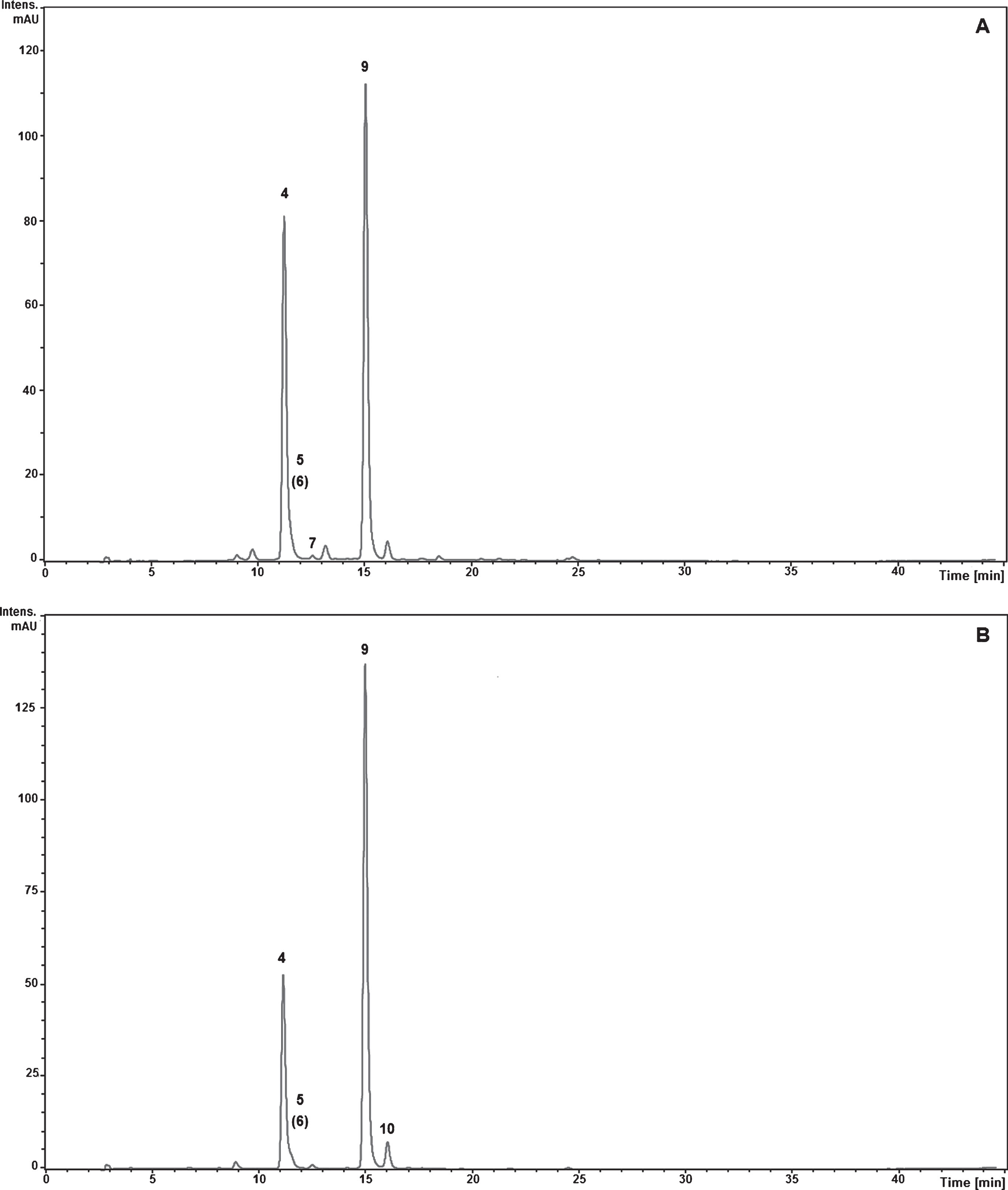
Chromatograms of fresh (A) and FD murta (B) showing the abundance of peonidin-O-hexoside (peak 9). Chromatograms were recorded at 520 nm with HPLC–MS. Peak numbers are described in Table 4.
This study shows that different drying methods have an impact on the antimicrobial activity, phenolic compounds abundance and composition and antioxidant capacity from murta berry extracts. All ethanolic extracts of dried murta possessed antimicrobial activity, whose MIC values classify them as weak inhibitors against E. coli and Penicillium sp. upon drying by FD and CD extracts show stronger antimicrobial activity (lower MIC) and can thus be classified as moderate inhibitors against S. aureus and S. cerevisiae. Compared with fresh murta, dehydrated samples showed significant losses of TPC, TFC and antioxidant capacity. Our evaluation of drying methods suggests that freeze and vacuum drying are the most reliable methods to maintain bioactive compounds with antimicrobial activity against all four tested microbial species. In conclusion, our results show that the selection of a drying method is of high importance to ensure a high content of bioactive substances that are vital for human health.
Conflict of interest
The authors have no conflict of interest to report.
Funding
The authors report no funding.
Footnotes
Acknowledgments
The authors gratefully acknowledge financial support of FONDECYT 1140075.