
Abstract
Digital retinal images are commonly used for hard exudates and lesion detection. An efficient segmentation method is needed to detect and discern the lesions from the retinal area. In this paper, a hybrid method is presented for digital retinal image processing for diagnosis and screening purposes. The goal of this research is to suggest a supervised/semi supervised approach for exudates detection in fundus images and it is also to investigate a technique to find the optimum structure. The image is first transformed into fuzzy domain after an initialization. A cellular learning automata model is used to detect any abnormality on the image which is related to a lesion. The automaton is created with an extra term as the rule updating term to increase the flexibility and capability of the cellular automata. The selection and updating of rule are implemented automatically We also performed allocating the score and penalty value for the cells toward the process of segmentation Three main statistical criteria are introduced as the sensitivity, specificity and accuracy. A number of 50 retinal images with visually detection hard exudates and lesions are the experimental dataset for evaluation and validation of the method. For STARE retina image dataset, for a neighborhood of 5 × 5, score of ϑ = 0.01, penalty of ξ = 0.01, ratio of state overall variation in three sequential cycles in cellular automata
Keywords
Introduction
Two main interesting issues for computer aided diagnosis in ophthalmology are as the automatic detection of blood vessels and detection of the optic disc. The shortcoming of detecting and counting lesions in the human retina like micro aneurysms and exudates are considered as the consuming and high possible to human error therefore a lot of works have been done on order to solve the problem of lesion detection automatically [1]. The most prevalent reason of visual deterioration and the modern overwhelming blindness among workers of industrialized countries is Diabetic retinopathy (DR) which is a visual complication of diabetes [2, 3]. Due to clinical importance and epidemical problem, the researchers had a lot of efforts to improve the methods for diagnosis and medical medication by developing and implementing algorithms to perform retinal image analysis [4], fundus image enhancement [5] and monitoring [6]. Designing automatic image analysis algorithms had special significance to detect hard exudates (HEs) (7). HEs represent the best distinguished markers for the being of retinal oedema which is the main problem of visual loss in non-proliferation forms of DR [2]. Moreover, during early stages of DR, HEs are one of the widespread lesions during initial stages of DR [2].
Many techniques have been presented for HE detection in fundus images considering a variety of methods [7], the methods includes the usage of image contrast and brightness analysis [8–15], Bayesian classifier [16, 17], neural network [18, 19]. In order to segment a candidate bright exudates area, system was developed by Zhang et al. [20] that used local contrast enhancement, Fuzzy C-mean clustering in LUV color space, but the major problem with FCMC is to estimate the number of clusters to use. In order to detect contours typical of exudates, a new mathematical reconstruction was applied by Walter et al. [21]. The results showed the productivity values of 92.4% and sensitivity value of 92.8% with a set of 15 abnormal retinal images. But this method couldn’t distinguish between exudates from cotton wool spots. Gardner et al. [22] used the back propagation neural network in order to perform the segmentation of exudates. If we compare the result of Gardener et al. [22] method with similar expert ophthalmologist, they achieved a sensitivity of 88.4% and specificity of 83.5%. In order to detect the exudates, but it had a drawback which it couldn’t get good result on low quality images.
Many of past techniques have used retinal features which were quite obvious on acquired imagery. To acquire high quality and apparent visible retinal features is time consuming and very difficult and troublesome for patient. Also sometimes there are a number of exudates with different sizes apart or stuck which requires a model based method for detection. Therefore, in this paper the cellular learning automata in fuzzy domain is used for automated and accurate detection of exudates on retinal images. The final objective is to propose a method and system to automatically detecting of exudates, to provide decision support and to reduce workloads for expert ophthalmologists’ by a supervised method which can also act as automation system.
Cellular automata (CA) consist of a regular grid of cells, each of which can be in only one of a finite number of possible states. The previous states of a region of surrounding neighborhood of cells will determine the state of a cell. The state of a cell will be updated in discrete time steps synchronously. The rule which identically contained in each cell is basically a finite state machine It normally forms a rule table with an input for every possible neighborhood configuration of states. Cellular automata are discrete dynamical systems. These are very helpful for implementing and analyzing the phenomena ordering, turbulence, chaos, symmetry breaking, etc. and a lot of applications have been designed in modeling systems in some domains such as biology sociology, Physics Though only a few simple rules are attributed to each cell, one of the main usage of cellular automata is that the combination of matrix of cells with their local interaction leads to more sophisticated emergent global behavior. That is, although each cell has an extremely limited view of the system (just its immediate neighbors), localized information is propagated at each time step, enabling more global characteristics of the overall CA system [27].
In this paper, fuzzified cellular learning automata is introduced for segmentation of digital retinal images. A retinal image dataset consisting 50 retinal images with hard exudates are used to assess the proposed method. The paper is organized as follows: Section 2 presents the dataset and the methodology. Results are given in Section 3 and in Section 4 the results are discussed. Finally, some conclusions will be shown in Section 5.
Materials and method
Image database
Subset of STARE project’s dataset was used in order to obtain main dataset [23‖26]. The subset consists of 81 retinal images for assessing the proposed exudates detection method. The captured images were taken using a TopCon TRV-50 fundus camera, FOV(field of View) = 35° and then it was digitized at 605×700, 24-bits pixel The dataset is used for both the evaluation of the proposed method and also comparison to some other methods. The dataset consists of a total of 50 color retinal images which are taken without pupil dilation with a KOWA-7 non-mydriatic retinal camera with a 45° FOV. The image size is 768×576 pixels at 24 bits per pixel in RGB format. All retinal images analyses are performed on a Core2 Duo 2.2 GHz Laptop using MATLAB 7.6.0 for all implementations.
Transformation of retinal images into fuzzy domain
Let supposed to have an image of size M × N with L gray levels g ranging from 0 to L - 1. The image γ can be viewed as an array of fuzzy singletons [19]. Each element of the array is the membership value μ
γ
i
(g
xy
) of the gray level g
xy
, corresponding to the (x, y) th pixel, regarding to an image property such as brightness, edginess, homogeneity, etc. Using the fuzzy sets notation image γ can be represented as:
To enhance the gray levels across the edges and to make better the contrast of image, gray levels of the pixels are normalized to unit interval [0, 1] corresponding to the whole gray level range [0, 255]. The primary motion matrix is obtained by calculation of Ω function over
Matrix Ω
i
is obtained as the primary motion detected in ith image of the image set Γ. Parameter T1 is the threshold value for motion detection in corresponding
The matrix Ω
i
is then undergone normalization in the [minimum, maximum] range as
Therefore matrix Ω i is normalized to the range [0, 255]. Equation (5) represents also a defuzzification formula though some elements of the matrix are out of the unit interval. Now, Cellular Learning Automata is used to detect the object of interest in the primary motion matrix Ω i which has been converted to gray scale image format.
A retinal image fuzzy automaton in the Fuzzy Cellular Learning Automata is created as
States in the automaton corresponds to decision variables of the problem. The rules are set as transition functions:
The abbreviations which are used in formula (20), (21) and (22)
Where ρ
i
is the accordance factor for the ith rule, state rules for this equation at above the conditional term can be considered as similar to process of searching within a look up table. Threshold ρ
i
selection directly specifies the sensitivity to the rules selection and to the state changes. Very low ρ
i
values lead to significant score assignments and it may result to enlarging the state values and also vice versa. Therefore, this parameter can be justified in each time interval of the cellular automata and this updating process leads to a learning scheme. Threshold value ρ
i
is updated in each cycle according to this formula:
For lesion detection in retina images, a binary image comprising two segments of healthy area of retina as background and the detected lesion(s) as foreground is desired. Therefore, since after the cycle t = τ, the resulting image is in the fuzzy domain and its pixels are in unit interval, a non-sensitive threshold ς ranged between 0.1 and 0.9 is selected to produce is binary image I
b
as
Segmentation quantitative criteria
To report the performance of the classifier, sensitivity, specificity and accuracy as three common criteria are reported and calculated as follow:
A number of 50 retinal images with hard exudates and lesion detected visually are considered as the dataset to evaluate the segmentation method which was proposed. Implementations of the proposed method were done in MATLAB software suing image processing toolbox. All codes were developed in MATLAB M-files while a set of the images are copied in the same directory as the M-file. The program consists of four main parts as the denoising step, initialization part, segmentation method and image binarization and assessment part.
In our experiments, we first evaluate our methods to find the optimum parameters values and the most suitable structure. Then, the results of the retina exudates detection for the proposed method are compared to the results of some other methods in recent literature. All detections are executed for lesion based images with no limitations in the number and regions of the lesions. If a lesion overlaps at least in part with the ground truth then it be called true positive (TP), if in the automatic segmentation there can be not found any corresponding lesion then it be called a False Negative (FN), If here be an exudate in automatic segmentation but there is no corresponding lesion has been manually segmented and True Negative is the true assignment to the non-lesion regions. In the first part of the experiments, the effects of score and penalty values for two iteration values are studied. All runs have been done for two neighborhood sizes of 3×3 and 5×5. Samples of the original retinal images with hard exudates are shown in Fig. 1. Figure 2 shows the fuzzifed images for which all pixels’ grayscale are normalizedto (0,255).

Original Retinal Images with hard exudates.
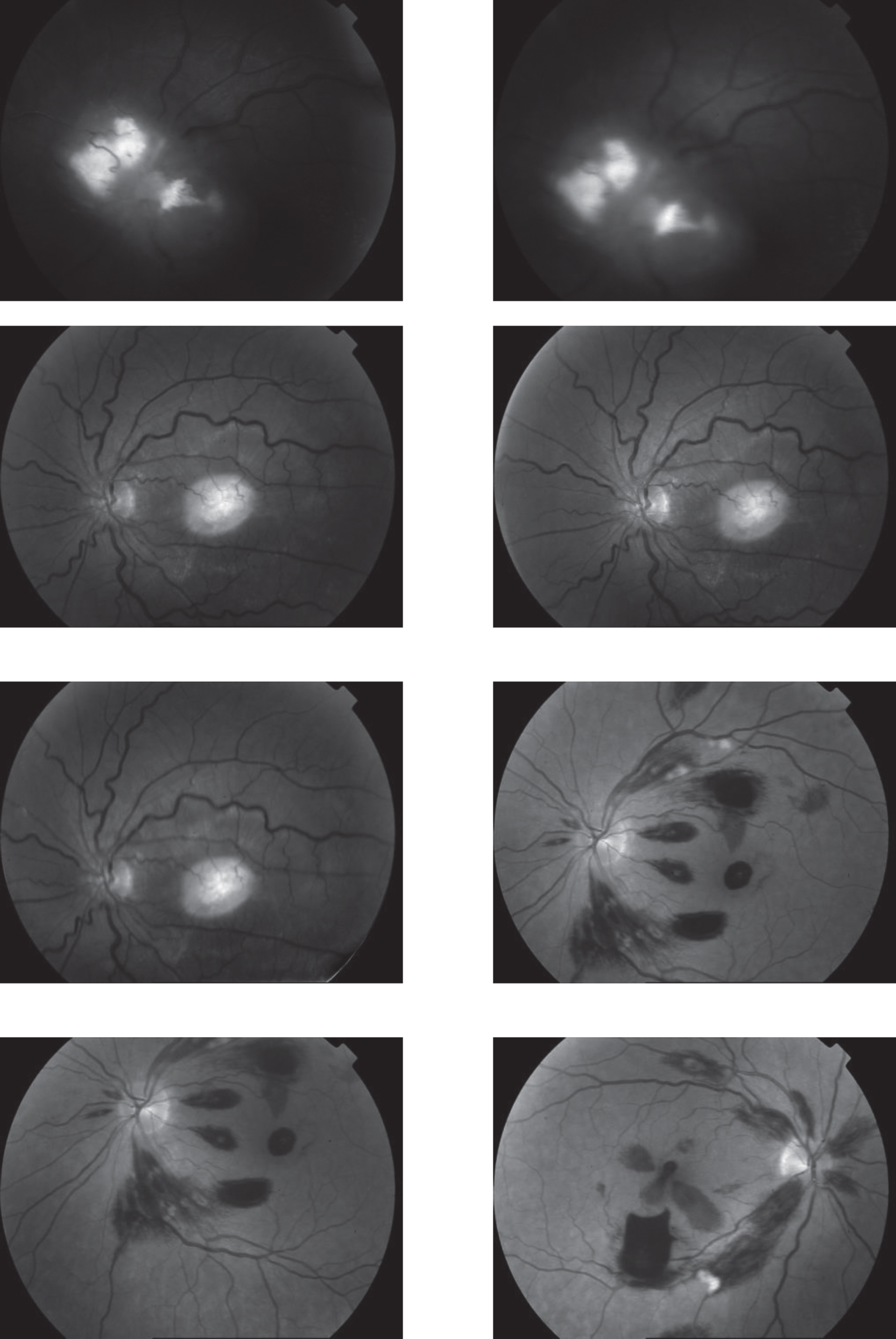
Fuzzified Retinal Images normalized in (0,255) grayscale.
Segmented images of retinal images are shown in Fig. 3. The images in Fig. 3 correspond to the implementation of the proposed method with a marginally optimum set of parameters while their numerical results are given in Table 2. The results of statistical analysis on score and penalty assignments are given in Table 2 while the proposed method. It is shown that the optimum score and penalty values stands about 0.01 and 0.02 while the product of score/penalty values and the number of iterations is important. It is also shown that for our dataset images the neighborhood size of 5×5 which is an extended Moore neighborhood leads to better results compared to simple Moore neighborhood. Table 3 presents the results of a brief comparison between the Fuzzy cellular automata and fuzzy cellular learning automata. For this part of the experiment, the rules are updated for two values of
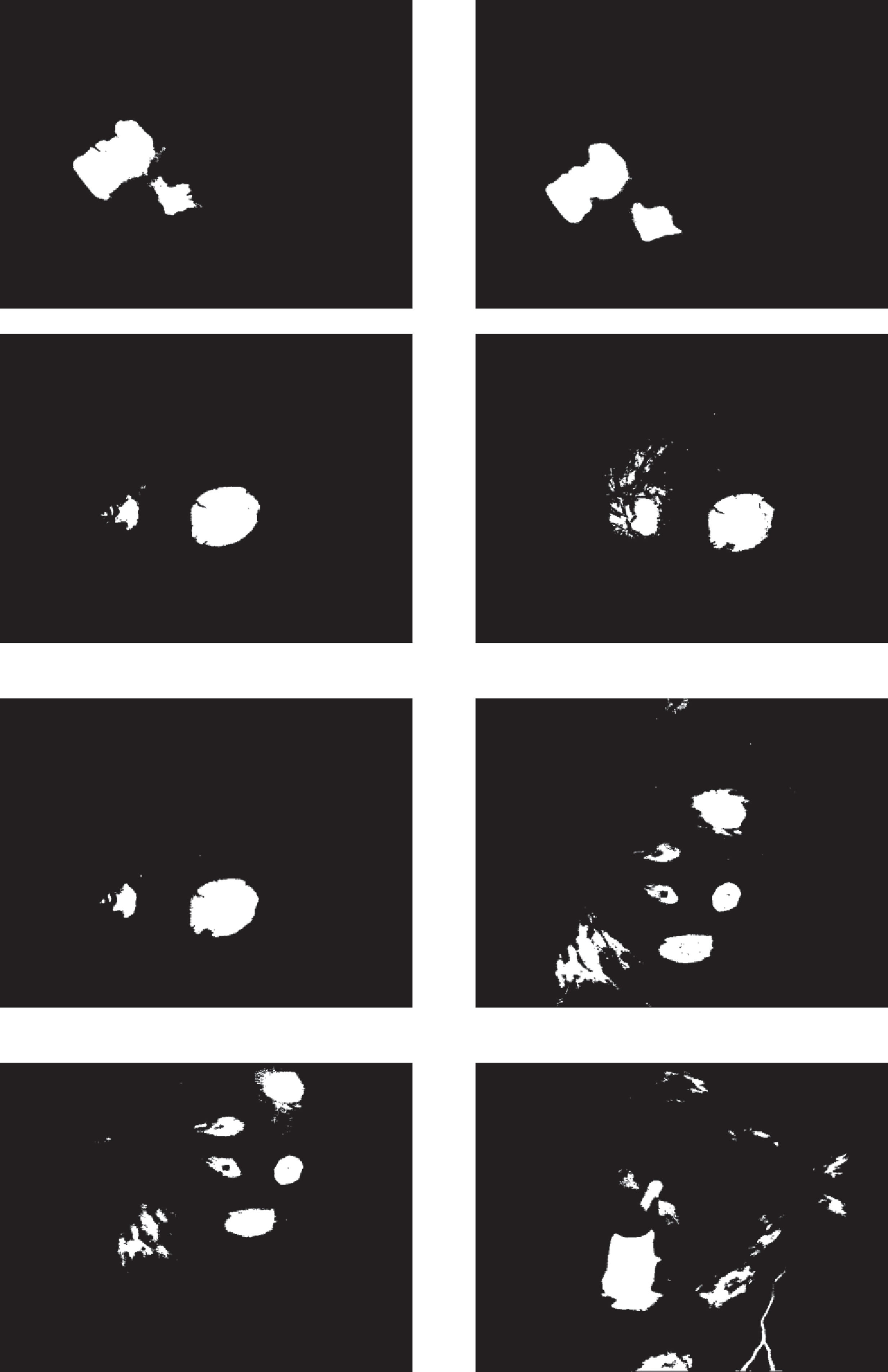
Segmented Retinal Images by the FCLA method.
Statistical parameters for exudates detection process by the proposed method (τ = 50) averaged over 50 images
Statistical parameters for exudates detection process by the proposed FCLA method for different rule updating parameters when N δ = 5 ×5, ϑ = 0.01, ξ = 0.01 & τ = 30
The final state of the FCLA iterative method gives out a retina image with segmented exudates. Based on the number of iterations, defined rules and the values of score and penalty the final state maybe an image in fuzzy domain its pixels ranged in the unit interval. Hence, a threshold parameter for image binarization ς is proposed to assign the pixels of the images either 0 or 1 according to their fuzzy value. If appropriate rules are defined and suitable values for ϑ, ξ & τ are set, the sensitivity to parameter ς is too low. Then the values in the range [0.3, 0.9] will lead to slightly different binary images. The differences between the statistical parameters for three different values of ς are demonstrated in Table 4.
Statistical parameters for exudates detection process by the proposed FCLA method for different threshold values of image binarization when N
δ
= 5 ×5, ϑ = 0.01, ξ = 0.01,
A comparison to some other methods is performed in terms of the statistical parameters. To evaluate the efficiency of the proposed FCLA method, the results of the six methods of references [], K-Nearest Neighbor method are listed in Table 5. The results for the proposed method are attained with the optimum parameters found in the previous parts of the experiment. These parameters are N
δ
= 5 ×5, ϑ = 0.01, ξ = 0.01, τ = 50,
Statistical parameters for exudates detection process by the proposed FCLA method compared to some other recent methods. For FCLA method N
δ
= 5 ×5, ϑ = 0.01, ξ = 0.01, τ = 50,
In this research, a hybrid method was presented for digital retinal image processing for diagnosis and screening purposes. A supervised/semi-supervised method for detecting of exudate in fundus images was presented and the optimum structure was found in a comprehensive analysis. The images were first transformed into fuzzy domain. A cellular learning automata model was used to detect any abnormality on the image which is related to a lesion. STARE retina image dataset, for a neighborhood of 5 × 5, score of ϑ = 0.01, penalty of ξ = 0.01, ratio of state overall variation in three sequent cycles in cellular automata