
Abstract
Recognition of finger vein patterns is essential technique that analyses the finger vein patterns to enable accurate authentication of an individual. A proper, accurate and quick learning of patterns is essentially required for improving the classification pattern. It is essential in developing an intelligent algorithm to effectively study and classify the patterns. In this paper, we develop an improved deep learning hybrid model for feature extraction and classification. A dimensional reduction deep neural network (DR-DNN) model has included a dimensional reduction model for extracting the essential features by reducing the dimensionality of feature datasets. A convolutional neural network (CNN) helps in classifying the benign vein patterns from the malignant vein patterns. The effectiveness is compared against existing deep learning classifiers to measure how effective the deep learning model is used for classifying finger vein patterns for biometric authentication. The results shows that the proposed method achieves an accuracy rate of 97.16% for the proposed method, where the other existing methods including CNN, Recurrent Neural Network (RNN) and Deep Neural Nets (DNN) has an accuracy rate of 86%, 80.66% and 88.31%, respectively.
Keywords
Introduction
Biometrics became a fast paced personal identity authentication. Biometrics made it easier to identify the biological features than existing identification systems [1]. In case of security systems, the user authentication and monitoring access requires an optimal biometric sign that includes fingerprint, face, iris and hand geometry. These signatures can be altered in traditional biometrics since they are superficial to the human body [2]. In contrast to the traditional biometric methods, a future alternative can be vein identification. The characteristics associated with vein identification protects systems from being manipulated [3, 30–32].
Conventionally various methods are developed on vein recognition systems that includes palm vein [4], dorsal vein [5, 6], and finger vein [1]. Recognition methods for finger veins can be divided into three categories, traditional methods [7–10], machine-learning [11–15] and hybrid methods [16–20]. A conventional system tends to operate on identification or verification mode that entirely depends on the context of application. The classical process of recognition consists of steps: preprocessing, feature removal and comparison [21].
At present, deep learning systems have replaced traditional systems with a mechanism from the end-to-end. In comparison with conventional algorithms, CNN is characterized by its speed and accuracy of classification [22]. In this paper, a deep learning classifier is designed with a dimensionality reduction feature extraction method over various datasets. The novelty of the article involves state-of-art model in the field of finger vein recognition, where at times the parameters tend to yield high recognition rate during the process of testing. In order to resolve such constraint, the study uses high computing loads (input images) with single parameter estimation.
The contribution of the paper is given below: The authors developed a deep learning hybrid model for feature extraction and classification. The authors develop a novel solution using dimensional reduction Convolutional Neural Network (DR-CNN) model to reduce the dimensionality associated with input data for extracting the essential features by reducing the dimensionality of feature datasets. A convolutional neural network (CNN) classifies the benign vein patterns from the malignant vein patterns. The experiments are conducted to test the efficacy of the model that includes accuracy, specificity, sensitivity, f-measure, recall and other metrics. The effectiveness is compared against existing deep learning classifiers to measure how effective the deep learning model is used for classifying finger vein patterns for biometric authentication.
The outline of the paper is given below: Section 2 provide the methods. Section 3 validates the proposed DR-CNN. Section 4 concludes the entire work with possible directions of future work.
Related works
The adaptive k-nearest centroid neighbour (akNCN) classifier was proposed by Rosdi, B. A., et al. For the first time, two new adaptive sampling rules are introduced. Finger Vein database results show good results in which the akNCN classification time is greatly lowered to 51.56%. An effective finger vein recognition system developed by Kapoor, K., et al. [34] includes the Local Phase Quantization (LPQ) for feature extraction and Grey Wolf Optimization (GWO) based SVM to compute the ideal parameter combination of SVM for optimal binary classification results. After extracting the LPQ and LDP features, which are invariant to motion blur and deformation, they are combined to enhance the recognition and reduce the computing time of the algorithm. As a last step, GWO-SVM is utilized to identify the best SVM parameters to enhance classification accuracy.
Before developing a pixels-based graph, Lei, et al. [35] produced a region-based graph. Using this novel graph, the study can drastically lower the complexity. To further reduce computing costs, an initial constraint is given based on the acquisition equipment modelling. A Gaussian model is then used to automatically establish the labels of both the background and object, taking into account that the finger is typically situated in the centre of an image. Finally, the MAP-MRF algorithm is used to update the labels during the iterative procedure.
Wen, et al. [36] developed a simple inter-class data augmentation strategy that can increase the training classes. In order to solve the problem of data shortage, we combine it with standard intra-class data augmentation methods, and so accomplish extremely diverse expansion. The study created a fusion loss that incorporates both the classification loss in order to better distinguish between deep features. There are two penalty signals that may be fused to improve the generalization of learnt features, resulting in a better tradeoff between the similarities between classes and differences across classes. The performance and model complexity of various network designs for FVR applications are also examined. As the digital world becomes more and more intertwined, Sharma and Agrawal [37] examine the vascular pattern-based FVP as a possible authentication method. Various other models in this field includes Multi-feature fusion partitioned local binary pattern [41], Forehead vein and Periocular Pattern-based Biometric System [42] and Exception convolutional neural network [42].
Proposed method
In this section, an improved deep learning hybrid model is developed for feature extraction and classification. A dimensional reduction deep neural network (DR-DNN) is used for dimensionality reduction and it helps in extracting the features from the input datasets. Finally, a CNN classifies the benign vein patterns from the malignant vein patterns.
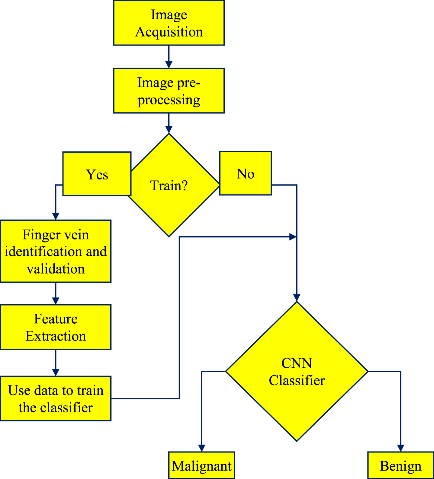
The automated finger vein pattern classification framework is illustrated in Fig. 1(a) that helps in classification of finger vein patterns image composed five layer model. The proposed model Fig. 1(b) is designed with four different modules that involves the following: Acquisition of input images Pre-processing of input data Feature Extraction and Image Classification.
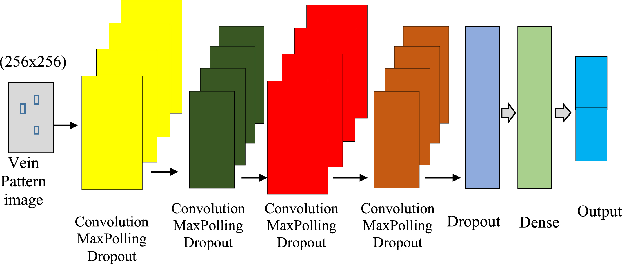
(a): Architecture for finger vein biometric recognition.
(b): Architecture of finger vein classification.
The CNN classifier is designed for the classification of a benign and malignant finger vein patterns.
Figure 1b shows the model of the proposed system that involves data collection, preprocessing, and then followed by the training and testing. The study uses vein identification and validation at the first stage of training and then feature extraction is conducted to extract the labels for training the proposed classifier. During the process of testing, the labels are supplied to the CNN classifier for testing the input test images in order to identify the malignant and benign.
These real-time images are captured using a digital camera at various orientations, different dimensions, illumination, backgrounds and poses.
b. Pre-processing
The study find a finger vein images from the database (captured images are stored in database) of both local and global repositories. Each image contains three RGB channels. We will test the applicability of our approach both with our RGB images and gray images of our experiments. Each image in our data set will become a grid in a pre-processing step with 256×256 pixels.
The noise is taken from the collected pixel images when the pixel is selected to clean or corrupt. When the pixel is saved, a pixel in the ratio of 0 to 255 is coded for corruption. The width of the window will be increased to 5×5 following the removal of the damaged pixels from the starting stage frame. The window size is enlarged so that all damaged pixels in this specific system can be processed.
The corrupted pixels create a median value, which reduces the window width to the median value. In the current window the changed median value(s) will be replaced with corrupted pixel(s). Furthermore, a previous pixel number replaces this pixel if all pixels in the window were distorted. After the first window is completed, turn the current window into the next pixel processing component. When the pixel is saved, the pixel is encrypted from 0 to 255.
After removing damaged pixels from the first step frame, the window width will be extended. The window size is increased in order of allowing the corrupted pixels processing. These corrupted pixels produce a median which lowers the width of the window to the median value. In the current window the changed median values for the corrupted pixels are replaced. Moreover, when all pixels in the window are detected, the current pixel is replaced with the previous pixel number. Just move the current window to the pixel component after the first window has been completed.
c. Feature Extraction
The extraction of features involves Gray Level Co-occurrence Matrix (GLCM) from the pre-processed images of 256×255 pixel size, where the position of matrix is counted using Scale Invariant Feature Transform (SIFT) for optimal extraction of features in a scaled image.
d. Classification
The CNN classifier involves the following components:
Pre-Training Phase: We train CNN at this stage on a large dataset. For the next phase, the aim is to establish network weight.
Training: From the first phase the resulting network is improved. We have a new output level, which supersedes two classes of finger vein models to the output layer of pre-trained networks.
Finger Vein Classification: The user uses a finger image in this method to determine the patterns of the finger veins with CNN.
Visualization: The user can visualize the leaf image regions which characterize the finger veins after classification. The user also has an instrument for estimating the exact pattern of the finger veins through the visualization of the patterns.
e. CNN Classification
CNN is used for the classification of the finger vein patterns.
The finger veins after the feature extraction is extracted using convolutional layers with increased stability using reduced dimensional features. After the training the CNN classifier with Image-Net, the finger vein patterns are extracted from the convolution layer in real-time.
1. Convolutional layer carry out a linear processing operation when weight series are multiplied by the video input data array. In order to detect the features present in the input image, the study uses filter to scan the image and this process is called as translation invariance. A functional map of two-dimensional is then created, where the translation invariance filters are used repeatedly on an input image pixel array.
2. Pooling layers are capable of carrying out a non-linear down sampling process. The most common operation is to divide the input into uncut sets and generate the maximum sets of output from each group. This helps to reduce the complexity of the convolutional network in spatial representation, parameters, over fitting and computation.
3. Activation function used in the study is of rectified linear unit one where the function is defined as f(x) = max (0, x).
4. Permutation layers confirm the finger veins classified using the convolution layer that finds the reference features in the input images. The permutation function is used to estimate the similarity.
5. Fully Connected Layers consists of neurons, which are fully connected with entire previous layer activations and it is applied after the convolutional and permutation and pooling layers.
Neural networks are composed of weight, bias of neurons and learning parameters. The weights are determined based on the labels of input data. The calculation of errors between representative inputs and their expected results adjusts these weights.
Results and Discussions
The DR-CNN is compared with conventional classifiers: DNN [23], CNN [25], RNN [24] and DR-CNN. The study uses increased workload i.e. 6000 images for training and testing over each parameter that includes: activation function, CL numbers, kernel size, and max-pooling layer numbers. The experiments are conducted with 80% of the training data and remining 20% of the data is used for evaluating the performance of the classifier. The study used a ReLU activation function on a simple CNN model and the results are estimated.
a. Datasets
MMCBNU_6000 [26] with 6000 images, PLUSVein-FV3 Finger Vein Data Set [27] with 21600 images, University of Twenty Finger Vascular Pattern Database [28] with 720 and Idiap Research Institute VERA Finger vein Database [29] with 440 images.
b. Performance metrics
The experiment is carried out with set of finger vein images in a five-fold cross validation. The proposed method is validated in terms of various metrics that includes the following:
P is considered as the sensitivity and R is considered as the precision
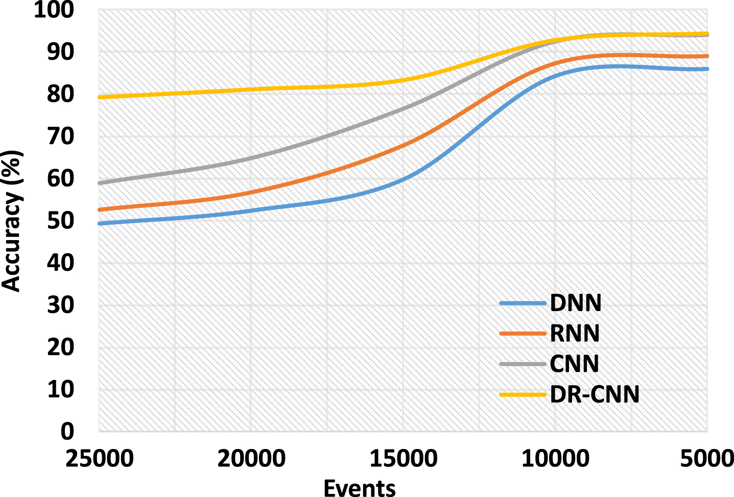
In Fig. 3 (Table 1) shows the results of detection accuracy between the DR-CNN and DNN, RNN and CNN. The result of DR-CNN has higher accuracy in vein pattern classification. With increasing number of number of images datasets, the accuracy degrades due to vein patterns complexity in computation. From the simulated results, it is seen that the error associated with classification has reduced to a greater extent than the existing methods.

CNN framework.
Accuracy.
Accuracy
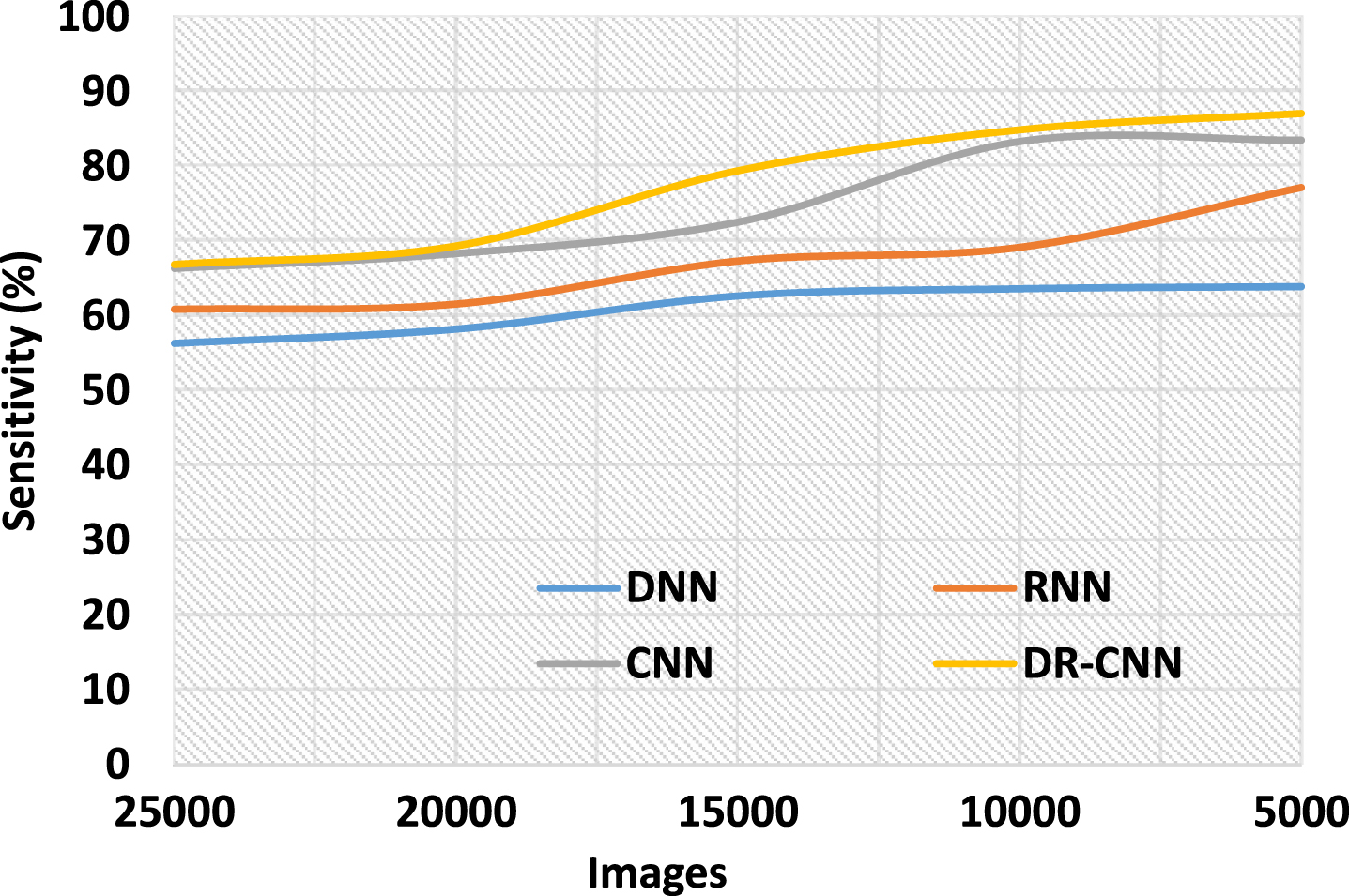
The Fig. 4 (Table 2) shows the results of sensitivity between the DR-CNN and DNN, RNN and CNN. The result of DR-CNN has higher sensitivity in vein pattern classification. The accurate detection from the rule set provides optimal detection of finger vein patterns than DNN, RNN and CNN. The sensitivity reduces in DR-CNNs as well as existing methods with the increasing vein images. The Fig. 5 (Table 3) shows the results of specificity between the DR-CNN and DNN, RNN and CNN. The result of DR-CNN has higher specificity in vein pattern classification. The accurate detection provides optimal detection of finger vein patterns than other existing methods.
Sensitivity.
Sensitivity
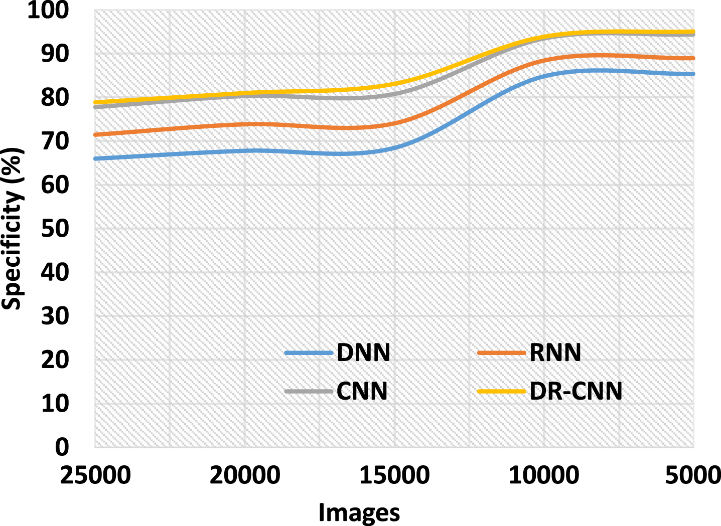
Specificity.
Specificity
The Fig. 6 (Table 4) shows the F-measure between the DR-CNN and DNN, RNN and CNN. The result of DR-CNN has higher F-measure in vein pattern classification. The accurate detection provides optimal detection of finger vein patterns than DNN, RNN and CNN. With increasing test images, the accuracy degrades as the complexity of computation gets increased. The Fig. 7 (Table 5) shows the Execution Time between the DR-CNN and DNN, RNN and CNN. The result of DR-CNN has reduced execution time in vein pattern classification. With increasing images, the execution time increases with increasing time complexity.
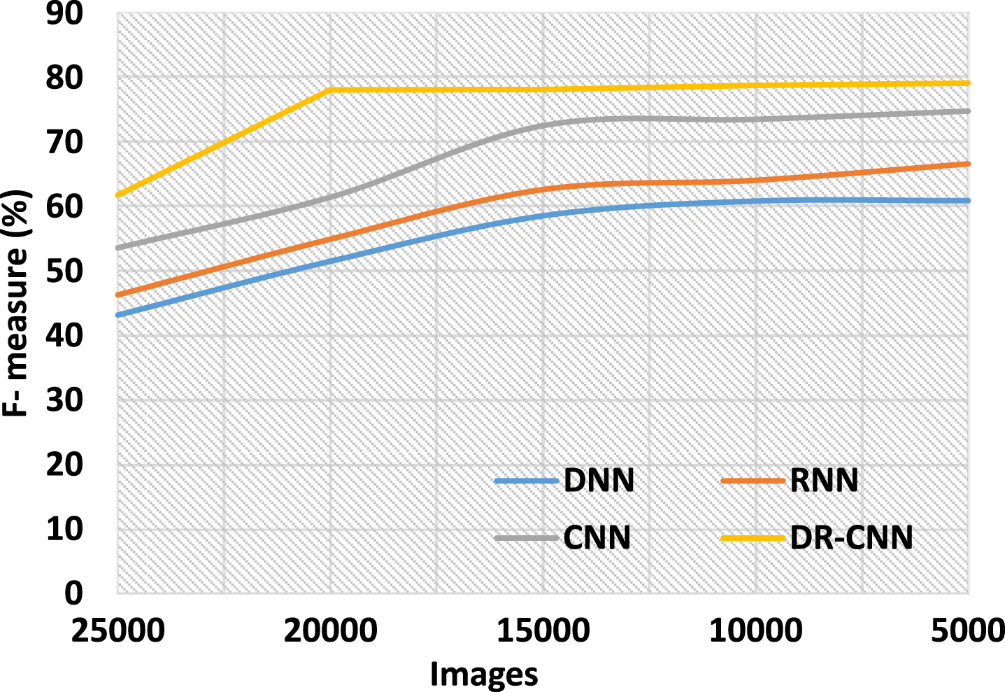
F-Measure.
F-measure
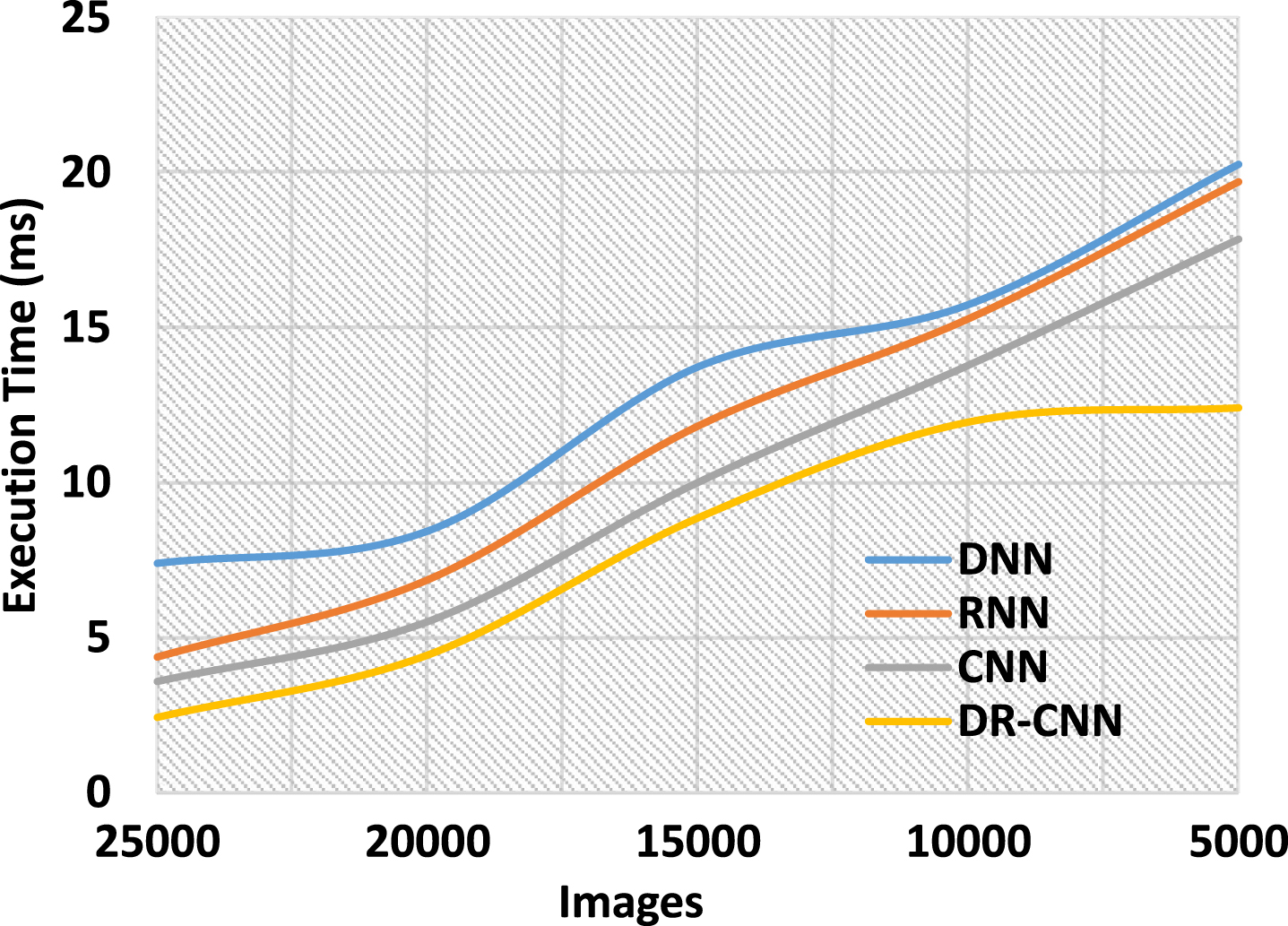
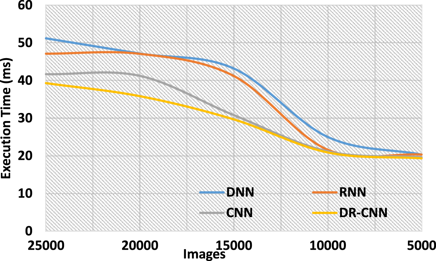
Execution time (ms).
Execution time (ms)
The Fig. 8 and Table 6 shows the MAE between the DR-CNN and DNN, RNN and CNN. MAE tends to grow as a result of the complexity with which the vein patterns of other images are calculated. In the DR-CNN, the removal of minor features to improve the MAE rate is said to be largely important. In a database that contains many different obstacles mode shown in Table 7, such as data set with accuracy rate, specification, recognition rate, and error rate. The low contrast and noise testing processes utilized in the database have an impact on the performance of a finger vein detection system verified. Experiments on the proposed system will be carried out using data from three public databases containing varying amounts of training images in order to demonstrate its reliability.
Mean Absolute error.
Mean Absolute error
Performance comparison of proposed and existing work model
However, only one test image and one training image are included in each class, despite the fact that there are over 25,000 images in this database. However, while the database of 25,000 photos gave reliable results, our strategy decreases classification errors and increases the accuracy of the results. With 98% accuracy, the detection accuracy was higher than that of previous methodologies that used this database.
The high quality of the training data is what distinguishes CNNs from other types of neural networks. In order to recognize finger veins, small datasets and basic vein forms are used, both of which have an impact on the training process. A newly developed network design, the CNN operates in an optimal manner to attain increased results. An alternative approach was to use a DR-CNN; however, instead of using a single image, the outputs of multiple distinct finger vein images were combined.
In this paper, DR-CNN is designed for vein pattern classification and enables accurate authentication of individuals. It uses optimal labels to enable the supervised learning to improve the pattern on classification. This intelligent CNN algorithm with additional dimensionality reduction of features enables the extraction of optimal features. The DR-CNN finally classifies the benign and malignant vein patterns. The simulation results concludes that the proposed method has higher accuracy, specificity, sensitivity, f-measure, recall and reduced MAE than existing CNN, RNN and DNN classifiers. In future, the system can utilize the advances in deep learning methods enabling the dimensionality reduction at the pre-processing stage. Thus, it is seen that the proposed method is efficient in classifying the vein pattern from input models. The study has a limitation on increased number of images, where the model leads to overfitting, however, in future the study may use an exclusive model to avoid the problem of overfitting.