
Abstract
Brain tumor is an anomalous growth of brain cells. Segmentation of brain tumors is currently the most important surgical and pharmaceutical procedure. However, manually segmenting the brain tumor is a challenging task due to the complex structure of brain. In recent years, artificial intelligence techniques with the fuzzy logic have shown better results in the field of medicine. In this work, a novel deep learning classification network with fuzzy hexagonal membership function (DLC-FHMF) model has been proposed for accurately segmenting brain tumors. The different MRI modalities namely T1, T1-c, T2 and Flair images are preprocessed using a fuzzy hexagonal trilateral and median filter to eliminate the Rician noise. Afterwards, the DLC-FHMF model is used for segmenting the tumor portion by using the multimodal composition of MRI as input. The fuzzy weights are determined with hexagonal membership functions and convoluted with the corresponding MRI images. The quantitative examination is carried out using the performance metrics namely accuracy, specificity, precision, sensitivity, incorrect segmentation, under-segmentation, and over-segmentation. In addition to the above metrics, the pre-processing metrics include PSNR, RMSE, and SSIM. The experimental fallout portrayals that the proposed DLC-FHMF approach attains a better accuracy range of 99% for detecting brain tumors using the BRATS 2013 dataset. The proposed DLC-FHMF model improves the overall accuracy by 15.1%, 11.1%, 3.0%, 21.2% and 0.5% better than ANN, SVM, NB, DNN and DAE respectively.
Introduction
The brain tumor is an anomalous abnormal growth of tissues in the brain of the human beings. Other tumor spreads by local rarely to the all over parts of the human being. But the brain tumor spreads to other parts of the body due to the development of multiple abnormal tissue cells [1]. Doctors are identifying the tumor initially based on the symptoms including severe head ache, loss of sense, partial or total loss of realization, fatigue, vomiting and sleeping problem. In medical image processing, to detect the brain tumor using image processing techniques there is a need to study on the anatomy of human brain [2]. MRI is the non-invasive technique as it is free from radiation modalities from other medical techniques. Even though it is a versatile technique; the quality of the image is affected during the acquisition process [3]. The artifacts in MRI are associated with hardware, software, patient and physics. These artifacts are handled by the Magnetic Resonance (MR) scanner but still, noise/artifacts remain in the MRI which needs to be removed as it affects the diagnosis process [4]. The medical images are affected by noises which occur during acquisition of MR images, transmission of the data through medium, image quantization and due to the causes of radiation from source. The corrupted image with noises results in the degradation of visual feature of the MRI images. In present, diverse set of noises like impulse noise, gaussian noise, uniform noise, Poisson noise, gamma noise, speckle noise, Rayleigh noise, Rician noise etc. [5]. The primary source of noise in MRI occurs during the acquisition and transmission is the thermal noises in the form of complex data occurs during acquisition. The thermal noise is complex in nature with the real as well as imaginary parts of the MRI images with the mean and variance of zero. The anatomical structural details and functional details of the MRI human brain can be retrieved and deal with in an enhanced way, only when it is represented as magnitude data. During acquisition the data will be as gaussian distribution, the transformation data into magnitude converts into Rician distribution which leads to Rician noise [6]. The Rician noisy image only acquire through the machine while acquisition and the other types of noises occur in MR image are due to the movement of the person while taking MRI scanning. Hence there is a need to remove the Rician noise in MR image for efficient processing, since the denoising problem is an inverse problem used to rebuild noise-less image which can be further easy for the process of image segmentation, disease identification etc. The major contributions of this research are summarized as follows, The different modalities of MRI images are collected from the BRATS 2013 dataset for detecting brain tumor. The noisy MRI is given an input to fuzzy hexagonal membership function with trilateral filter and median filter for eliminating Rician noise. It convolutes the fuzzy weights with the corresponding restored MRI and then summed up to obtain the restored MRI, which will be further processed for segmentation. The deep learning classification network with fuzzy hexagonal membership function (DLC-FHMF) is used for segmenting the tumor portion. The efficiency of the proposed DLC-FHMF model is evaluated using various parameters for image pre-processing and segmentation.
The rest of this work was pre-arranged into following sections. Section 2 briefly discussed about the background of the study. Section 3 includes the proposed Deep learning classification method with Fuzzy hexagonal membership function (DLC-FHMF) for identifying the brain tumor from different MRI modalities. Section 4 encompasses with the results and discussion, and finally the Section 5 enfolds with conclusion.
Background
Image restoration is the pre-processing of the corrupted or noisy data to obtain the original image. Noise removal technique in medical applications plays a dynamic issue in identifying the disease. Different kinds of noise were present in the medical images, namely Rician, impulse, salt and pepper, gaussian, speckle, gamma, uniform, Rayleigh, poison etc. [7]. The MRI is predominantly pretentious due to the presence of Rician noise. The intricacy in performing restoration of the MRI due to the presence of the Rician noise with high resolution increases and leads to the reduction in intensity and dissimilarity of the image [8, 9]. The pre-processing of the Rician noisy MRI by restoration without corrupting the structural data plays a vital concern in MRI investigation; still there is a need to develop the restored MR image algorithm for better accuracy [10]. A non-parametric restoration method for brain MR image has been built to obtain restored image with intensity uniformity and increases the robustness of the anatomy of the human brain [11]. In the Linear minimum mean square Error method (LMMSE), the Rician noise is estimated which helps to increase the restoration efficiency due to more like and tough information but produce the larger structure [12].
Deep learning-based approach for segmenting the brain tumours was introduced in [13] to identify the particular location of the tumour. There was a wide variety of tumour tissues present in the occurrence of a variety of cases. In most cases, the similarity between normal tissues makes this process challenging. This system was developed on the basis of Berkeley’s wavelet transformation (BWT) and deep learning algorithm to enhance efficiency and streamline medical image segmentation. The Grey-level co-occurrence matrix (GLCM) approach was employed for extracting the relevant characteristics from each segmented tissue, and a genetic algorithm was used to optimise the features. Super Resolution method have been implemented for reconstructing the Low Resolution and High-Resolution images, as it is an inverse problem. The regularization is used to obtain the enhanced reconstructed MR image by preserving the edges and maintain the robustness [14]. Total Variation (TV) with intersecting cluster is formed for image restoration applications leads to failure, and solves the corresponding minimization problem using anisotropic TV with overlapping group sparsity [15]. The noises in the MRI are sensed and detached using the directional method filtering system however it receipts with increased complexity burden [16]. Non-Local Mean (NLM) filter works well for denoising applications but fails due to over smoothening while selecting the optimal parameter. The problematical view of the anatomical features of the brain fails to view the precise structure of the boundary among the dissimilar tissues and shows as its absence leads to the difficulty while performing the segmentation [17]. Deep learning for brain tumor identification has been used for segregating the normal and non-tumor tissues in the images with spatial consistency, can also be extended in noisy environment by integrating with the Fully Convolutional Neural Network (FCNN) along with Conditional Random Field (CRF) [18]. In tumor region, the details of the structural data are low associated with the non-tumor region cause to loss of information with data unevenness problem, to obtain the solution for this problem dictionary-based segmentation is used [19]. Detection of brain tumor in low intensity boundary area is a difficult task. Non-Sub Sampled Contourlet Transform (NSSCT) system enhance the brain MRI categorization by extracting the texture features which can be applied for training and testing to classify the tumor portion [20]. Spatial-filtering process by mapping the input image, taking the reference image from histogram, and histogram matching the image with histogram are effectively applied for MR image segmentation with neural networks. The less processing time and high recognition rate creates the system as portable device [21]. In brain tumor detection using the deep learning with auto encoder performs classification to segments the tumor portion; still there occurs the problem of getting spatial information during segmentation. Cascading Handcrafted method used for MRI segmentation fails to provide supplementary formerly information about the contour, borders and spatial location of the tumor [22].
A hybrid deep learning approach [23] was suggested for classifying the various kinds of brain tumours, including pituitary, meningiomas, and gliomas. A hybrid DeepTumorNet technique was created by removing the final five GoogleNet layers and inserting fifteen additional layers. In the feature map, leaky ReLU function was used to make the model more expressive. Based on the CE-MRI dataset, the suggested DL classification approach exhibits the highest accuracy compared to the current state-of-the-art approaches. The deep learning based YOLOv5 object detection method was developed in [24] to create a transportable micro-wave head imaging framework capable for automatic diagnosing and classifying human brain disorders. Initially, the constructed MWHI system is used to collect 400 RMW images, including tumors and non-tumors. A 3-dimensional wide-band antennae array was used in the developed imaging framework to create reconstructed micro-wave (RMW) brain images, which are investigated with a tissue-imitating head phantom. Brain tumor segmentation based on whale optimization algorithm needs to be improved based on identifying the false positive e and false negative rate to detect tumor. To overcome this issue another method based on image adaptive algorithm customizes the NLM in MR image for better edge preservation and restores MR image by suppressing the noisy image [25]. To overcome these challenges, in medical image application like identifying the diseases to lessen the operator dependency and achieve greater accuracy in diagnosis using MRI and machine learning model like fuzzy logic-based model are broadly utilized for resolving the difficulties in various domains of medical image application and here the proposed method uses the fuzzy logic for MRI restoration and deep learning segmentation to detect brain tumor effectively [26, 27].
Proposed method
In this section, DLC-FHMF model was proposed for classifying and segmenting the brain tumors using different MRI modalities as shown in Fig. 1. The Noisy MRI is given an input to fuzzy hexagonal membership function with trilateral filter and median filter for Rician noise restoration by utilizing the hexagonal membership function to attain the fuzzy weights of the median and trilateral filter. It convolutes the fuzzy weights with the corresponding restored MRI and then summed up to obtain the restored MRI [27, 28] which will be further processed for segmentation.

Schematic representation of the proposed DLC-FHMF model.
The MR images are the raw data in nature during acquisition is given in frequency domain by means of k-space. After taking inverse Fourier transformation of the k-space information, the subsequent information become more complicated with real and imaginary form is still distorted by the noise. The magnitude form, which performs non-linear operations, gets converted to Rician by attaining the probability distribution function (PDF) of image noise and it is derived as,
The proposed brain tumor detection method to detect brain tumor in the MRI using the Deep Learning Classification-Fuzzy Hexagonal Membership Function (DLC-FHMF). The restored MR image is given to the DLC-FHMF for MR image segmentation and segments the tumor portion in the MRI by obtaining the fuzzy weights of the different modalities of MRI using fuzzy hexagonal membership function and convoluting with the input image of the various modalities of the MRI [29].
Fuzzy Hexagonal Membership Function along with Trilateral and Median Filter
The proposed system utilizes the fuzzy hexagonal membership function to preprocess Rician MRI to identify brain tumors, with a trilateral filter and median filter to improve the denoising performance at a variety of noise levels. The outcome will preserve fine edges and structures while enhancing the denoising performance. First, the trilateral filter operation is performed for noisy MRI using the formula from (2) to (5) and the output is stored in TTrilateral. Secondly, the statistical characteristics are determined such as local average (μi) and global average (μg) and is applied to the hexagonal membership function to obtain fuzzy weight for trilateral filter as WTrilateral. Then 1 is subtracted from the WTrilateral to obtain the fuzzy weight for median filter as WMedian. Thirdly, the median filter operation is performed for noisy MRI and the output is stored in MMedian. Fourthly, the convolution is performed between the WTrilateral and TTrilateral Similarly, the convolution is performed between the WMedian. and MMedian. Finally, the result of both the convolution is summed to form the restored MRI and the overall flow of the MRI restoration is shown in Fig. 2. The proposed DLC-FHMF model uses the median filter and trilateral filter along with fuzzy hexagonal membership function works better than the other filtering techniques with added parameter like laplacian filter, range and spatial filters to attain better edge-preserving and smoothening of the image. This laplacian filter, range and spatial filters used here measures the difference between the gradient of the MRI and correspondingly, adjusts the weights of the neighboring pixels (2). The trilateral filter fines the neighboring pixels in high gradient portions by contrasting them to the modification in the center pixel. Furthermore, in the high-frequency portions of the MRI are conserved and produce better restoration.

Fuzzy hexagonal membership function with trilateral and median filter.
Where f (i, j) defines the MRI images as input, wP, denotes the normalization parameter, wD, wL and wR defined as the domain filter’s weights, Laplacian filter, and range filter respectively. Ω is the neighborhood of a central pixel. σ d and σ r regulates the decay of two weight factors, D (P - S) denotes the Euclidean distance between the present pixels (P) and neighboring pixels (S), (IP - IS) denotes the variance between the two values of intensity.
The motivation of the DLC-FHMF is used for segmentation by determining the weights of the different modalities of the MRI using Fuzzy Hexagonal Membership Function (FHMF) method to obtain the enhanced efficiency and produce the effective output. First, the fuzzy weights using FHMF is given from (6)-(9) and it is calculated by taking each block of an input image as a delegation for the input data content of the block for the input image for different modalities of MRI. Secondly, convolute the fuzzy weights with the corresponding input MRI of the various MRI modalities like T1, T1-c, T2 and flair images. Thirdly, the output of the convolution of the various modes of the MRI are summed to form a new image. Finally, the new image is fed as input to the deep CNN to perform segmentation. The aids of choosing the fuzzy weights for MRI segmentation is to maintain the segmented portion with essential information such as structural, edges, better pixel classification etc to classify the tumor portion in brain MRI effectively.
Figure 3 shows the work flow of the DLC-FHMF for segmentation. The weight of the MRI W1, W2, W3 and W4 for the flair, T1, T1 C and T2 of MRI different modalities were determined using FHMF. Then the convolution and summation are performed between the weights and inputs of the flair, T1, T1-c and T2 MRI to attain the new images which is taken as an input for the conventional DCNN, then it segments the MRI brain. Similarly, the fuzzy weights have been obtained for different modalities of the MRI using FHMF. Then convolution operation between the weights obtained with the corresponding input MRI as shown in Fig. 3 i.e. I1 x W1, I2 x W2, I3x W3 and I4 x W4. Then perform summation of the different MRI modalities convolution operation is given in I. The new image is given input to the DCNN, which performs conventional DCNN to perform classification to segment the tumor portion of the MRI.
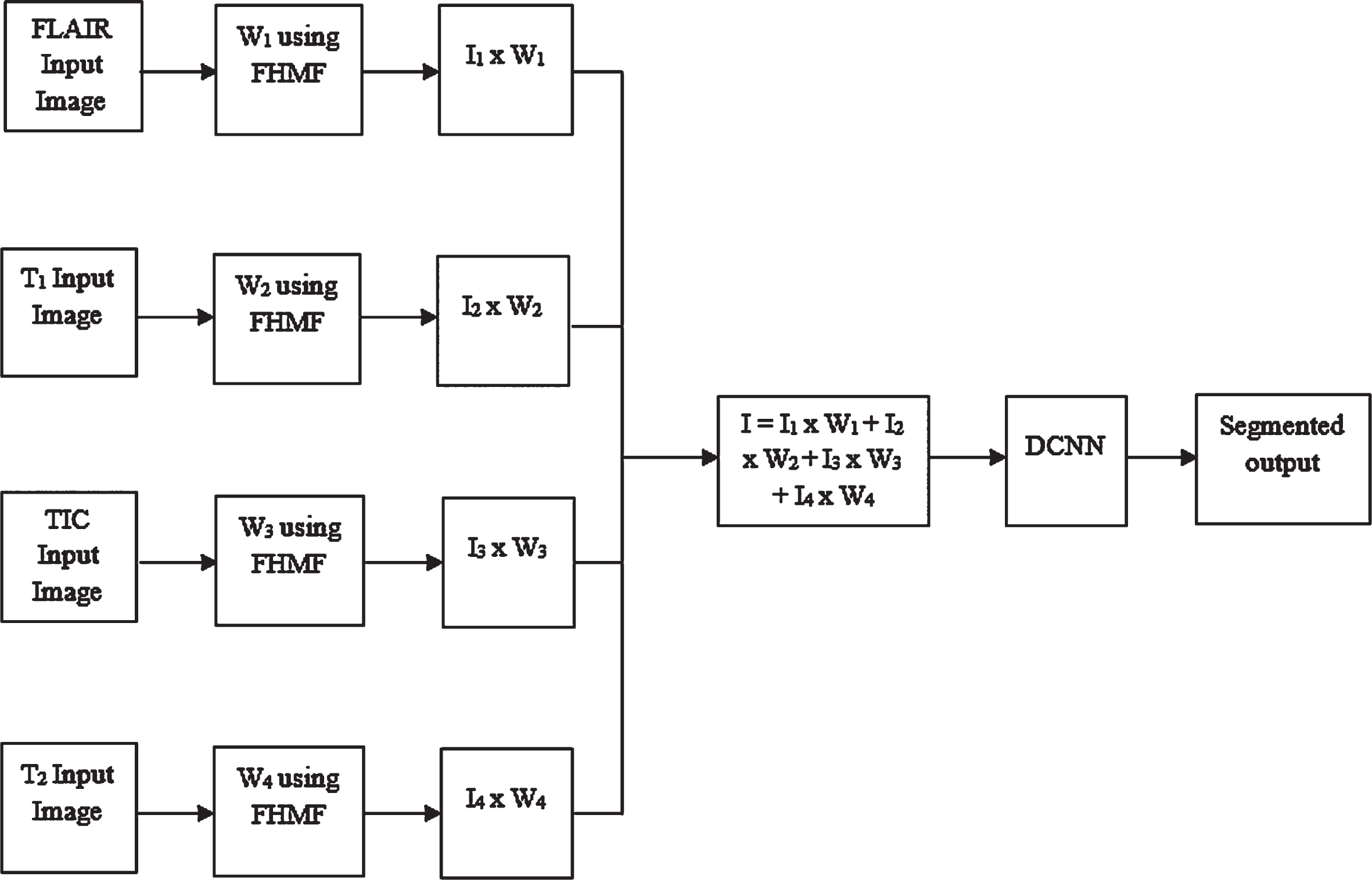
Flow diagram of the DLC-FHMF.
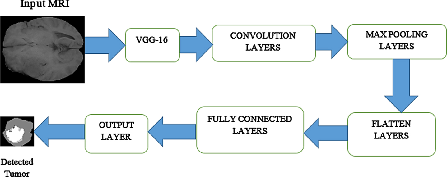
The proposed method uses the Visual Geometry Group (VGG) DCNN architecture for segmentation process to detect the brain tumors. The proposed DCNN structure entails with seven layers as depicted in Fig. 4. First, the input layer, the new image formed after applying FHMF is taken as the input image. In this input layer, the image features are specified as input and given to this layer. Secondly, convolutional layer: 32 filters are used with the kernel size of 5×5. This convolutional layer is used to perform operation for eliminating the redundant structural information’s by keeping the significant information along with the sigmoid activation function is utilized in this layer. Third, is the max-pooling layer which is used to help the training process simple manner by reducing the use of limited parameters and it performs max operation for resizing the into half of its output from previous layer that is convolutional layer. Fourth, the flatten layer is used for the purpose of converting the output of the pooled features to a single column which is taken as an input for the succeeding layer. Fifth layer is the fully connected layer comprises of features that are utilized to detect the target class. Sixth layer, output layer, which gives the final segmented output, to identify the MRI image encompasses of tumor or not along with the softmax activation function.
Proposed DCNN architecture with their layers.
The structure of the VGG 16 model is depicted in Fig. 5 VGG DCNN works better for segmentation by replacing with large size filters with multiple 3 x 3 filter size one after another and max pooling layers 2 x 2. The architecture follows with the sequentially arranged convolution and max pooling layer consistently. Consequently, the fully-connected layer and the output layer with softmax activation function was employed to extract the features as output. Spatial information is retrieved efficiently by using this VGG 16 while segmenting the tumor portion from the MRI images.

VGG architecture with their layers.
The effectiveness of the DLC-FHMF approach was evaluated using the different quantitative measures. For visual perception, it has been investigated qualitatively. The widely used quantitative parameters were used to evaluate efficacy of the DLC-FHMF model quantitatively for MRI restoration. Peak Signal to Noise Ratio (PSNR), Image Enhancement Factor (IEF), Root Mean Squared Error (RMSE), Structural Similarity Index Measure (SSIM) and Normalized Absolute Error (NAE) were used for analysing the restored MRI results. Similarly, for segmentation the Under Segmentation (UnS), Over Segmentation (OvS), Incorrect Segmentation (InS), Specificity (Spec), Accuracy (Acc), Precision (Prec), Sensitivity (Sens), and F-score are used respectively.
Dataset description
The BRATS2013 dataset (Sharif et al. 2020) includes both simulated and original data. This database contains data on 30 cases of High-Grade Gliomas (HGG) / Low-Grade Gliomas (LGG), that divided into 20 HGG cases and 10 LGG cases [32]. In the simulated dataset, there are training datasets and testing datasets, while the original dataset contains challenge datasets, leaderboard datasets, and testing datasets. A training dataset contains detailed information about 30 patients, a testing dataset includes images, and a challenge and leaderboard dataset provide details about 25 patients.
Simulated MR image
The simulated MR images from BRATS 2013 data are considered to detect tumor. Tables 1–3 gives the quantitative investigation of the proposed DLC-FHMF method for the different simulated MR images. Tables 1–3 analyzes the efficiency of the MR image pre-processing experimentally and compared with the existing system, proposed brain tumor detection method obtains high PSNR and SSIM value. The RMSE error is reduced effectively. From Table 1, it is observed that the PSNR value for the DLC-FHMF model is improved 33%, 33.9% and 43% for FLAIR MRI compared to adaptive hexagonal fuzzy hybrid filter, NLM filter and fuzzy trapezoidal NLM filter respectively. Similarly, the RMSE is reduced for the proposed method is improved 78.5%, 78.56.9% and 85% for FLAIR MRI compared to adaptive hexagonal fuzzy hybrid filter, NLM filter and fuzzy trapezoidal NLM filter respectively. Also, the SSIM value for the proposed method is improved 18%, 20% and 20% for flair MRI compared to adaptive hexagonal fuzzy hybrid filter, fuzzy trapezoidal NLM filter and NLM filter respectively.
PSNR analysis of simulated BRATS MR image for pre-processing with PSNR, SSIM and RMSE of the proposed DLC-FHMF model with the existing models
PSNR analysis of simulated BRATS MR image for pre-processing with PSNR, SSIM and RMSE of the proposed DLC-FHMF model with the existing models
RMSE Analysis of Simulated BRATS MR Image for Pre-processing with PSNR, SSIM and RMSE of the Proposed DLC-FHMF model with the Existing Methods
SSIM analysis of simulated BRATS MR image for pre-processing with PSNR, SSIM and RMSE of the proposed DLC-FHMF model with the existing models
Table 4, analyzes the efficiency of MR image classification and is observed that the proposed brain tumor detection method has no UnS, less OvS and InS. The Spec, Sens, Acc, Prec and F-score obtained during segmentation are efficient leads better classification to detect tumor.
Analysis of simulated BRATS MR image classification with UnS, OvS, InS, Spec, Sens, Acc, Prec and F-score for the proposed DLC-FHMF model
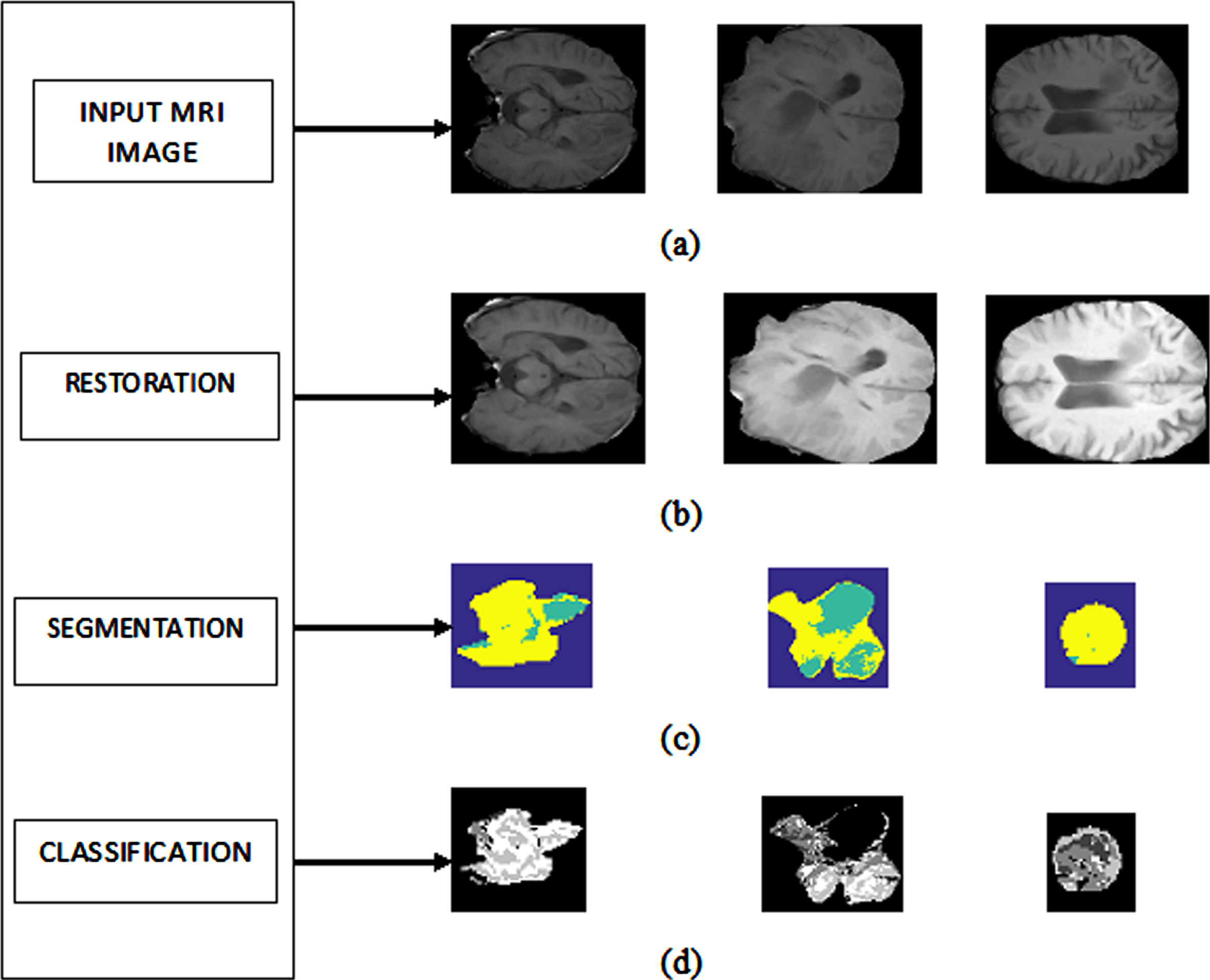
Figure 6 displays the qualitative evaluation of the proposed DLC-FHMF model with different simulated MR images. Figure 6 (a), (b), (c), and (d) shows the different MR images, restored MR image, segmented MR image and classified MR image to show the detected tumor with better resolution and improved classification rate.

Brain tumor detection for the simulated MRI images (a) Input image (b) Restored image (c) Segmented image (d) Tumor portion.
The original MRI images from BRATS data are used for analysis to detect brain tumor. Tables 5–7 gives the experimental study of the proposed DLC-FHMF model for the different real MRI images. Similar to the simulated MRI images, real MRI images are analyzed to show the efficiency of MR image pre-processing and classification. It is perceived from the Tables 5–7, the proposed DLC-FHMF model obtains high PSNR, SSIM value and low RMSE error. Also, the proposed brain tumor detection method obtains no UnS, less OvS and InS. The Spec, Sens, Acc, Prec and F-score obtained during segmentation shows the effectiveness of the proposed brain tumor model.
PSNR analysis of simulated BRATS MR image for pre-processing with PSNR, SSIM and RMSE of the proposed DLC-FHMF model with the existing models
PSNR analysis of simulated BRATS MR image for pre-processing with PSNR, SSIM and RMSE of the proposed DLC-FHMF model with the existing models
RMSE analysis of simulated BRATS MR image for pre-processing with PSNR, SSIM and RMSE of the Proposed DLC-FHMF model with the Existing Methods
SSIM Analysis of Simulated BRATS MR Image for Pre-processing with PSNR, SSIM and RMSE of the Proposed DLC-FHMF model with the Existing Models
In Tables 5–7, the PSNR, RMSE and SSIM values are taken for the different real MRI from BRATS 2013 database. The PSNR value is measured for analyzing the resolution of the image and in the proposed DLC-FHMF model it is found to be improved. The RMSE value is measured to analyze the error rate and it is found to be reduced. The SSIM value is improved and the structural information are restored better while utilizing the adaptive-fuzzy hexagonal bilateral filter.
In Table 8, the UnS, OvS, InS, Spec, Acc, Sens, Prec and F-score are given for the various Real BRATS MR images. The proportion of false negative segmentation (UnS) is 0, OvS and InS are found to be very low for the proposed brain tumor method. The Acc, Prec Sens, and Spec are assessed to evaluate the performance efficiency for the various MRI images. Spec detects the rate of false segmentation, Sens recognizes the TP rate of the segmented MRI, Acc, Prec and F-score value are obtained for the proposed brain tumor detection method and found to be with better values. These parameters are used to detect the absence and presence of tumor in the patients. These metrics are utilized to calculate the rate of retrieved true and false positive samples of the original samples.
Analysis of Real BRATS MRI images for Classification with UnS, OvS, InS, Spec, Sens, Acc, Prec, and F-score for the Proposed DLC-FHMF model
here TN and TP denotes the true negative and positive of the MRI, FN and FP denotes the false negatives and positives of the MRI.
Figure 7 illustration the outcomes of the DLC-FHMF model for detecting brain tumors. The different real MR images are shown with the results. Figure 7 (a), (b), (c), and (d) shows the different MR images, restored MR image, segmented MRI image and classified MRI scan to show the detected tumor. The visual perception shown in the Fig. 7 indicates that the proposed DLC-FHMF model detects the tumor portion separately in an efficient manner without the loss of structural details in the MRI.
Detection of Brain Tumor Detection for the Real MR Images (a) Input Images (b) Restored Image (c) Segmented Image (d) Tumor portion.
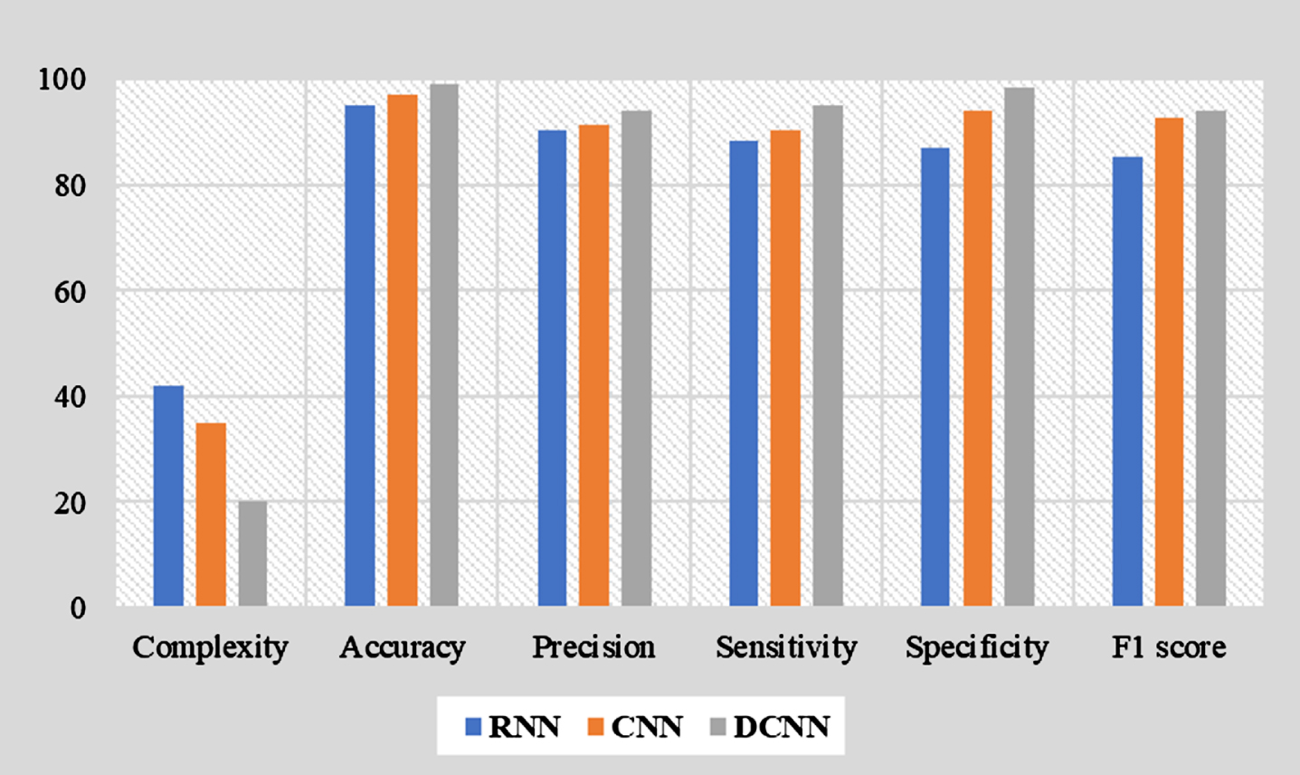
Figure 8 shows the comparative assessment of deep learning networks such as RNN, CNN and DCNN in terms of accuracy, specificity, sensitivity, precision and F1 score. To achieve high accuracy, the traditional networks like RNN and CNN employ a large number of parameters such as 56890k and 7554k with low level of accuracy. Since the proposed DCNN uses only 4816k parameters, so the complexity is reduced while maintaining 99% accuracy ranges. The complexity of the DCNN is extremely low compared to other classic models as represented in Fig. 8. The proposed model improves the overall specificity, sensitivity, precision and F-score by 9.67%, 10.8%, 5.26% and 7.60% better than RNN. The proposed network enhances the overall specificity, sensitivity, precision and F-score by 2.06%, 5.24%, 4.34% and 3.19% better than CNN. The proposed DLC-FHMF model improves the overall accuracy by 3.63% and 1.91% better than RNN and CNN respectively.
Performance comparison of traditional deep neural networks.
The proposed DLC-FHMF model for detecting brain tumor were analyzed for the BRATS database. The quantitative metrics such as Spec, Sens, Acc, Prec, F-score, UnS, OvS and InS are evaluated for the proposed DLC-FHMF model with the existing models. From Table 9, Spec, Sens, Acc, Prec and F-score are improved by 0.1%, 5%, 0.2%, 0.2%, 0.2% and 0.3%, 3%,1%,24%, 37%, 44%, and 54%, 0.5%, 8%, 6%, 11%, 15%, and 21%, 2%, 0%, 18%, 31%, 39%, and 49%, 1%, 2%, 13%, 22%, 29%, and 36% than the existing methods such as respectively [30–34]. UnS, OvS and InS are evaluated for the DLC-FHMF method with the existing frameworks. The existing method performs more UnS, OvS and InS than the proposed methods leads for the false prediction. UnS, OvS and InS are increased by 75%, 51%, 97%, 98%, 99% and 99%, 28%, 94%, 34%, 38%, 41% and 47%, 33%, 89%, 86%, 92%, 94% and 95% for respectively. The experimental outputs of the proposed DLC-FHMF model are enhanced and perform better classification leads to the detection of brain tumor in an efficient way.
Comparison of proposed DL model with the existing models
In the proposed DLC-FHMF model, the fuzzy membership functions with the deep convolutional neural networks are combined to identify the tumor portion with the appearance and spatial consistency. Artificial intelligence techniques namely fuzzy logic and deep CNNs have been employed for detecting the brain tumor. The FHMF, along with the Trilateral and Median filters, allows for efficient tumor detection during the pre-processing step. By combining a trilateral filter with a median filter and fuzzy hexagonal membership function, the MR image is smoothed to preserve the edges while retrieving structural information effectively. Fuzzy Hexagonal Membership Function with the Deep Learning network classifies tumors in MR images by segmenting them with a higher degree of accuracy. The DLC-FHMF model effectively segments the different modalities of MR images with better classification rates, without loss of image details. In comparison with the existing techniques, the proposed DLC-FHMF method has excellent results based on well-established metrics. As a result, the results are close to the point at which they can be used for practical pre-processing and higher-level segmentation, In future, the research will be carried out with different kinds of noise and using other databases. Moreover, swarm intelligence methods will be used for optimizing the proposed network to increase accuracy. The proposed method will increase the level of accuracy in the diagnosis of brain tumors by precisely segmenting the stages of cancer.