
Abstract
Brain diseases is a wide range of disorders and diseases that affect the brain. They can change a person’s behavior, personality, and capacity for thought and function. CT images are more essential than conventional clinical tests for detecting brain hemorrhage accurately. MRI images of the brain can reveal even small abnormalities in the cranial region, helping providers diagnose a wide variety of conditions, ranging from brain stroke, cancers, aneurysms, and Alzheimer’s. This paper proposes a novel Fused dual neural (FDN) network for detecting brain cancer, stroke, aneurysms, and Alzheimer using Brain Medical Images (BMI) the combination of MRI and CT. In BMI, the adaptive bilateral filter reduces noise artifacts. Google Net is used to extract features from pre-processed MRI images, and Mobile Net is used to extract features from pre-processed CT images. The integration of extracted features from Google Net and Mobile Net is fused by the Wrapper method. Finally, the Deep Belief Network is employed for classifying brain stroke, cancer, Aneurysm, and Alzheimer’s diseases using BMI images. The quantitative analysis of the suggested method is determined using the parameters like specificity, recall, precision, F1 score, and accuracy. The proposed FDN achieves a high classification accuracy rate of 98.19%, 97.68%, 94.31%, and 93.82% for detecting stroke, cancer, Aneurysm, and Alzheimer respectively. The proposed FDN model improves the overall accuracy by 5.35%, 3.14%, 9.48%, 5.33%, and 0.55% better than Faster R-CNN, CNN, Inception-V3, DCNN, and Fine-tuning Network respectively.
Introduction
The detection of brain tumors is crucial to the treatment of patients. Tumor segmentation is crucial before any form of treatment to protect healthy tissues during therapy and destroy tumor cells. The process of tumor segmentation requires locating, precisely identifying, and separating tumor tissues [1]. A qualified radiologist can assess whether a patient has had a stroke or not using tools like CT and MRI, but if that radiologist makes a poor decision, the patient may miss the greatest opportunity for treatment. Enhancing the diagnostic image’s consistency is also essential for assisting the doctor in formally diagnosing or identifying the image [2]. Alzheimer’s disease has reportedly emerged as one of the world’s leading illnesses, according to the WHO. There are roughly 50 million persons with various forms of dementia and Alzheimer’s disease worldwide. Nearly 10 million new dementia cases are recorded worldwide each year, which equates to one instance every three seconds [3]. The history of the condition and the existence of neurological and psychiatric symptoms are used to make the diagnosis of AD. Family members may be asked about the patient’s medical history, and the patient’s conduct may also be evaluated [4]. The term “intracranial aneurysm” refers to a central anomalous expansion of the internal cerebral artery’s wall. When an intracranial aneurysm develops, it might squeeze adjacent nerves or tissue since it usually happens around the base of the skull where the artery travels into the subarachnoid space. A brain aneurysm occurs in roughly 3–5% of the general population over their lifespan [5]. The structural neuroimaging modalities CT and MRI are frequently used. MRI is the preferred modality for assessing brain atrophy and volumetric quantification using automated methods because of its superior soft tissue contrast [6]. The body’s natural configurations can be examined using MRI, which is an important technique. It produces superior images of the brain and contrasts malignant areas for various therapeutic imaging procedures, like X-ray or CT, which is commonly employed. MRI is widely used and is similar to a non-intrusive method [7]. The diagnosis process comprises doing a head CT and having a radiologist evaluate the results to make the diagnosis. For 3D cross-sectional views of the body’s blood, bone, and soft tissue, CT scans combine several X-ray images collected at various angles [8]. The approaches used to segregate brain tumors can be broadly categorized into three groups: those based on machine learning, those based on DL, and those based on conventional image algorithms. Due to its great accuracy, deep learning has lately been the preferred solution for difficult problems [9]. Deep learning (DL) approaches have become popular in recent years as a promising strategy with exceptional performance on par with or sometimes even outperforming that of human specialists. In recent years, DL algorithms have been applied to the classification of brain disorders, addressing the issues of generalizability and subjectivity posed by ML algorithms. When employing unsupervised learning techniques to automatically extract pertinent characteristics, DL systems should require as few human involvements as possible. They transmit 19,900 features into a deep neural network (DNN), attaining the average accuracy using two stacked denoising autoencoders [10]. A hierarchical structure with various levels of complexity and non-linear transformations with various levels of abstraction offered by hidden layers are advantages of high-performing DL classifiers. This enables us to define the consistent connection patterns or “fingerprints” associated with autism that can be used to distinguish brain illness patients from healthy controls. The numerous hyperparameter decisions one must make, like learning rates and batch sizes, make it difficult to choose the proper model configuration settings. For using DL algorithms in reality, an automatic hyperparameter tuning approach is essential. In this study, a three-layer DBN model with automatic hyperparameter tuning is proposed, and by addressing this problem, the accuracy is further improved [11]. The main contributions of this research are as follows:
The overall process of the suggested method (FDN network. This paper proposes a novel FDN network for detecting brain cancer, stroke, aneurysms, and Alzheimer using BMI images. In BMI images, the adaptive bilateral filter reduces noise artifacts. Google Net is used to extract features from pre-processed MRI images, and Mobile Net is used to extract features from pre-processed CT images. The integration of extracted features from Google Net and Mobile Net is fused by the Wrapper method. Finally, the Deep Belief Network is employed for classifying brain stroke, cancer, Aneurysm, and Alzheimer’s diseases using BMI images. The quantitative analysis of the proposed method is determined using the parameters like accuracy, precision, recall, F1 score, and specificity.
The following five divisions were made for the remaining sections of this work. Part 2 presents the literature review, Section 3 defines the suggested approach, Section 4 defines the results and discussion, and Section 5 presents the conclusion and resources for further research.
A brain tumor is one of the most common and deadly brain disorders that has impacted and destroyed many lives around the world. Cancer is a disease that develops when cancerous cells multiply in the tissues of the brain. This section gives an overview of a few of those study papers.
In 2021 Gharaibeh, M et al., [11] has suggested a novel method that built a digital subtraction angiography image that provides satisfactory information about a new biomarker for brainy blood flow. Angiograms from patients with AD and normal control samples were digitally eliminated from the database. As compared to current medical standards and other state-of-the-art methods, the trial results are 99.14% accurate.
In 2022 Lehnen, N.C et al., [12] had suggested the TOF-MRA method for reporting the software can assist radiologists. Although the software was extremely effective at identifying saccular aneurysms, fusiform or thrombosed aneurysms still needed to be improved. The software’s overall success rate in identifying cerebral aneurysms was 82.6%.
In 2022 Din, M et al., [13] had presented several AI CAD tools that have been developed and show promising diagnostic accuracy for automatically detecting cerebral aneurysms. The usefulness of the AI approach in predicting cerebral aneurysms using DSA or CT, and MRI was extensively evaluated and meta-analyzed.
In 2021 Zhang, S et al., [14] had presented a two-stage detection architecture for the Faster R-CNN method. The ROI is created using the RPN, and the resulting ROI is then categorized and regressed. The MRI scans of 300 ischemic stroke-affected people from two respected hospitals were acquired to be able to describe the traits of stroke lesions and carry out comprehensive intelligent automatic identification. The predicted accuracy of 3D CNN can reach up to 93.16%, which is higher than both 2D CNN and conventional manual feature extraction.
In 2020 Herzog, L et al., [15] had developed a comprehensive paradigm that incorporated Bayesian uncertainty into the analysis process for detecting patients with ischemic stroke. On 2D MR images, a Bayesian CNN generates a stroke lesion probability along with accuracy uncertainty information. Bayesian CNN attained an accuracy of 95.33% in a group of 511 patients.
In 2020 Khan, H. A et al., [16] had developed a CNN method employing data augmentation and image processing to categorize brain MRI images as non-cancerous or cancerous. The effectiveness of our reworked CNN model using models from ResNet-50, Inception-v3, and VGG-16 that have been previously trained. Despite using a tiny dataset, the experiment’s findings indicate that our model is 75% accurate for Inception-V3, 89% accurate for ResNet-50, and 96% accurate for VGG-16.
In 2021 Ahmadi, M et al., [17] has presented a DL approach for segmenting brain tumors. A CNN was utilized in this study to segregate tumors in seven different forms of brain diseases, including, sarcoma, Alzheimer’s disease, Alzheimer’s plus, glioma, meningioma, Huntington’s, and Pick’s disease. The accuracy of the suggested method is 96%.
In 2021 Irmak, E. [18] has suggested employing a CNN to classify various types of brain tumors for the goal of early diagnosis CNN. CNN is used to classify MRI images of brain tumors, and the grid search optimizer is used to alter almost all of the parameters. Brain tumor identification is accomplished utilizing the first designed CNN model with extremely high levels of accuracy is 99.33%.
In 2020 Bhanothu, Y [19] had suggested a Faster R-CNN DL algorithm that was identifying the tumor and highlighted its location using RPN. Three primary brain tumors chosen for the MR imaging dataset are glioma, meningioma, and pituitary. The suggested method uses VGG-16 as the basic layer for both the classifier network and RPN.
In 2020 Khagi, B. and Kwon, G.R., [20] had proposed CNN models are utilized in PET and MRI classification tasks for AD prediction, highlighting the disease’s characteristics by adjusting and changing several parameters. The proposed architecture, known as “divNet,” was tested in several ways to see how well it performed in terms of memory consumption, parameter count, computation time, classification error, and generalization error.
All existing methods are using multi-modality images to detect a single disease but our proposed method detects multiple diseases in real-time.
Proposed method
In this paper, a novel FDN net has been suggested to detect brain cancer, stroke, Aneurysm, and Alzheimer using BMI images. The adaptive bilateral filter is pre-processed to reduce the noise artifacts in BMI images. Using Google Net, the features are extracted from the pre-processed MRI images, its designed to discover Signiant hidden features from denoised images to identify affected patients from HC subjects.
The Mobile net is used to feature extraction from the pre-processed CT images. The integration of extracted features from Google Net and Mobile Net is fused by the Wrapper method. Finally, the Deep Belief Network is employed for classifying brain cancer, stroke, Alzheimer and Aneurysm diseases using BMI images.
Adaptive bilateral filter
In this study, ABF was used during preprocessing to remove noise from BMI images. Pre-processing is a crucial and initial stage in improving the quality of the brain’s BMI images. Impulsive noise reduction and image scaling are crucial pre-processing stages. Initially, create a corresponding gray-scale image from the brain BMI scans. The ABF approach is used to reduce unwanted noise by eliminating the distorted noises that can be seen in brain images. This raises the classification and enhances the diagnosis and accuracy rate. Using a nonlinear grouping of nearby image pixels, bilateral filtering preserves edges while smoothing images. This filtering method is straightforward, focused, and local. It creates a degree of grey based on their similarity and symmetrical closeness and assigns closer values to farther ones in both range and domain. The adaptive bilateral filter was therefore proposed as a new smoothing and sharpening algorithm.
Segmentation of MR images with the help of threshold information, which allows more precise identification of broken regions. It used to be a popular theory that objects placed near together can have comparable properties and traits. The intensity is taken into account as the most important factor in this case for noise removal.
A variable MS is used to evaluate and define the inceptive threshold value between the two pixels k q andk p . If the k p pixel is maintaining a darker insensitivity than the assessed threshold, it is assigned to the appropriate segments most effectively. The objective function is used to determine the adjacent segment when the pixel is brighter than the intensity value of k q .
Using the following equation, the initial threshold value is approximated by taking into account the intensities m
y
and m
z
of pixels k
p
and k
q
. An Automated Brain MR Image Segmentation.
The pre-processed image has been smoothed out, the edges may not be kept, and the image will look dull even if the pre-processing is finished and the image is noise-free. Use the edge detection method known as Sobel filtering to get around all of these.
In this research, a novel FDN network has been suggested to detect cancer, stroke, Aneurysm, and Alzheimer’s using BMI images. Using Google Net, the features are extracted from the pre-processed MRI images, and Mobile Net is to extract the features from the pre-processed CT images. The integration of extracted features from Google Net and Mobile Net is fused by the Wrapper method. The Deep Belief Network is employed for classifying brain stroke, cancer, Aneurysm, and Alzheimer’s diseases using BMI images. The structure of FDN is shown in Fig. 2.
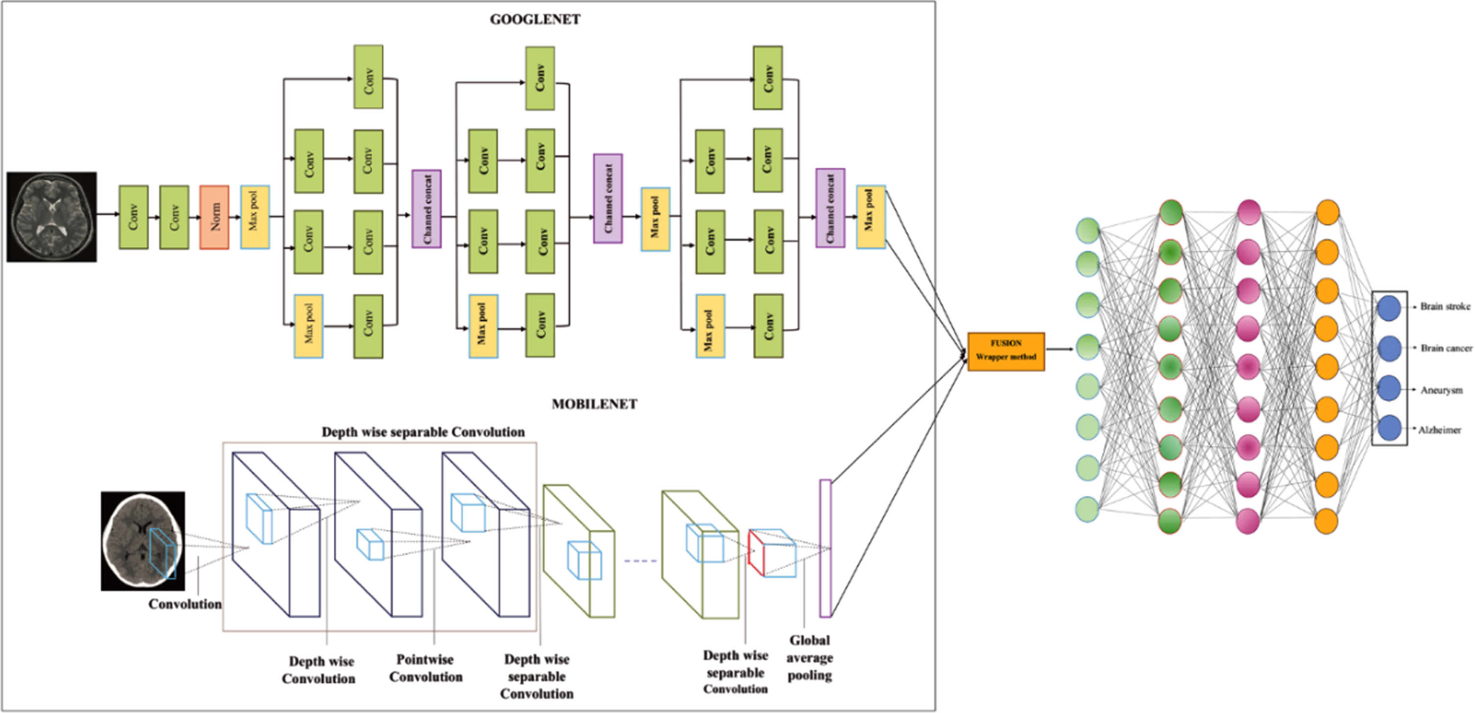
Architecture of fused dual neural network.
Using Google Net, the features are extracted from the pre-processed MRI images. Its goal is to find significant hidden features in denoised pictures so that affected patients may be distinguished from healthy controls. The proposed intrusion detection method extracts the features using Google Net rather than a traditional technique. For control tests utilizing alternative feature extraction strategies, a training set and the same parameters were employed. The loss function was built from the cross-entropy loss that was observed through the training phase. In the case of conventional feature extraction, the cross-entropy loss converges to zero after about 200 iterations. When Google Net is used, however, it quickly converges to zero after only about 100 iterations. This suggests that the model built using Google Net is more responsive to the retrieved features. The contrastive loss function-based features are described in
Where λ is a hyperparameter that stands for the margin and
Mobile net is used to feature extraction from the pre-processed CT images. In this section, the depth-wise separable filters form the foundational layers of the Mobile net. Then, discuss the Mobile net before describing the resolution multiplier and width multiplier, the two-model shrinking hyperparameters.
Depth-wise separable convolution
The depth-wise separable convolutions serve as the foundation of the Mobile net model. These factorized convolutions convert a conventional convolution into a depth-wise convolution and a 1 x 1 convolution known as a pointwise convolution. After the PWC, the DWC outputs are combined using a 1 x 1 convolution. When using Mobile nets, each input channel receives a single filter during the DWC procedure. A 1x1 convolution is used to combine the results of the pointwise and depth-wise convolutions. A typical convolution process filters the inputs in one stage before combining them to create a new group of outputs. The DWSC is used to build a filtering layer and a combining layer. This factorization considerably decreases the processing and model size.
A convolutional layer accepts an E F × E F × N feature map F as input and outputs an E F × E F × M feature map H, where a square input feature map’s spatial width and height are designated as E F , number of input channels N and square output feature map’s spatial width A and height are represented by E F , number of output channels M.
Convolution kernel K of size E
k
× E
k
× N × M, the spatial dimension of the kernel E
k
, a quantity of the input and output channels are N and M, is the parameter for the conventional convolutional layer. Assuming the output feature map for regular convolutional is generated as follows:
The computational cost of typical convolutions is:
A DWSC is divided into two layers: PWC and DWC. DWC should be used as the single filter applied to each input channel. PWC, a traditional 1×1 convolution process, is used to combine the output of the depth-wise layer linearly. Both layers of Mobile nets use batch norms and ReLU nonlinearities. DWC with one filter per input channel represents input depth as follows:
Where the r
th
channel in F produce r
th
, feature map’s channel in the filtered output
DWC is far more effective than traditional convolutional. However, it does not combine input channels to produce new features; it just filters the input channels. Cost of DWSC,
Where E
k
. E
k
. N . E
F
. E
F
+ N . M . E
F
. E
F
is a depth-wise separable convolution cost. Convolution can be expressed as a two-step filtering and combining process, which results in a computation reduction of,
In comparison to conventional convolutions, Mobile Net uses 3×3 DWSC, which needs 8 to 9 times less computation but has a slightly reduced accuracy.
The integration of extracted features from Google Net and Mobile Net is fused by the Wrapper method. Therefore, choosing a feature subset is crucial for classifying brain diseases because employing a high number of features could degrade the classifier’s performance. Additionally, a smaller feature set will lead to a less complex classifier structure, preventing the data from being overfitting. Wrapper approaches alter the feature subset by improving the fitness function of the original feature domain in response to the performance of the classifier. On the other hand, filter approaches use some proxy measures, like variance, to order the features according to their relevance. Numerous analyses of these feature reduction techniques have demonstrated that no single methodology outperforms all others. For feature subset selection, two widely used wrapper methods are forward feature selection and backward feature removal. To reduce the original feature domain, a wrapper with a backward elimination strategy starts with all the features and eliminates the least important feature at each iteration. The forward selection scheme, on the other hand, begins with a null set and continues to add features with the highest relevance at each iteration to shrink the original feature domain. The process is then repeated for the addition or removal of features until no improvement is seen in either the forward or backward schemes. The extracted features from the dual neural network are given as input to the fusion model. In the wrapper method, the training images’ feature vectors are merged with feature vectors derived from other numerical data, so that as many features as possible can be utilized for further classification. By using the wrapper method, the accuracy of the suggested method will be enhanced.
Deep belief network
DBN is employed for classifying brain stroke, cancer, Aneurysm, and Alzheimer’s diseases using BMI images. The Deep Belief Network is a subclass of deep neural networks based on restricted Boltzmann machines (RBM). DBN can be used for unsupervised learning tasks to reduce the dimensionality of feature space as well as supervised learning tasks to create classification or regression models. The two steps in training a DBN are layer-by-layer training and fine-tuning. After completing the unsupervised training, the parameters of the DBN are fine-tuned using error back-propagation methods. Layer-by-layer training is the term for the unsupervised training of each RBM.
In DBN learning, there are two phases: pretraining, and fine-tuning. The BP approach, which modifies the starting and biassing samples, is used to infer supervised learning, while hierarchical stacked RBM training’s inverse divergent properties are primarily essential for unsupervised learning. Unsupervised DBN training is highly wanted for enhancing RBMs and extracting features from the data. The applied energy function E (v, h|θ) and set of (v, h), is obtained.
In an RBM, the joint distribution q (v, h|θ) over the visible units v and hidden units h, given the model parameters, is defined in terms of an energy function E (v, h|θ) of
The probability distribution from the visible and hidden neurons in the case of Gibbs sampling is as follows:
The definition of an active state is the possibility hx. Given that RBM is made up of uniform features for processing the hidden layer h, the option of each neuron in a visible layer activating may be determined using the above equation. The RBM hierarchy is trained to reach the starting weights W = w1, w2...wl using the preset learning technique, which also applies unsupervised learning from the DBN.
The use of MATLAB, a DL toolbox, enabled this study’s innovative setup. In this result analysis, to detect the brain diseases are classified as stroke, cancer, Aneurysm, and Alzheimer’s from the BMI images in the Kaggle datasets. Figures 3, and 4 describe the visualization results of the proposed FDN network model.
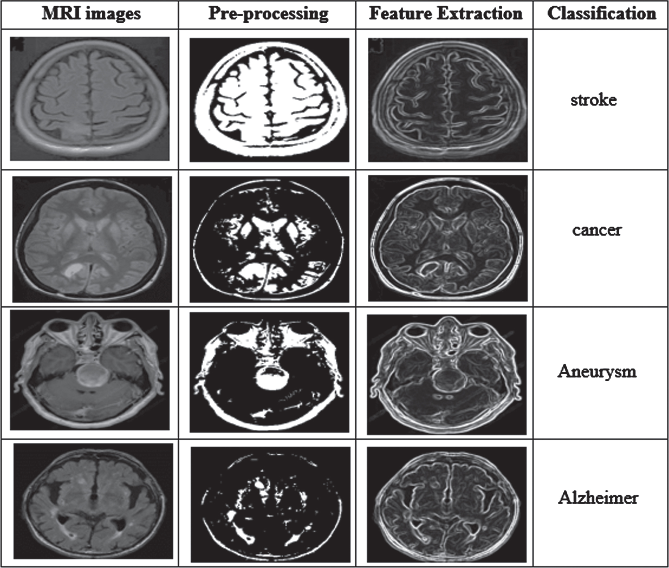
Results of the proposed FDN model of MRI images.

Results of the proposed FDN model of CT images.
The specificity, accuracy, recall, precision, and F1 score were used to calculate the performance analysis in this study. Performance analysis of the suggested method is shown in Table 1.
Performance analysis of the suggested method
Performance analysis of the suggested method
Were,
TP- true-positives
TF- true-negatives
FP- false-positives
FN- false negatives.
Figure 5 represents the performance of the proposed model for four classes including stroke, cancer, Aneurysm, and Alzheimer’s. The proposed method achieved higher accuracy of 0.981,0.976, 0.943, and 0.938 for stroke, cancer, Aneurysm, and Alzheimer’s. The proposed method achieved higher specificity of 0.973, 0.965, 0.912, and 0.874 for stroke, cancer, Aneurysm, and Alzheimer and precision is 0.901,0.928, 0.897, and 0.923 for stroke, cancer, Aneurysm, and Alzheimer. The proposed approach achieved a higher recall of 0.906, 0.948, 0.975, and 0.929 and the F1 score is 0.918, 0.959,0.912 and 0.939 for stroke, cancer, Aneurysm, and Alzheimer that can be measured via TPR and FPR parameters.
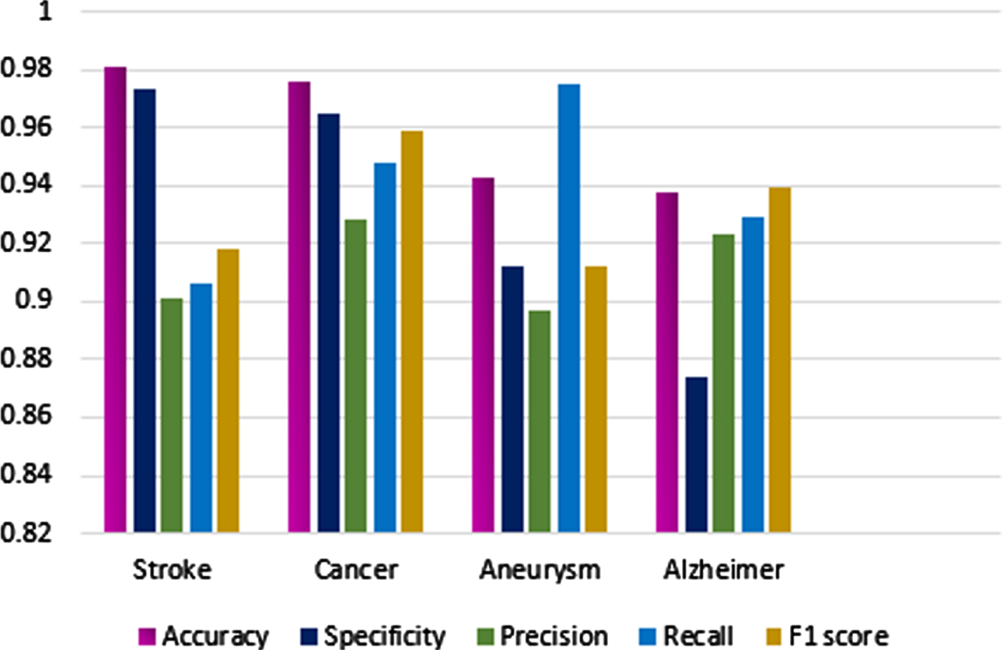
Performance metrics for four classes.
Figure 6 shows the high accuracy of the proposed model during training and testing, and Fig. 7 shows the loss. The performance is based on the accuracy, specificity, precision, recall, and F1 score and the accuracy obtained by the proposed model is 98.19 %.

Training and Testing accuracy of the proposed method.

Training and Testing loss of the proposed method.
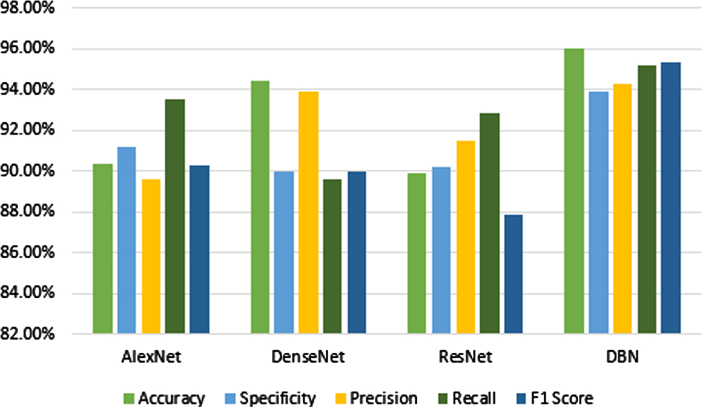
In this division, a comparison between the proposed model and traditional neural networks is also made. Based on a comparison of this technique’s performance with existing approaches, it is more productive than those ways. Performance is assessed using accuracy, specificity, precision, recall, and F1 score. This comparison analysis compares the proposed model to three existing deep learning methods.
Table 2 shows the outcomes in terms of the total accuracy rate. From Table 2 the traditional networks like Alex Net, Dense Net, and Resnet obtain less accuracy compared to the DBN. DBN achieves a high accuracy range of 95.98%. From Fig. 8 shows that the accuracy obtained by Alex net, Dense net, and Resnet is 90.34%, 94.39%, and 89.92% respectively. The specificity obtained by Alex Net, Dense Net, Resnet, and DBN is 91.19%, 89.98%, 90.18%, and 93.89%. Precision is obtained by Alex net, Dense net, and Resnet and DBN is 89.58%, 93.87%, 91.49%, and 94.29%. The recall is obtained by Alex net, Dense net, and Resnet and DBN is 93.51%, 89.59%, 92.87%, and 95.18%. F1 score is obtained by Alex net, Dense net, and Resnet and DBN is 90.31%, 89.98%, 87.89%, and 95.34%. In comparison to existing models, the DBN provides a higher accuracy rate.
Comparative analysis of deep learning networks with the proposed model
Comparative analysis of deep learning networks with the proposed model
Comparison of traditional deep learning models.
According to Table 3, the suggested FDN model improves the overall accuracy by 5.35%, 3.14%, 9.48%, 5.33%, and 0.55% better than Faster R-CNN, CNN, Inception-V3, DCNN, and Fine-tuning Network respectively. According to the comparison above, the proposed FDN model is more accurate than the existing models. In the future, the accuracy of the suggested method will be enhanced.
Comparison between the suggested and the existing models
An ablation study was made to assess the efficiency of the wrapper method utilized in the feature fusion stage. In this experiment, with wrapper and without wrapper according to a comparison, the classification accuracy for brain stroke, cancer, Aneurysm, and Alzheimer’s diseases was illustrated in Table 1. According to our findings, classification using ablation models was typically less accurate than feature fusion using wrapper method, which proves that the usefulness of the wrapper method in the feature fusion process.
Performance comparison in ablation study of with and without a wrapper
Performance comparison in ablation study of with and without a wrapper
Initially, we evaluated the effectiveness of the proposed FDN, and the ablation study is performed with and without the Wrapper method in terms of different brain disease classes. There is a possibility that the model without the wrapper had the lowest accuracy in classification. The classification model the brain diseases use high-level semantic and contextual features. The FDN model with the wrapper method had accuracy in classifying different brain diseases.
In this paper, a novel FDN network has been proposed to detect various types of brain diseases using BMI images. The adaptive bilateral filter is pre-processed to reduce the noise artifacts in BMI images. Using Google Net, the features are extracted from the pre-processed MRI images, and the Mobile Net is used to features extraction from the pre-processed CT images. The integration of extracted features from Google Net and Mobile Net is fused by the Wrapper method. Finally, the Deep Belief Network is employed for classifying brain stroke, cancer, Aneurysm, and Alzheimer’s diseases using BMI images. The proposed FDN achieves a high classification accuracy rate of 98.19%, 97.68%, 94.31%, and 93.82% for detecting stroke, cancer, Aneurysm, and Alzheimer respectively. The proposed FDN model was compared with other traditional models like Alex Net, Dense Net, Resnet, and DBN. The proposed FDN model improves the overall accuracy by 5.35%, 3.14%, 9.48%, 5.33%, and 0.55% better than Faster R-CNN, CNN, Inception-V3, DCNN, and Fine-tuning Network respectively. In future work, we plan to increase both training and testing datasets based on diversity and size for accurately detecting brain diseases.