
Abstract
Over the past two decades, diagnosis of tooth caries or cavities is considered as one of the emerging research topics. So far, a number of methods are introduced to diagnose the tooth decaying, tooth demineralization and re-mineralization as well. However, the sophistication against the tooth decaying diagnosis arises when the environs are relatively complex. With all this in mind, this paper introduces the caries diagnosing model. Here, the feature extraction is based on Multilinear Principal Component Analysis (MPCA). Further, the classification is done by utilizing renowned classifier named Neural Network (NN). The proposed model is compared with other conventional methods such as the Principal Component Analysis (PCA), Linear Discriminant Analysis (LDA), Auto Correlation-NN (AC-NN), Gray-Level Co-Occurrence Matrix (GLCM AC-Support Vector Machine (SVM)), and Independent Component Analysis (ICA), and the performance of the approach is analyzed in terms of measures such as Accuracy, Sensitivity, Specificity, Precision, False Positive Rate (FPR), False Negative Rate (FNR), Negative Predictive Value (NPV), False Discovery Rate (FDR), F
Introduction
Image processing is one of the popular technologies that find numerous applications [6, 10, 12, 14, 15]. Among such applications, the medical field is the most useful real-life application, where researchers pay huge interest. The medical image processing mainly adopts various image segmentation algorithms [9], registration and classification algorithms for diagnostic purposes. The processes include image analysis, tracking and segmenting. Advanced methodologies are also adopted to improve the computational efficiency of the image processing applications [11, 13].
The medical image processing applications include tumor detection, segmentation of lung portions [21], investigation on cervical cytological images [22] and much more. The applications are also extended to estimating the growth stages of cells by determining the intracellular patterns, cell size and other parameters that have an impact on cell growth [8]. Techniques on computed tomography (CT) images of the chest can lead to estimate the heartbeat rate [7]. The diagnostic procedures adopt a variety of technologies on the images, associated features, and their contents. For instance, artificial intelligence techniques such as neural network are adopted as a classifier to determine the grades of cancer [5].
Photoacoustic (PA) imaging is emerging as a new modality for non-invasive medical imaging and diagnosis, while tooth decay, often called as caries or cavity, remains as one of the most common oral diseases. Moreover, the health of dental tissue is diagnosed by the PA imaging. Current methods for detecting tooth decay employing the dental explorer and X-ray radiography are subjective. The former researchers mainly discuss extracted tooth decaying diagnosis by ANN (Artificial Neural Network) technology and rule-based system [5]. However, it requires adequate improvement and precision for promising results. Few of the earlier research works are briefly reviewed further.
The main contribution of this paper is to detect the caries on the basis of the advanced image processing algorithms in order to ensure detection accuracy. In the proposed caries detection approach, the acquired dental images are subjected to enhancement followed by required pre-processing. Here, the enhancement algorithm mainly adopts the contrast enhancement to distinguish the white intensity of the teeth. The pre-processing steps dynamically adjust the grey level of the image so that the regions of interest are well focussed. Subsequently, it adopts feature extraction algorithms and a supervised classifier. The proposed feature extraction algorithm describes the images, which are two dimensional, in multidimensional space. Thus, the contents of the images are projected into multi-dimensional format and so the useful information from the images can be required. Then, NN with enhanced learning algorithm will be used to classify the grade of caries.
The paper is organized as follows: Section 2 reviews the literature work; Section 3 describes the framework of the proposed caries detection model. Lastly, Section 4 discusses the results that were obtained, and Section 5 concludes the paper.
Literature review
Kang et al. [1] have proposed new methods to measure the tooth demineralization and re-mineralization. It uses Optical Coherence Tomography (OCT) to measure the depth and severity of early lesions. After the OCT, the difference in the depth and integrated reflectivity are detected after demineralization. Moreover, the OCT images of dental decay are exploited to program a scanned laser beam in order to remove decays only. Also, the OCT images are obtained before as well as after removal, and the scans demonstrate the extremely selective removal. As a result, cross-polarization-OCT is best suited for the non-destructive assessment of early demineralization.
Hughes et al. [2] have proposed a laboratory method for the early detection of the tooth decay using ultrasound, which is done by recording B-scans of teeth which exhibit caries lesions with high-frequency ultrasound. This is because of the difference in the relative acoustic impedances of the caries lesion as well as the surrounding healthy enamel. The method also makes use of the signal processing algorithm. The demineralized enamel will produce a moderate reduction coefficient, and it produces a larger difference in the ultrasound.
Li et al. [3] have determined the tooth decay by using the Support Vector Machine (SVM) based diagnosis method. The same can also be done by the rule-based system and Artificial Neural Network (ANN) methods. However, SVM provides better results. Moreover, the SVM is exploited for one step forward by means of dealing with clinical X-ray image on the basis of the tooth decay diagnosis. Finally, the simulation outcome exhibits that the gray-level co-occurrence matrix are better than autocorrelation coefficient.
Sampathkumar et al. [4] proposed a non-contact optical technique for imaging and detection of early-stage dental caries. They have developed a fine-resolution ultra-broadband all-optical photoacoustic imaging (AOPAI) system to image the early stages of tooth decay. AOPAI system provides a non-contact method for early detection of white-spot lesions with a high detection bandwidth. Moreover, AOPAI system presents a non-contact method for early detection of white-spot lesions with a high detection bandwidth, which presents advantages over previously demonstrated ultrasound methods.
Yu et al. [5] have proposed Artificial Neural Network (ANN), to detect the tooth decay. By using the back-propagation (BP) neural network, the X-ray image of patient’s teeth is analyzed. The network attained considerable superior performance in making differential diagnoses between decayed as well as normal teeth with inter-pixel autocorrelation coefficients as its input feature vector. In comparison with the other method, the tooth decay detection accuracy was significantly improved.
Palmier et al. [43] presented the International Caries Detection and Assessment System (ICDAS) and the Post-Radiation Dental Index (PRDI), which is limited viability for the estimation of Radiation-Related Caries (RRC) lesions due to their criteria it does not take into consideration qualitative clinical features of RRC, which are very prevalent in their clinical presentation and patterns of progression.
Zain et al. [44] presented the accuracy of Optical Coherence Tomography (OCT) in order to detect naturally occurring non-cavitated fissure caries (NCFC) in totality as well as at different loci by visually assessing cross-sectional OCT scans (B-scan) with an interpretation criterion. Moreover, the agreement between dimensions of NCFC is evaluated and measured with OCT and polarized light microscopy (PLM).
The PS-OCT technique that used in [1] is a non-invasive method for detecting the flaws. It is a simple method in which the output is shown in a three-dimensional manner. However, the images obtained will be less contrast, and it is not realistic in nature. B-scan is a diagnostic test, which produces a cross-section of the output images is used in [2]. The performance of this method is stable, and it provides accurate output. At the time of process execution, the waves have only limited penetrations, and the resolution of the output image is less. The support vector machine used in [3] has been experimentally demonstrated for its flexibility, and it also provides the unique solution. Nevertheless, the support vector machine suffers from its lack of transparency. Moreover, 3D PA [4] method has shown its advantages over image formation in a direct manner and also provides high detection sensitivity. However, the method fails in terms of accuracy. On the other hand, the neural network [5] method is easy to detect the complex nonlinear relationship.
Architecture diagram of the proposed methodology.
The schematic diagram of the proposed model of dental caries recognition is briefly illustrated in Fig. 1. The image
Pre-processing
This is the initial stage, which enhances the input image by following three steps.
Contrast enhancement
The resized gray level input image (it is necessary to increase or decrease the total number of pixels of the image) is given for enhancing the contrast of the image, where input image is
The gray thresholding by Otus’s thresholding [40] process determines the threshold for the image which is used to convert grey pixel to either white or black based on the gray intensity. In other words, if the image intensity
Active contour [39]
Active contour model also referred as snakes it is exploited to create a contour or mask of the objective, a set of coordinates must be initialized. The formed contour is relocated over the image by the driven forces of the image to the boundaries of the particular objective. This process exploits two kinds of driven forces namely internal energy and external energy. The main aim of the internal energy term is to control the deformations made to the snake, and the use of the external energy term is to control the fitting of the contour onto the image. Hence, a set of coordinates
In contour, the control points’ normalized index is represented as
where the interior energy forces of the curve are represented as
Hence the resultant pre-processed image via the active contour model is represented as
Preprocessing stage (a) input images, (b) images after contrast enhancement, (c) images after grey thresholding, (d) images after active contour.
From the pre-processed image, the features are extracted using MPCA [42]. MPCA is a multi-linear extension of PCA. MPCA is employed in the analysis of arrays i.e., a cube or hypercube of numbers, also referred to as ‘data tensor’. MPCA has better generalization ability than PCA in image reconstruction. With this approach, the image is rearranged into a 3D tensor as
where index
Diagrammatic illustration of multi-linear projection.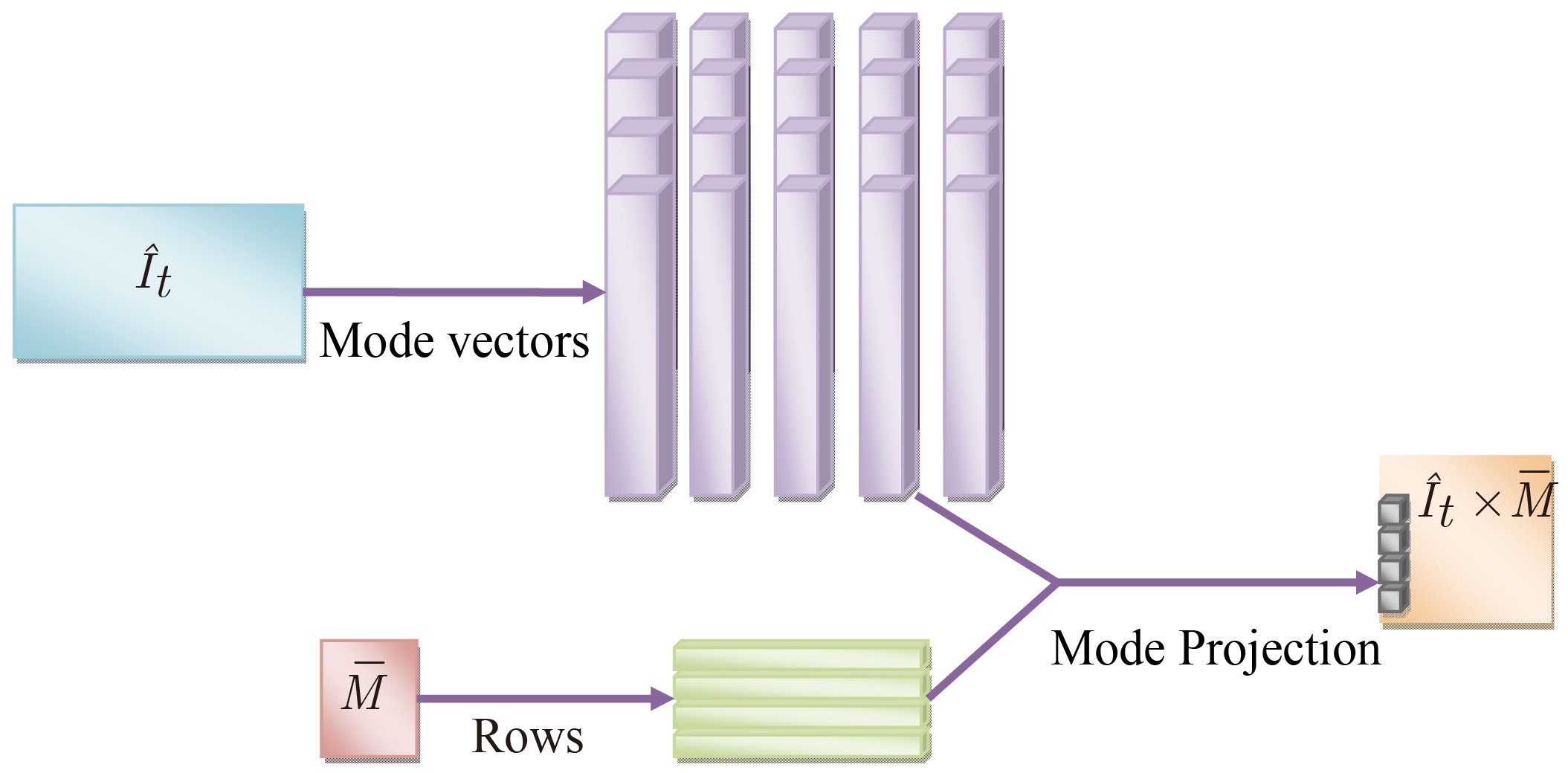
Since the neural networks are established to be prominent for classification in various applications due to its flexibility than any other classifier, in this paper, the chosen features are applied to the neural network for recognizing caries. Here, the back propagation algorithm model is used for classification. With known classifications, the back propagation trains a given feed-forward multilayer neural network for a given set of input patterns. While each entry of the sample set is presented to the network, the network observes its output response to the sample input pattern. The output response is then compared to the known and desired output as well as the error value is computed. On the basis of the error, the connection weights are adjusted. The back propagation algorithm is on the basis of the Widrow-Hoff delta learning rule in which the weight adjustment is performed through Mean Square Error (MSE) of the output response to the sample input.
The feature set is represented in Eq. (10), where
The weight
To provide the training to the network, weight
The classifier results in the detection of caries or non-caries dental images.
Performance measure of the classifier with varying threshold ranges (a) accuracy of NN for test cases 1–3, (b) sensitivity of NN for test cases 1–3, (c) specificity of NN for test cases 1–3.
Performance measure of the classifier (a) accuracy of NN for test cases 1–3, (b) sensitivity of NN for test cases 1–3, (c) specificity of NN for test cases 1–3.
The experimentation was carried out in MATLAB 2015a. The database was downloaded from [45], which was categorized into three sets (test cases 1–3) randomly for the analysis purpose. Such test case divisions are required to ensure fair experimentation. Each test case consists of 40 caries images. Here, 28 images are employed for training data and the residual 12 images are employed for test data. Further, the performance of the proposed model was investigated in correspondence with ten measures like accuracy, sensitivity, specificity, and precision, FPR, FNR, NPV, FDR, F
where, TP represents the True Positive; TN represents the True Negative; FP represents the False Positive; FN represents the False Negative.
The performance of the proposed method is compared to the conventional methods namely (PCA) [37], LDA [36], ICA [38], AC-NN [5] and GAC-SVM [3] in terms of afore-mentioned measures by analyzing varied training percentages including 25%, 50%, and 75% respectively for each test case. The performance comparison of the proposed method over conventional methods for the test case 1 is tabulated in Table 2. From the table, it is observed that the accuracy of the proposed model for 25% training is 35.5% better than ICA, 50.9% and 60% better than LDA and PCA. For 50% training, the proposed model is 44.4% superior to ICA and LDA. Similarly, for 75% training, the introduced method is 60.7% better than ICA, 55.5% and 77.7% superior to LDA and PCA. It is also observed that the FDR of the proposed method is minimized gradually whereas the other methods have increased value. It is observed that, for 25% training, the proposed approach is 51.1% better than ICA and 58%, 68.6% better from LDA and PCA, for 70% training, the introduced approach is 66%, 74.6% and 83% superior to ICA, LDA, and PCA.
The performance analysis of the proposed model for the test case 2 is tabulated in Table 2. From the table, it is clear that the accuracy of the proposed method for 25% training is 75%, 70.1%, and 51.9% better than ICA, LDA, and PCA. For 75% training, the proposed method is 60.7% and 28.3% superior to ICA and PCA. FDR of the proposed method is decreased gradually, and for 25% training, the proposed method is 38.8%, 78%, and 51.1% better from the conventional methods like ICA, LDA, and PCA and for 50% training, the proposed model is 86.6% and 41.1% better than both LDA and PCA. Table 3 shows the performance analysis of the proposed method over existing approaches for the third database. It is observed that for training percentage, 25%, the accuracy of the proposed method is 35.5% superior to ICA, 70.2% better than both PCA and LDA. For FNR measure, the proposed model is 72.7%, 80.6%, and 73.9% better than ICA, PCA, and LDA for 25% training. For 50% training, the proposed model is 50% better than ICA, 72.7% better from LDA.
Performance investigation of proposed model over conventional methods for test case 1
Performance investigation of proposed model over conventional methods for test case 1
Performance investigation of proposed model over conventional methods for test case 2
Performance investigation of proposed model over conventional methods for test case 3
The advantages of MPCA over PCA are that it has the potential to serve the same function for analyzing tensor structure data. Further, it performs the feature extraction by determining the multi-linear projection that captures most of the original tensorial input variation, which also avoids the initial resizing step. Due to the multidimensionality projection, the features are distinguished well in multidimensional and so classification can be precise.
In this Section, the performance of the classifier is determined by varying the contrast threshold ranges including 0.1–0.9, 0.2–0.8, 0.2–0.7, 0.3–0.7, and 0.3–0.8 respectively in terms of accuracy, sensitivity, and specificity for the first, second, and third test cases, which is illustrated in Fig. 4. From Fig. 4a, it is observed that, for the first test case, the accuracy of the NN classifier using the proposed MPCA is more at the ranges like 0.2–0.7, 0.3–0.7, and 0.3–0.8 respectively, for the second database, at the ranges like 0.1–0.9, 0.2–0.8, 0.3–0.7, and 0.3–0.8, the NN classifier with MPCA has higher accuracy rate, whereas the other methods have poor accuracy. For the third test case, the accuracy of the NN using the proposed model is more precisely increased at the ranges such as 0.1–0.9, 0.2–0.8, 0.2–0.7, and 0.3–0.8. Similarly, the performance of the NN classifier is analyzed in terms of measures like sensitivity and specificity, which are illustrated in Fig. 4b and c. Overall results have shown the efficiency of the developed model, whereas the conventional methods have shown its poor performance.
Classifier configuration
The performance of the NN classifier is analyzed in terms of measures like accuracy, sensitivity, and specificity of varying hidden neurons for the first, second, and third test cases, which is illustrated in Fig. 5. From Fig. 5a, it is observed that the accuracy of the NN classifier using the proposed MPCA model for the test case 1 with 10 hidden neurons is 77.7% and 60% better than ICA and PCA. For 40 hidden neurons, the proposed model is 65%, 29.4%, and 17.8% better than ICA, LDA, and PCA. Similarly, for 50 hidden neurons, the proposed model is 58.3%, 18.7% and 69.6% superior to ICA, LDA, and PCA respectively. For the second test case, the accuracy of the NN classifier using proposed MPCA model for 10 hidden neurons is 90%, 5.5%, and 68.4% better than ICA, LDA, and PCA, for 20 hidden neurons, the accuracy of NN using MPCA is 75% better from ICA, LDA and PCA, and for 30 hidden neurons, NN using proposed model is 2.9% and 27.2% superior to both the LDA and PCA. For 40 hidden neurons, the accuracy of NN using MPCA is 69.0%, 24%, and 56.9% better from ICA, LDA, and PCA respectively. The same investigation is carried out for both sensitivity and specificity for all the three test cases, which are illustrated in Fig. 5b and c respectively. From the figures, it is observed that the performance of NN using proposed MPCA model is more efficient while comparing other conventional methods.
Conclusion
This paper has proposed the caries diagnosing model. The features were selected on the basis of Multilinear Principal Component Analysis (MPCA). Further, the classification was done by utilizing renowned classifier named Neural Network (NN). The proposed model was compared with the other conventional methods, and the performance of the approach was analyzed. The analysis was done by varying the training percentages, which have resultant the promising results. The proposed method has attained better detection accuracy for 25% training in test case 1, and it is 48.8% superior to the conventional methods, and this shows the superiority of the developed model.