
Abstract
A new Long-chain, di-Schiff base ligand (N2O2 donor, L) has been synthesized by the reaction of dodecylamine with, 1,5-bis(2’-formylphenyl)-1,5-dioxapentane in molar, 2:1 ratio (yielded 82%) and its Ru(II) complex have been prepared (yielded 78%) in solvent-free conditions. The newly synthesized compounds have been verified by ATR-IR, UV-vis., mass (LC-MS and MALDI-TOF) and NMR (1H,13C) spectra, elemental and thermal analysis. The DNA binding studies of the ligand and complex were examined by absorption method. The results indicated that the ligand and Ru(II) complex could interact with calf thymus DNA (CT-DNA) through electrostatic or/and groove binding (minor/major). The apparent binding constant values (Kb) of the ligand and Ru(II) complex at room temperature were calculated to be 5.9×10⁵ M-1 and 7.9×10⁵ M-1, respectively. Antioxidant study of the ligand and complex showed significant antioxidant activity against lipid peroxidation. The ligand and complex also have moderate radical scavenging properties.
Introduction
Avoiding the use of solvents in synthesis can reduce environmental contamination and even be more convenient than using solvent-based synthesis. It has also been said that “the best solvent is no solvent”. It is sometimes actually higher yield, easier and faster to synthesis some ligands and metal complexes by avoiding the use of solvents [1].
Schiff base are the containing -HC=N- group. They are condensation product of ketones or aldehydes with primary amines and were reported by Hugo Schiff 1864. Metal complexes of Schiff base having played a central role in the development of coordination chemistry [2–4].
Schiff bases show biological applications including antibacterial, antifungal and antitumor activity [5]. In contrast to the synthesis of aromatic Schiff bases, the preparation of long chain aliphatic Schiff bases is known to be relatively difficult [6–10].
In the present study, we report to the synthesis of a new long-chain N-alkyl di-Schiff-Base ligand (DFD), containing, di-salicylaldehyde and etheric groups and its Ru(II) complex in solvent-free Conditions. The ligand and Ru (II) metal complex can be used drug substances. The production of reaction the etheric dialdehyde with N-alkyl amine, that is Schiff’s base, often has the property of luminescence, especially, the ligand has structure of rigidity plane and rich-electronic conjugation [11].
Interaction of transition metal complexes with bio-macromolecules is an important area of research. Among bio-macromolecules, DNA is one of the main molecular targets in the design of anticancer drugs, because many small molecules exert their anticancer activities by binding with DNA, thereby altering DNA replication, blocking the division of cancer cells and resulting in the cell death. The platinum derivatives, such as “Cisplatin” [cis diammine dichloro platinum (II)], are the most effective and widely used anticancer drugs. However, it shows serious side effects such as nausea, kidney and liver failures. It has its own limitations because of the development of resistance in tumor cells [12]. These limitations have stimulated and extensive search for active metal based complexes other than platinum derivatives having more effective, less toxic and target spesific non-covalent DNA binding anti-tumor drugs. In the last years, ruthenium based organometallics have attracted much attention and represented one of the latest trends in metallodrug research. They have variable oxidation states, low toxicity, selectivity for cancer cells and ability to mimic iron ion binding to biomolecules [13–15]. It is also known that the uncontrolled production of reactive oxygen species such as superoxide anion radical (O2·-) and hyroxyl radical (OH·) can induce DNA damage in human. Such damage to DNA has been suggested to contribute to various diseases including cancer, cardiovascular disease and inflammation. For these reason, the investigation of the biological properties of the ligand and Ru(II) complex focused on the binding properties with calf thymus DNA (CT-DNA), antioxidant properties against DPPH, ABTS radicals, lipid peroxidation and B-carotene bleaching.
Experimental
Materials
The IR spectra were recorded in attenuated total reflection (ATR) technique on a Perkin Elmer Spectrum One Bv 5.0 spectrophotometer. Mass spectra (LC-MS/MALDI-TOF) were determined on HPLC and Agilent 530. Absorption spectra were recorded on an Agilent 8453 marka UV/VIS spectrophotometer. 1H-NMR ve 13C-NMR (CDCl3 and TMS) were determined on Varian Unity 500 spectrophotometer. TGA-DSC curves were obtained with a TA SDT Q 600 thermal analyzer apparatus using flowing nitrogen 100 mL min-1, temperature range 50–1200 C, at heating rate of 10°C min-1. Elemental analyses were obtained on a Thermo Finnigan Flash EA 112. Melting points were obtained with a Büchi Melting point B-540 apparatus in open capillaries. Ru(bpy)2Cl2·2H2O was synthesised and 1,5-bis(2’-formylphenyl)-1,5-dioxapentane were synthesised as literatüre [16, 17].
Methods
Preparation of the di-Schiff base Ligand (1,5-bis(benzylidene-N,N’-dodecylimine)-1,5-dioxapentane) (DFD) in solvent free condition
Schiff base ligand was prepared according to the following procedure. 1,5-bis(2’-formylphenyl)-1,5-dioxapentane (0.873 g, 0.1 mmol) was mixed to dodecylamine (1.320 g, 0.2 mmol), stirred and heated to 115 C for 1.5 hours under a nitrogen atmosphere in solvent free conditions. The product was cooled and washed with 2×40 mL methanol and extracted with 2×30 mL ether. The viscous long-chain the Schiff base is highly soluble polar and even in non-polar solvents such as petroleum ether, diethyl ether and n-hexane.; The ligand has luminescence properties. Yield 82%, ATR-IR: ν(cm-1): 3025, 3030 (Ar), 2920–2851 (CH2), 1637 (C = N), 1454–1488 (CH2), 1238–1110 (Ar-O-C), 750–721 (ar-sübst). 1H-NMR (500 MHz, CDCl3, δ / ppm): 0.80–1.75 (42 H, m, CH2), 2.35–2.49 (2 H, q, CH2), 4.15–4.45 (4 H, t, OCH2), 6.9–8.0 (4 H, m, Ar-CH), 8.65–8.75 (2 H, s HC = N); LC-MS: m/z = 619[M +]; Anal. Calcd for C41H66N2O2 (%): C, 79.56; H, 10.75; N, 4.53 Found: C, 78.52; H, 9.51; N, 4.59.
DFD- Ru(II) Complex
Ru(bpy)2Cl2·2H2O (0.714 g, 1.5 mmol) was added to the Schiff base ligand (DFD) (1.875 g, 3 mmol) stirred and heated to 90C for, 1 hour under a nitrogen atmosphere in solvent free conditions. The product was cooled and mixed with ether and precipitated. The Ru (II) complex was dark-brown. The long-chain viscous Schiff base Ru(II) complexes are highly soluble polar and even in non-polar solvents such as petroleum ether, diethyl ether and n-hexane. Yield 78%. (ATR-IR): ν (cm-1): 2920–2851 (CH2), 1671 (C = N), 1243–1162 (Ar-O-C), 727 (Ar-subst.) LC-MS: m/z=1026[M--], Anal. Calcd for C65H82N2O2Ru (%):C 76.21, H: 8.07, N: 2.73 Found: C 76.26, H: 8.68, N: 2.99.
Antioxidative activities
Determination of antioxidant activity with the β-Carotene Bleaching (BCB) Test
Antioxidant activity of the complex and the ligand was determined according to the β-carotene bleaching method [18]. The results were given as relative antioxidant activity (RAA). BHA was used as positive control sample.
Determination of total antioxidant capacity using thiocyanate method
The total antioxidant activity of the ligand and ruthenium complex was determined by the thiocyanate method according to the method described previously [19]. The results were calculated for inhibition of lipid peroxidation in percentage.
Inhibition of lipid peroxidation % = [(Ao – A1) / Ao×100]
Where A0 is the absorbance of the control reaction and A1 is the absorbance in the presence of the sample or standards.
Free Radical Scavenging Activity
The free radical scavenging activity of the complex and ligand was measured with 2,2-diphenyl-1-picryl-hydrazyl (DPPH) using the slightly modified methods of [20]. Briefly, 20 mg/L DPPH·solution in methanol was prepared and 1.5 mL of this solution was added to 0.75 mL of the sample and standard solution (20–100μg/mL). The mixture was shaken vigorously and the decrease in absorbance at 517 nm was measured at 30 min. The percent inhibition was calculated using the following equation:
Inhibition % = [(A0 – A1) / A0×100]
Where A0 is the absorbance of the control reaction and A1 is the absorbance in the presence of the sample or standards.
ABTS·+ Scavenging Assay
For the ABTS [2,2’-azino-bis(3-ethylbenzothiazoline-6-sulfonic acid) diammonium salt] cation radical scavenging assay were performed as described previously [21].
BHA (butylated hydroxyanisole), BHT (butylated hydroxytoluene), and epicatechin were used as standard antioxidants. The percent inhibition activity was calculated using the following equation:
Inhibition % = [(A0 – A1) / A0×100]
Where A0 is the absorbance of the control reaction and A1 is the absorbance in the presence of the sample or standards.
DNA binding studies
Electronic absorption titration
All experiments involving the binding of compounds with calf tymus DNA (CT-DNA) were performed in a buffer with tris-(hydroxymethyl)aminomethane (Tris, 5 mM) and sodium chloride (10 mM) and adjusted to pH 7.2 with hydrochloric acid. Concentrated stock solution of the compounds were prepared by dissolving the compounds in dimethyl sulfoxide (DMSO) and diluted suitably with buffer to required concentrations for all the experiments. Absorption spectral titration experiments were performed with fixed concentrations of the compounds (25μM) with varying concentration of DNA (0–50μM).
Results and Discussion
Preparation of the ligand and its complexes
The synthesis of linear alkyl groups containing Schiff bases is difficult due to the easy hydrolizes is less and yield is low. In this study, to prevent hydrolysis and with the aim of improving efficiency without the use of solvent, reagents and condensation melting at 115C for 1.5 hours with high efficiency by the reaction of long-chain alkyl group containing a new Schiff base and Ru(II) complex synthesized at 90C for 1 hour. Among the solvent tested for synthesis reaction in MetOH and yielded fort the ligand 15%, 4 hours and Ru(II) complex 11%, 8 hours. The best results (short reaction time and maximum yield of the product) were obtained under solvent-free condition, which is also economically and environmentally favorable.
In this study, first, di-aldehyde molecule is 1,5-bis(2’-formylphenyl)-1,5-dioxapentane was synthesized with salicylaldehyde of the alkaline environment and the 1,3-dibromopropane reaction in a nitrogen atmosphere with in the literature [17]. Then long-chain alkyl group containing a new Schiff and Ru(II) complex synthesized under solvent-free condition yield 55–82%. The Schiff base and its complex are highly soluble polar and even in non-polar solvents such as petroleum ether and chloroform.
The newly synthesized compounds have been verified by ATR-IR, UV-vis, 1H-NMR ve mass (LC-MS and MALDY-TOF) spectra and thermal analysis (TGA-DSC) techniques.
In this study, the synthesis of the molecules can be seen in the Scheme 3.1.
Synthetic route of the Schiff base ligand and Ru(II) complex.
FTIR spectrum of the ligand also provides additional data to confirm the structure given in Fig. 3.1. C = N stretching vibrations appear at 1637cm-1. In the FTIR spectrum of the Ru (II) complex C = N stretching vibration appeared at 1603 cm-1 (Fig. 3.2) In the FTIR spectrum of the complex, the shifts of the C = N to higher or lower frequencies. That indicates the formation of coordination bands between metals with N atoms at the ligand.
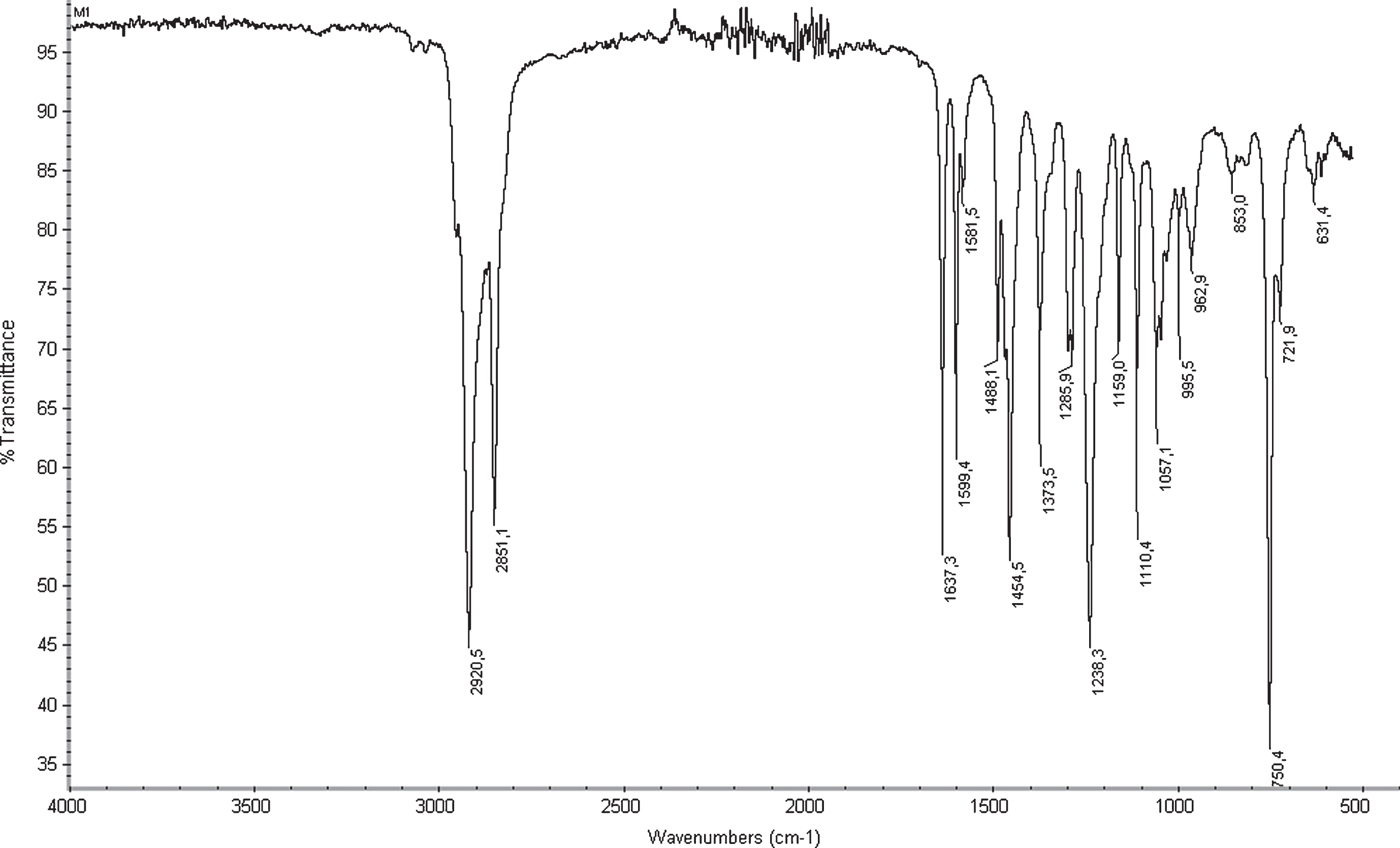
IR spectra of the ligand (400–4000 Cm-1).
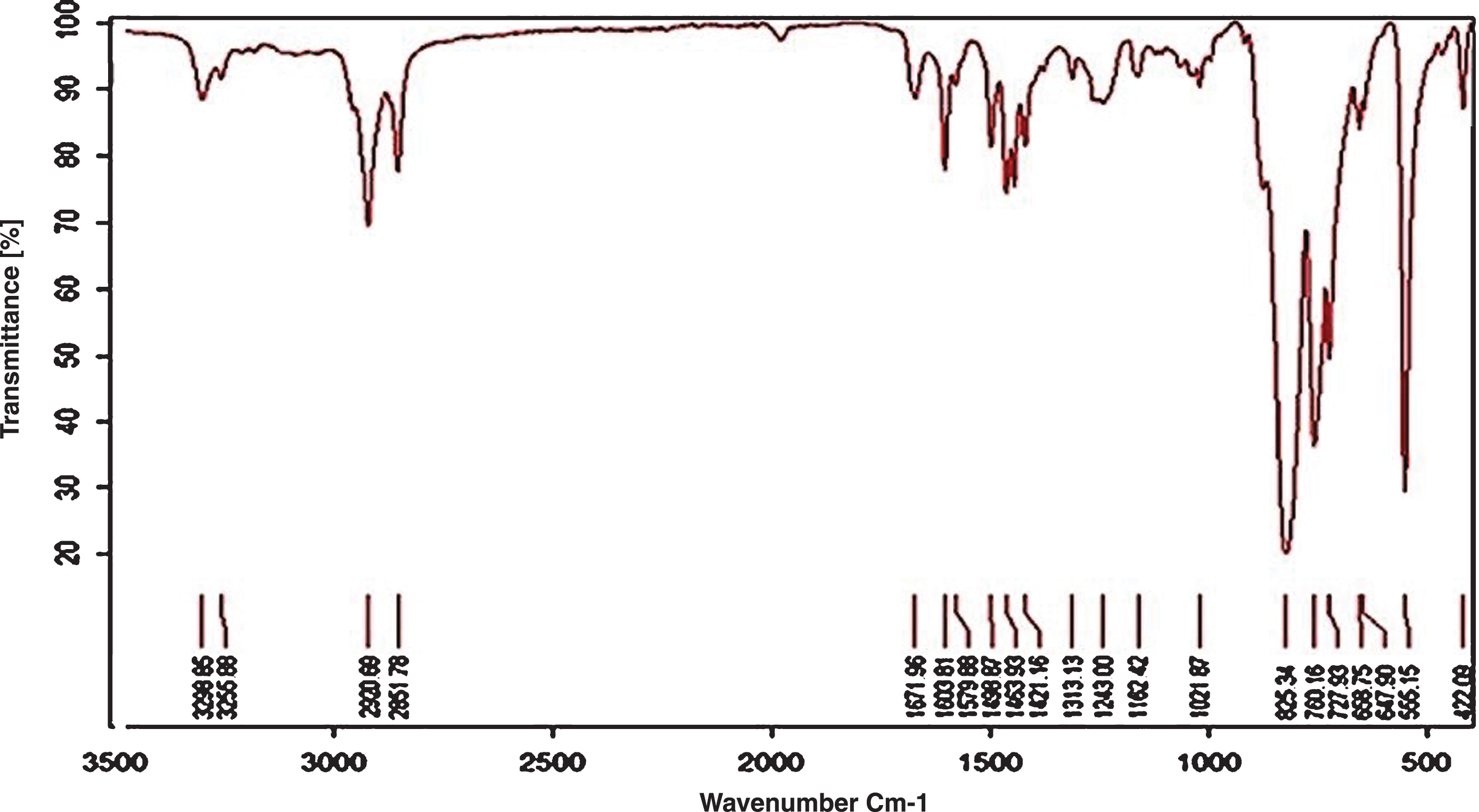
IR spectra of the Ru(II) complex (400–4000 Cm-1).
In the mass spectrum of the ligand and Ru(II) complex Shown in Figs. 3.3 and 3.4. Ion peak of the ligand and Ru(II) complex observed at m/z 619 and m/z 1026 can be assigned to the [M+1] and [M-2] ion peak respectively. The ligand and its Ru(II) complex were also identified by mass spectra.
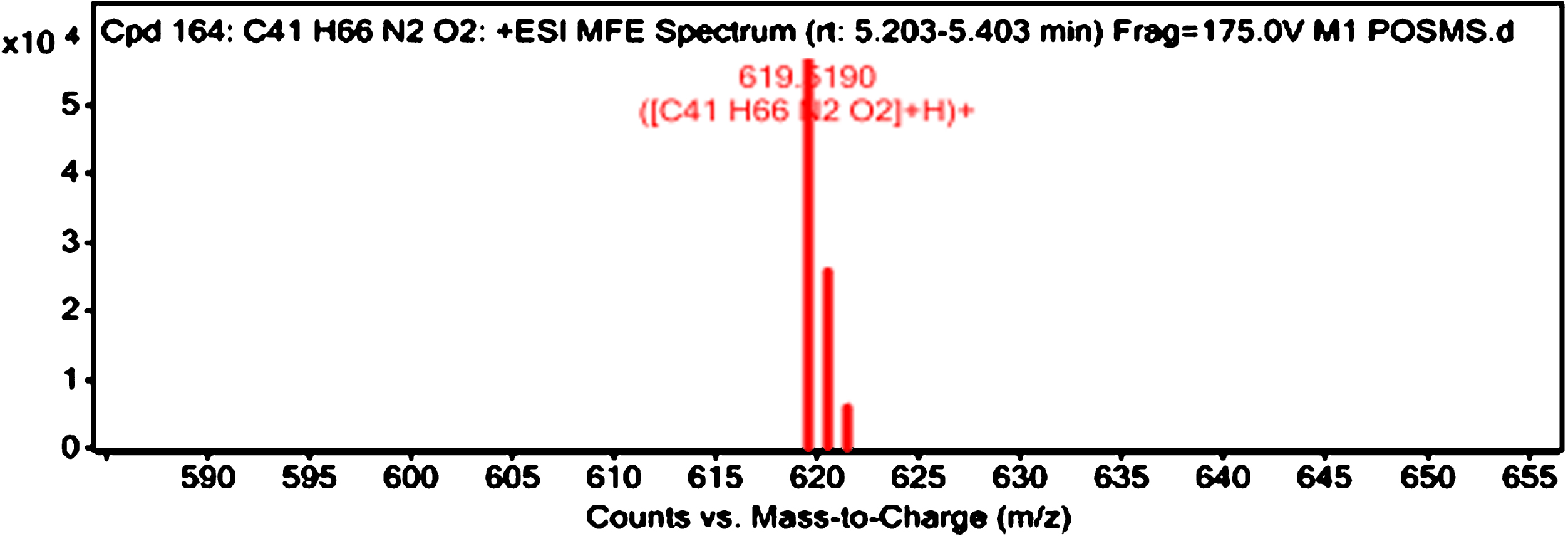
LC-MS spectrums of the Schiff base ligand.
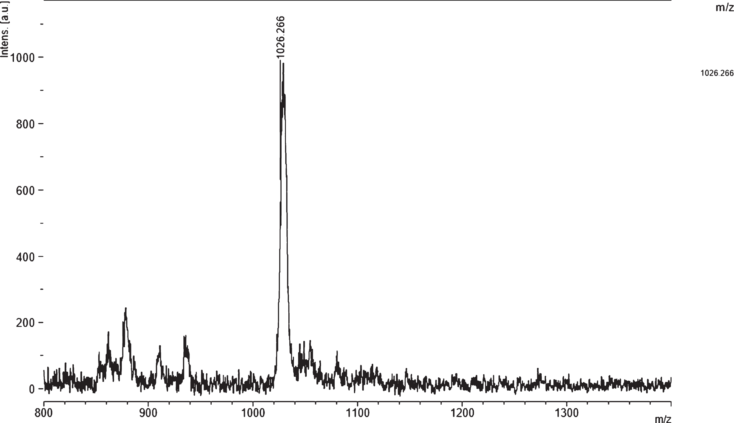
MALDI-TOF spectrums of the Ru(II) complex.
The electronic absorption spectral data for ligand and Ru(II) complex were dissolved in CHCl3 solutions (Fig. 3.5). The electronic spectrum of ligand showed two types of transition. The first one appeared at 240–270 nm which can be assigned to σ- σ* transition was due to transition involving orbitals located on the long-chain. The second type of transition appeared at 290–310 nm which can be assigned to π- π* and n- π* transition was due to transition involving molecular orbital of the C = N chromophore groups. The third type of transition appeared in complexes at 350–490 nm which can be assigned to d-d and ligand to metal charge transfer.
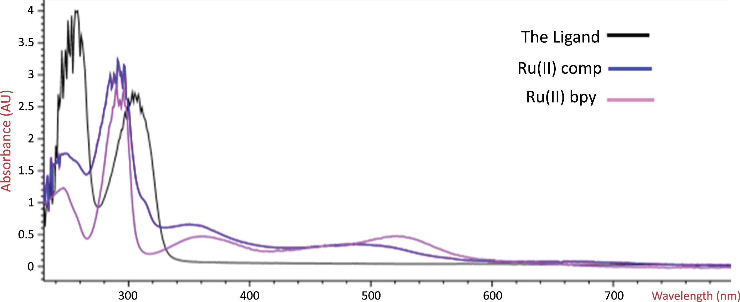
UV-vis spectra of the Ligand, Ru(II) complex, Ru(II) bpy. in CHCl3.
The TGA-DTG-DSC curve of the Ru(II)) complex was shown in Fig. 3.6. The complex heated from 50 C up to 1200 C. TGA curve of the complex shows two endothermic picks. The first is melting point (66C) was not appeared weight loss. The second was appeared at 227–303C in % 74 decomposition. Lost product ca 95% and the residue is black C at 1200C.

TGA-DTG-DSC spectra of the Ru(II) complex.
1H-NMR spectrum in CDCl3 of ligand confirmed the proposed structure showing 8.7 ppm as singlet peak for the N = CH (2 H), 6.9–8 ppm as multiplet for Ar (4 H), 4.7 ppm as triplet for the O-CH2(4 H), 3.7 ppm as triplet fort the N-CH2, 2.3 ppm as quintet for the etheric CH2 (4 H) and 0.80–1.75 ppm (42 H), for the N-alkyl groups (Fig. 3.7).
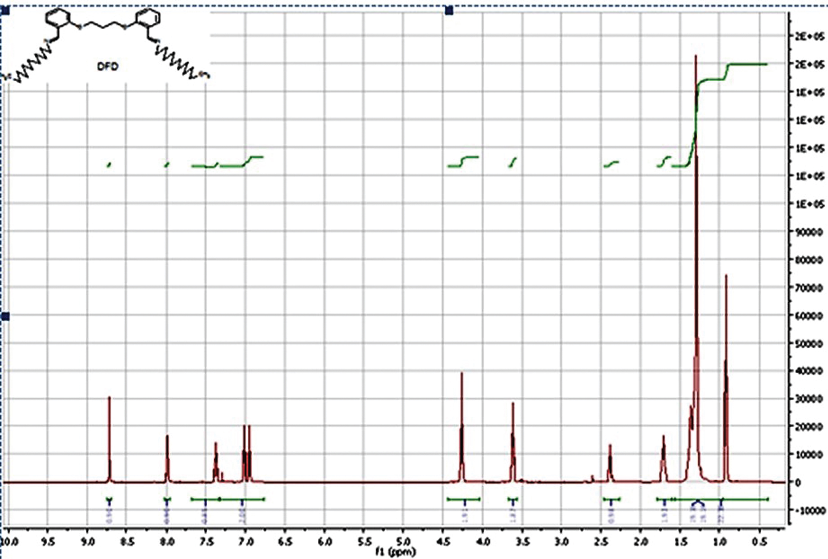
1H-NMR spectrum of the ligand (CDCl3).
1H-NMR spectrum in CDCl3 of Ru(II) complexes (Fig. 3.8) confirmed the new compound synthesised, but the spectrum isn’t clear.
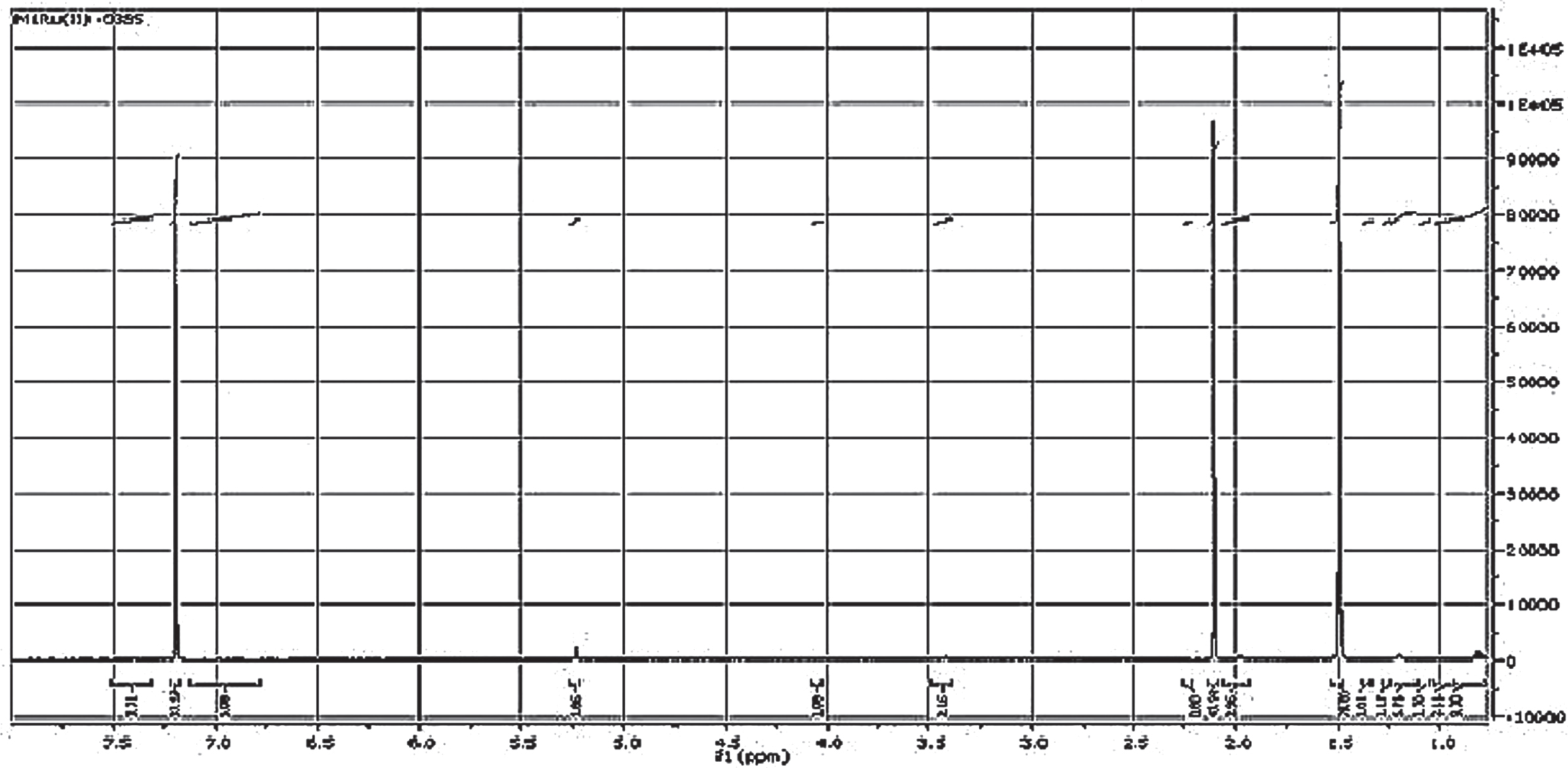
1H-NMR spectrum of the Ru(II) comp.(CDCl3).
The 13C NMR of the Schiff base in DMSO, shown in Fig. 3.9. NMR spectra of the ligand is characterized by 13–78 ppm aliphatic groups (N-alkyl) and very sharp peaks at 115–132 ppm corresponding of the aromatic ring. The downfield peaks at 155 and 158 ppm are attributed to Schiff C(C = N) atoms.
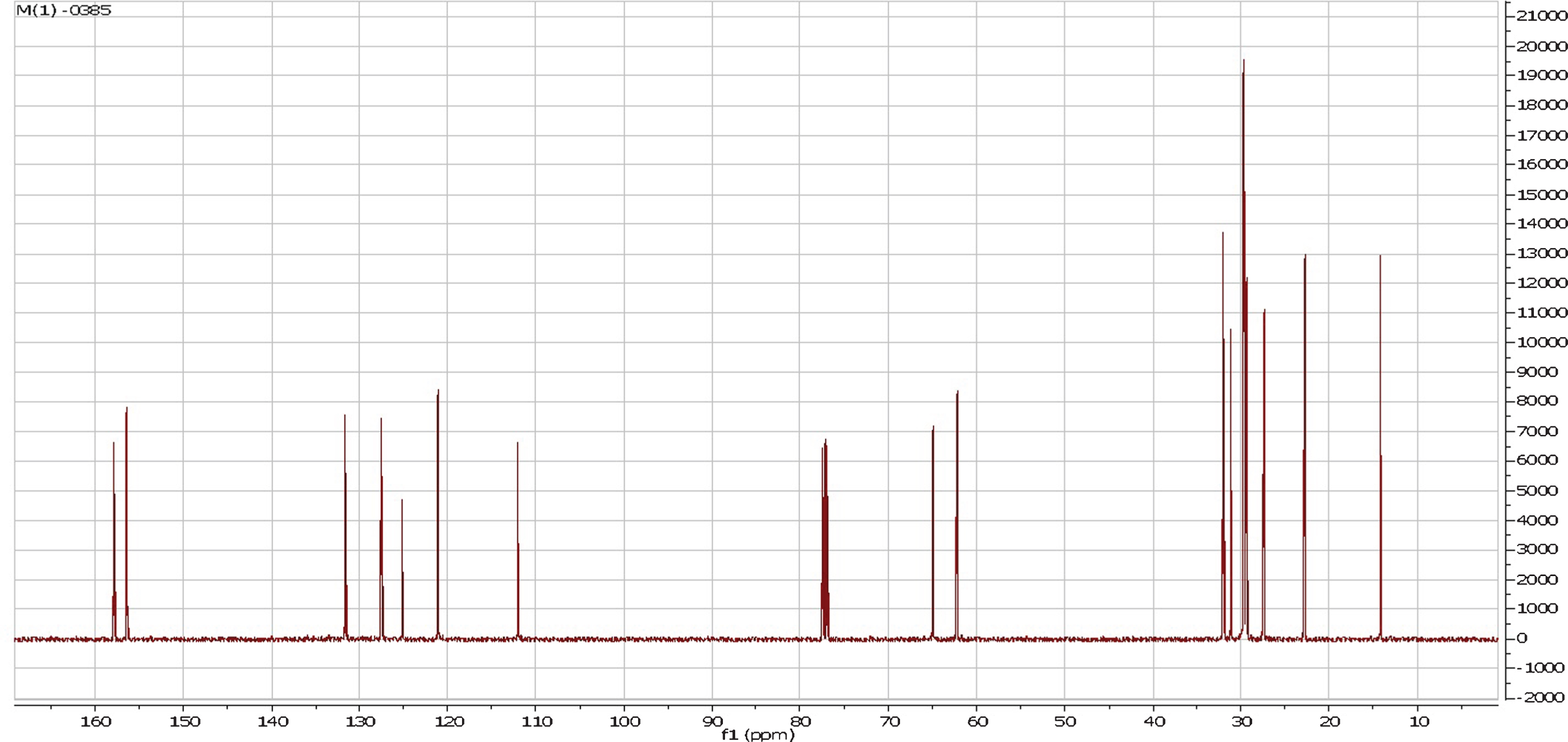
13C-NMR spectrum of the Lig.(CDCl3).
In the β-carotene bleaching test, the ruthenium (II) complex and the ligand were exhibited the similar activity, with antioxidant activity values almost equivalent and the activity was only slightly reduced after 120 min. These results (Table 1) showed that the complex and the ligand have a moderate activity in the β-carotene bleaching test.
β-Carotene bleaching test.a BHA was used as reference antioxidant
β-Carotene bleaching test.a BHA was used as reference antioxidant
aValues were the means of three replicates±standard deviation (SD). bRelative antioxidant activities. cNegative control: linoleic acid and β-carotene emulsion.
Total antioxidant capacity of the ruthenium (II) complex and ligand were determined by the thiocyanate method in linoleic acid emulsion. The effect of complex and ligand at different concentration on peroxidation of linoleic acid emulsion was given at Fig. 3.10. α-Tocopherol, BHA and BHT were used as standard antioxidants.
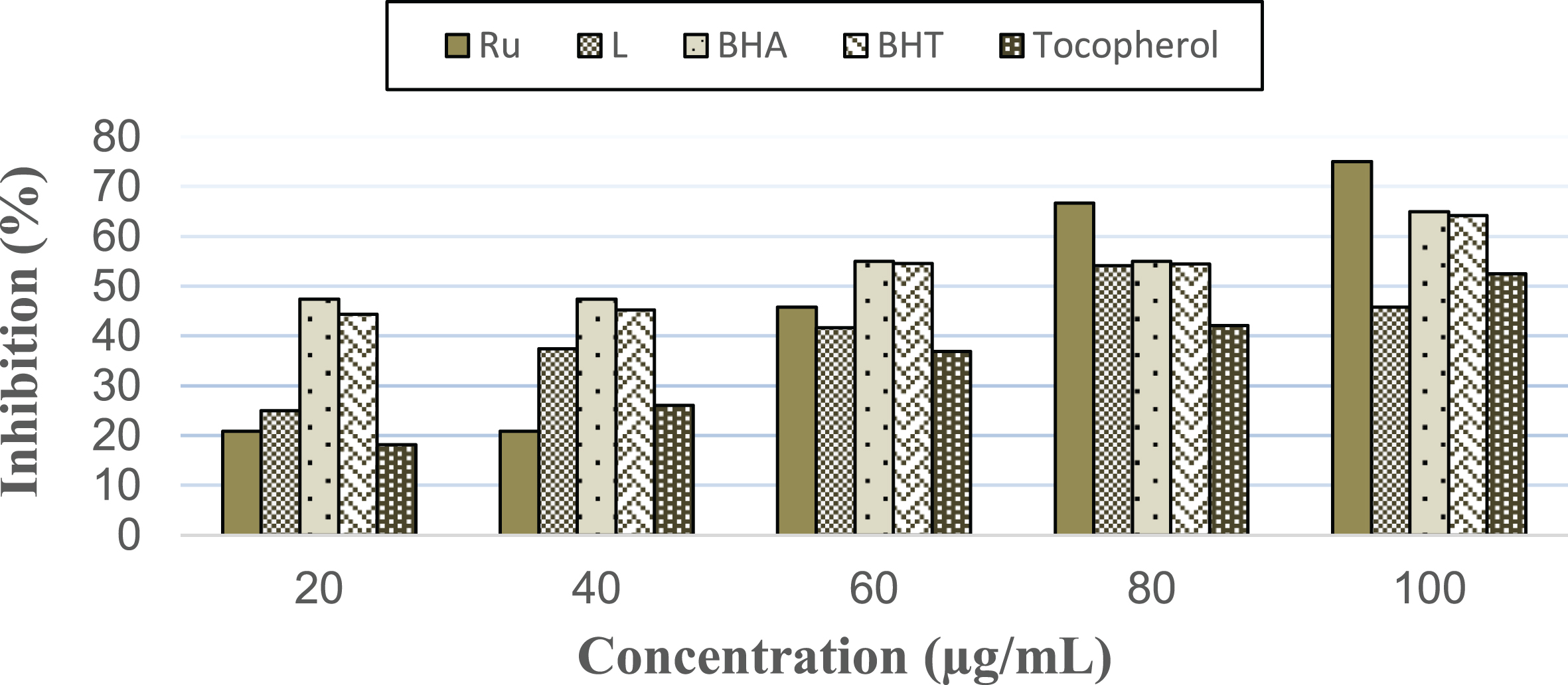
Total antioxidant capacity of Ru (II) complex and ligand measured by ferric thiocyanate method. α-Tocopherol, BHA, and BHT were used as reference antioxidants.
Ruthenium (II) complex exhibited higher antioxidant capacity (75%) than ligand (43%) at 100μg/mL concentration. Ruthenium (II) complex also showed higher activity than BHA (65.00%), BHT (64.22%) and α-tocopherol (52,47%) at 100μg/mL concentration.
The free radical scavenging activity of ruthenium (II) complex and ligand are presented in Fig. 3.11. All the compounds exhibited low free radical scavenging ability when compared the standard antioxidants. The scavenging effect of the free ligand is significantly similar with their corresponding ruthenium (II) complex.
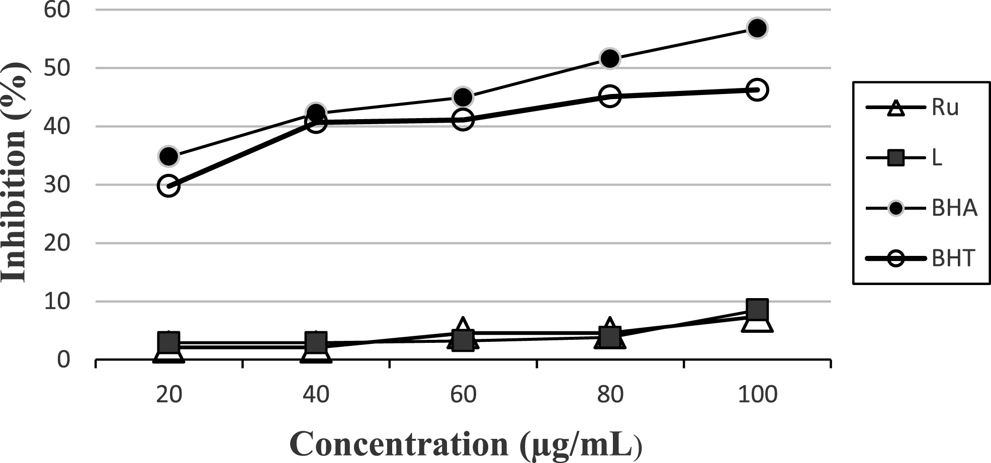
DPPH radical scavenging effect of Ru (II) complex and ligand at different concentration. BHA and BHT were used as standard antioxidants.
One of the most commonly used organic radicals for the evaluation of antioxidant efficiency of pure compounds and complex mixtures is the radical cation derived from ABTS. The ABTS·+ scavenging activity of the Ru (II) complex and the ligand were presented in Fig. 3.12. All compounds showed antioxidant activities proving their capacity to scavenge the ABTS+ radical cation. The new substances were exhibited moderate activity when compared BHA, BHT and epictechin.
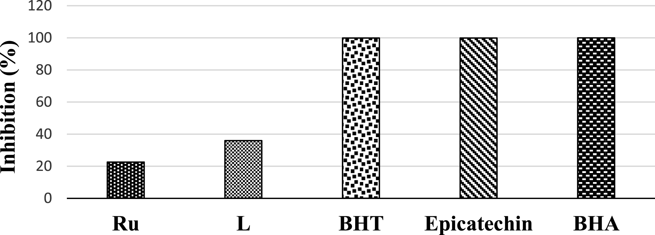
ABTS·+ scavenging activity of the Ru (II) complex and the ligand at 1 mg/mL concentration. BHA, BHT, and epicatechin were used as standard antioxidants.
For evaluating the anticancer property of newly synthesized complexes, DNA binding is the predominant property looked for in pharmacology and hence, the interaction between DNA and metal complexes is of paramount importance in understanding the mechanism. Thus, the mode and propensity for binding of the ligand and complex to DNA were studied with the help of electronic absorption technique. The change observed in the absorption spectra of the complex and ligand in the presence of increasing concentration of DNA is used for determining the interaction of complex and ligand with duplex DNA. The absorption spectra of complex and ligand in the absence and presence of CT-DNA (at a constant concentration of complex [Ru] = 25μM, ligand [L] = 25μM) are given in Fig. 3.13. Increasing concentrations of CT-DNA to a fixed concentration of complex and ligand exhibited an evident hyperchromism along with a slight blue shift of 4 nm. Compounds interact to DNA through electrostatic and major/minor groove binding results in hyperchromicity, hypsochromism (blue shift) [22] “Hypochromic effect” and “hyperchromic effect” are the spectral features of CT-DNA concerning its double helix structure. Hyperchromism results from the damage of the DNA double helix structure [23]. A plot of -[DNA]/ɛa-ɛf versus [DNA] gave a slope (Fig. 3.14). [DNA] is the concentration of DNA in base pairs, ɛa is the molar extinction coefficient of the complex at a given DNA concentration, ɛf is the molar extinction coefficient of the complex in free solution. From the absorption titration data, the intrinsic binding constants (Kb) of the ligand and Ru (II) complex with CT-DNA were calculated as 5.9×10⁵ M-1 and 7.9×10⁵ M-1, respectively. As a result, these compounds interacted to CT-DNA via electrostatic and/or groove binding due to hyperchromism, blue shift and higher Kb values.
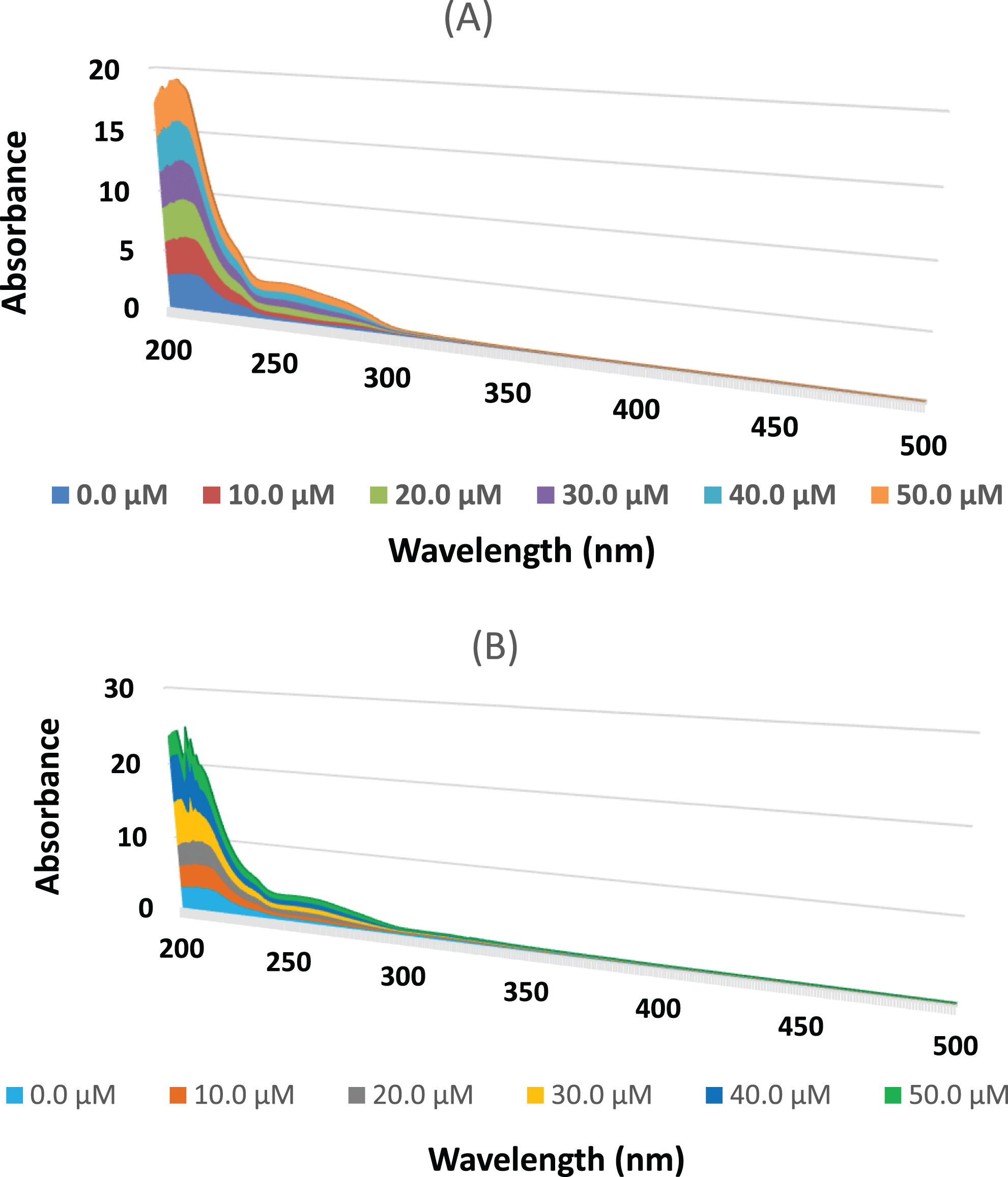
UV spectra of ruthenium complex (A) and ligand (B) in the absence and in the presence of CT-DNA in increasing amounts, [Complex] = 25μM, [Ligand] = 25μM, [DNA] = (0–50μM).

Plot of [DNA]/(ɛa-ɛf) versus [DNA] for the titration of DNA with ruthenium complex (A) and ligand (B), solid line is linear fitting of the data.
Footnotes
Acknowledgments
We wish to acknowledge the financial support by the “YTÜ BAPK Research Found” (Project No: 2015-01-02-KAP04).