
Abstract
Metal carboxylates possess wide range of intriguing structural topologies due to their diverse ligational behavior of carboxylate group. Two new polymorphs of monomeric complex, [Ca(dga)(H2O)5].H2O (
Keywords
Introduction
Over the past decades, metal complexes of carboxylic acid having N, O-donors with polymeric or network type fascinating structures have received much attention. An extended array of M-L-M connectivity formed of isolated metal atoms or clusters with polyfunctional organic ligands having porous architectures are termed as Metal Organic Frameworks (MOFs). The studies of MOFs have increasing interest in research fields due to their structural diversities, porous nature and the versatile coordination modes of MOFs create one, two or three dimensional structural topologies [1–3].
MOFs show many potential applications due to their large open spaces in its structures. MOFs show wide range of applications in the field of gas storage, gas adsorption, catalysis, molecular magnetism and photoluminescence [4–6]. The ligands are usually selected as polyfunctional ligands, which exhibit diverse coordination modes and variety of structural features [7–9]. Diglycolic acid is otherwise known as oxydiacetic acid (HOOCCH2OCH2COOH), is a polydentate ligand having four carboxylate oxygen atoms and one etherial oxygen atom, which act as bridging chelate coordination to central metal atom possessing increasing network topologies [10]. Metal complexes of diglycolic acid are employed for pharmaceuticals and have biological applications, which are further enhanced by some N-heterocyclic derivatives. Literature studies indicate that transition, rare earth or main group metals are used for generating the various metal carboxylates [11–13]. Among them,
The Cu(II) complexes of oxydiacetate ligand with 4-picolinic acid, [Cu(oda)(4-pic)H2O]2H2O and with 2,2’-bipyridine ligand, [Cu(oda)(bipy)(H2O)].4H2O exhibited considerable biocatalytic activity towards the dismutation of superoxide anion. The biological activity of metal oxydiacetate was further enhanced by the presence of auxiliary ligand especially N-heterocyclic derivatives such as picolinic acid. The copper, cobalt and iron complexes of oxydiacetic acid with picolinic acid: [M(oda)(4-pic)H2O].xH2O {M = Cu(II), x = 2; Co(II), x = 4} and [Fe(oda)(4-pic)].Cl shows remarkable antimicrobial activities against Escherichia coli, Bacillus subtilis, Staphylococcus aureus, Salmonella typhimurium, Candida albicans, Aspergillus fumigatus and Penicillium marneffei. The biological activity of the vanadium oxydiacetate, [VO(oda)(H2O)2] suggests that the compound is an inhibitory agent of osteoblast differentiation [11–13].
Alkaline earth metal carboxylates possess larger radii, high affinity for oxygen atom and show applications in material science [14, 15]. Majority of Ca(II) complexes exhibit higher coordination number due to the lack of d electron in Ca(II) ion. The higher ionic radii and oxophilic nature of Ba(II) ion results in the formation of inorganic-organic hybrid frameworks. The alkaline earth metal oxydiacetates, monomeric [Ca(oda)(H2O)5].H2O and polymeric [Ba(oda).H2O]n, [{Mg(oda)(H2O)2}.H2O]n, [Sr(oda)(H2O)3]n.nH2O (oda = –O2CCH2OCH2CO2–) have been reported by slow evaporation method [16–19]. Slow evaporation method is a technique in which a saturated solution of metal salts along with the ligand and suitable solvent is kept at a particular temperature and made for evaporation.
Taking these observations, herein we have grown two metal diglycolates of Ca(II) and Ba(II), [Ca(dga)(H2O)5].H2O (
Experimental
Materials and methods
Diglycolic acid (CDH), calcium nitrate (CDH), barium chloride (CDH) and acetic acid (CDH) of AR grade (99%) and were used without further purification. The FT-IR spectra were recorded in the range 4000-400 cm–1 using Thermo Nicolet, Avatar 370 Spectrometer in KBr pellets. The compositions of carbon, hydrogen, nitrogen in the compound were determined by using Elementar Vario-EL 111 CHN analyzer. The UV-Visible spectral studies were carried out using Varian Cary 5000 UV-Vis-NIR spectrometer in the range 200–1200 nm. Single crystal X-ray diffraction studies were carried out using Bruker AXS Kappa Apex 2 CCD diffractometer, with graphite monochromated Mo Kα radiation (λ= 0.71073 Å). The powder X-ray diffraction studies were conducted using a Bruker AXS D8 advance XRD with Cu Kα radiation (λ= 1.54056 Å). The thermogravimetric analysis of the grown crystals was carried out on a Perkin Elmer Diamond TG/DTG analyzer instrument with a heating rate of 10°C/min in nitrogen atmosphere. The photoluminescence of the samples are recorded using Fluoromax-3 spectroflurometer consisting of 150 W Xenon arc lamp, monochromator and a detector.
Growth Procedure of [Ca(dga)(H2O)5].H2O (1 ) and [Ba(dga)(H2O)] (2 )
Gel diffusion method was employed for the crystal growth of the compounds
Complex (
Complex (
X-ray Crystallography
Single crystal X-ray diffraction studies of the complex were collected using Bruker AXS Kappa Apex2 CCD diffractometer (Bruker, AXS GmbH, Karlsruhe, Germany) with graphite monochromated Mo Kα radiation (λ= 0.71073 Å). The unit cell dimensions and intensity data were recorded at 296 K. The programme SAINT/XPREP was used for data reduction and APEXT2/SAINT for cell refinement [21]. The structures were solved by direct methods using SIR92 and refinement were carried out by full-matrix least squares on F2 using SHELXL-97 [22, 23]. All the non-hydrogen atoms were refined anisotropically and the hydrogen atoms were refined isotropically. The structures were plotted by DIAMOND software version 3.1f [24]. The crystal data and structure refinement parameters for the complexes are given in Table 1.
Crystal data and structure refinement for [Ca(dga)(H2O)5].H2O (1 ) and [Ba(dga)(H2O)] (2 )
Crystal data and structure refinement for [Ca(dga)(H2O)5].H2O (
R1 =Σ||Fo| – |Fc|| /Σ|Fo|, wR2 = [Σw(Fo2-Fc2)2/Σw(Fo2)2]1/2.
Crystal growth
Transparent, needle shaped crystals appear at the gel interface within 1 week for 1 M diglycolic acid, 0.5 M CaNO3 solution, pH 6.5 and 1 M diglycolic acid, 0.5 M BaNO3 solution, pH 5.5 as its optimum crystal growth condition for compound 1 and 2 respectively. The crystals were separated, washed with doubly distilled water and dried. The photographs of crystalline complexes 1 and 2 are shown in Fig. 1(a) and (b).
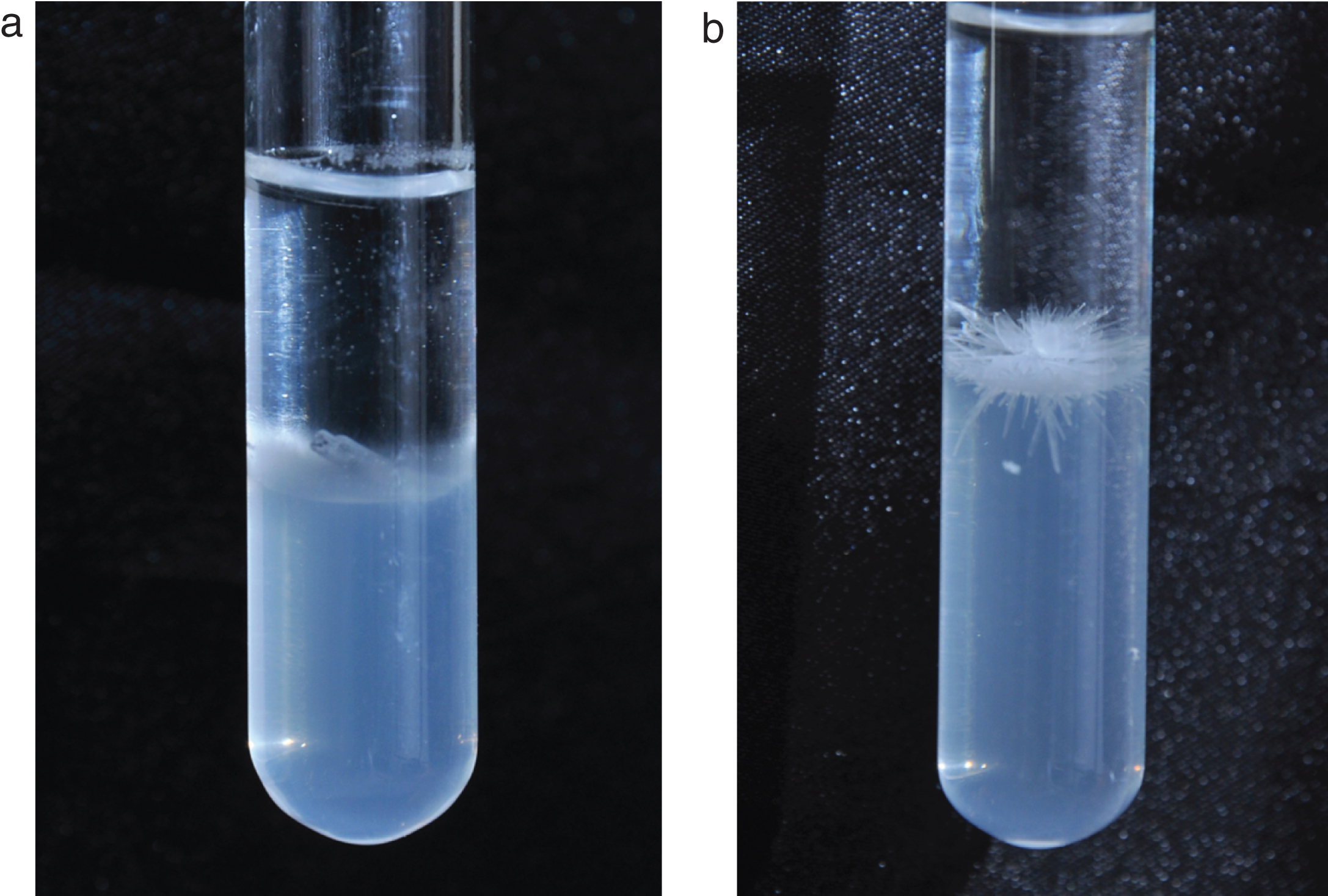
(a) Photograph of the crystalline complex 1. (b) Photograph of the crystalline complex 2.
In the present study, we report the new polymorphs of the complexes
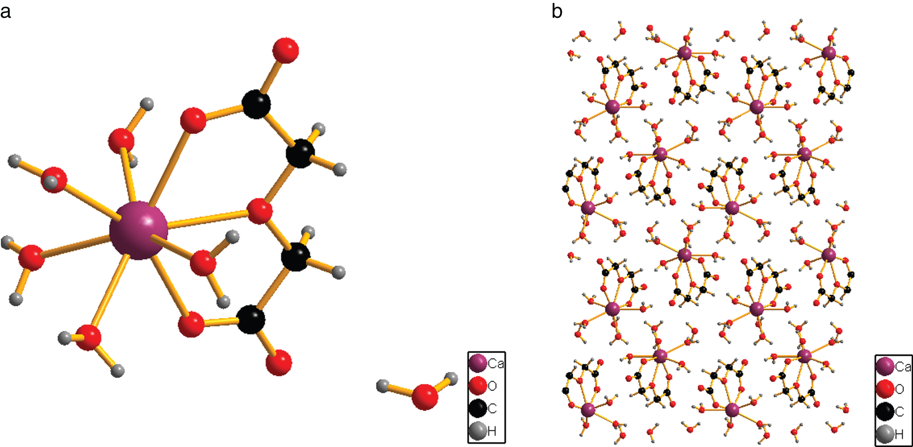
(a) Asymmetric unit of complex 1 with atom numbering scheme. (b) Packing diagram viewed along ‘a’ axis.
Bond lengths (Å) and bond angles (°) for [Ca(dga)(H2O)5].H2O
Symmetry transformations used to generate equivalent atoms: #1 x+1,y,z #2 x-1/2,-y+1/2,z-1/2 #3 x+1/2,-y+1/2,z-1/2. #4 -x-1/2,y+1/2,-z+1/2 #5 -x+1/2,y+1/2,-z+1/2. #6 -x+1,-y+1,-z+1 #7 x-1,y,z #8 -x,-y+1,-z+1. #9 x+1/2,-y+1/2,z+1/2 #10 -x+1/2,y-1/2,-z+1/2.
The asymmetric unit consists of one diglycolate and 5 water molecules coordinated to calcium metal ion. A lattice water molecule is also present in it. The diglycolate ligand in complex
Hydrogen bonds for [Ca(dga)(H2O)5].H2O [Å and deg.]
Symmetry transformations used to generate equivalent atoms: #1 x+1,y,z #2 x-1/2,-y+1/2,z-1/2 #3 x+1/2,-y+1/2,z-1/2. #4 -x-1/2,y+1/2,-z+1/2 #5 -x+1/2,y+1/2,-z+1/2. #6 -x+1,-y+1,-z+1 #7 x-1,y,z #8 -x,-y+1,-z+1. #9 x+1/2,-y+1/2,z+1/2 #10 -x+1/2,y-1/2,-z+1/2.
The crystal structure reveals that compound
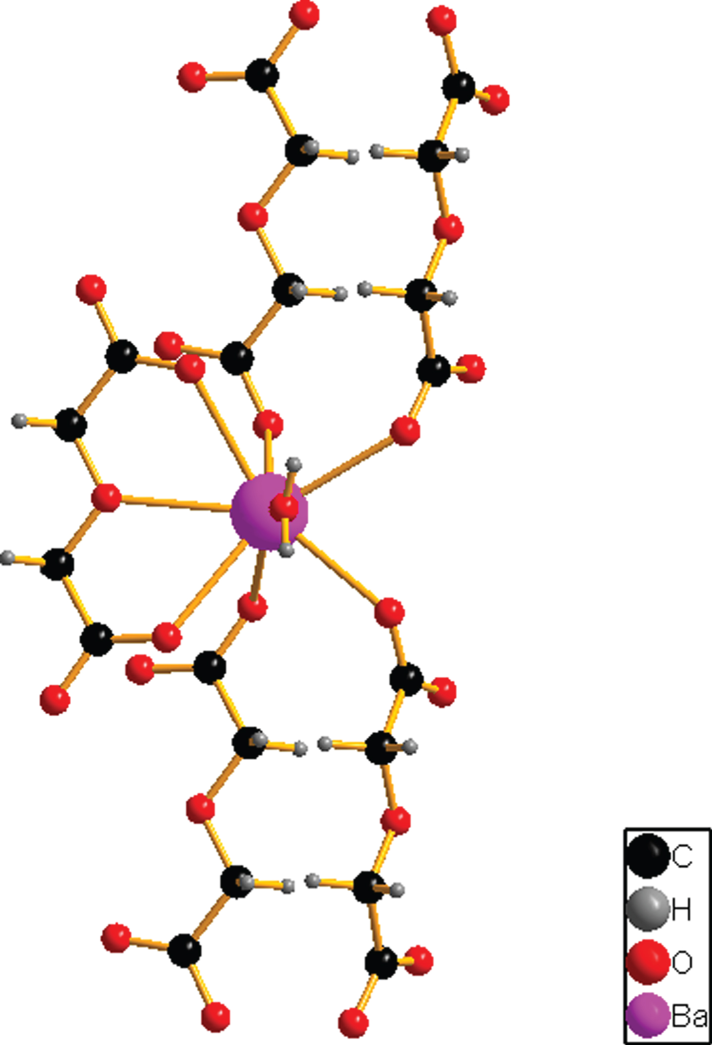
The molecular structure of complex 2.
Bond lengths (Å) and bond angles (°) for [Ba(dga)(H2O)]
Symmetry transformations used to generate equivalent atoms: #1 -x,-y+1,-z #2 x,-y+1/2,z #3 x-1/2,-y+1/2,-z+1/2. #4 x+1/2,-y+1/2,-z+1/2 #5 x+1/2,y,-z+1/2.
In the asymmetric unit of

H-bonded packing diagram of complex 2 viewed along ‘a’ axis.

(a) FT-IR spectrum of complex 1. (b) FT-IR spectrum of complex 2.
Hydrogen bonds for [Ba(dga)(H2O)] [Å and deg.]
Symmetry transformations used to generate equivalent atoms: #1 -x,-y+1,-z #2 x,-y+1/2,z #3 x-1/2,-y+1/2,-z+1/2. #4 x+1/2,-y+1/2,-z+1/2 #5 x+1/2,y,-z+1/2 #6 -x+1,y-1/2,-z #7 x+1,-y+1/2,z.
To determine the coordination modes of ligand to metal atom FT-IR spectral studies are carried out. The FT-IR spectrum of the complex 1 and 2 are interpreted on the basis of the various metal diglycolates [25–27]. The infrared spectrum exhibits several bands due to various coordination modes present in the complex. The broad band observed in the region 3400 to 3500 cm–1 in the complex 1 and 2 is attributed to O–H stretching vibration of water molecule. A strong band at 1706 cm–1 attributed to the ν(C=O) vibration of ligand is absent in the complexes and indicates that all carboxylic acid groups are deprotonated and carboxylate oxygen atoms are coordinated to the central metal atom. The corresponding asymmetric and symmetric stretching vibrations of COO– group appear at 1592 and 1441 cm–1 in 1 and 1592 and 1436 cm–1 in 2 support these observations. Δυ= 151, 156 cm–1 in complex 1 and 2 indicates that the coordinating carboxylic groups are bridging type. The very strong band at 1146 cm–1 in the free diglycolic acid is attributed to C–O–C stretching, which is shifted to lower wave number 1135, 1120 cm–1 in 1, 2 respectively. This suggests the coordination of etherial oxygen with the central metal ion. The M–O stretching vibrations are observed at 572, 556 cm–1 in 1 and 2. The FT-IR spectra of 1 and 2 are shown in Fig. 5(a, b).
Powder X-ray diffraction studies
The crystalline nature of the title compound was revealed from the well defined Braggs peak at specific 2θ angles. Inorder to check the bulk purity of the compounds, powder X-ray diffraction pattern was experimentally measured and compared with X-ray diffraction pattern obtained from mercury software using single crystal XRD data. These two results are in good agreement with each other which indicates that the compounds possess bulk purity. The powder X-ray diffraction pattern of compounds 1 and 2 are shown in Fig. 6(a, b).

(a) Experimental and simulated X-ray diffractograms of [Ca(dga)(H2O)5].H2O. (b) Experimental and simulated X-ray diffractograms of [Ba(dga)(H2O)].
The solid state absorption spectrum of complex 1 and 2 were recorded in a region of 200–1200 nm. The absorbance at 325 and 363 nm in 1 and 2 can be attributed to the intraligand charge transitions of the ligand. There is no absorption band is observed in a region of 400–800 nm in both the complexes, which shows the compounds are highly transparent in the entire visible region. The absorbance spectrum of 1 and 2 are shown in Fig. 7(a, b).
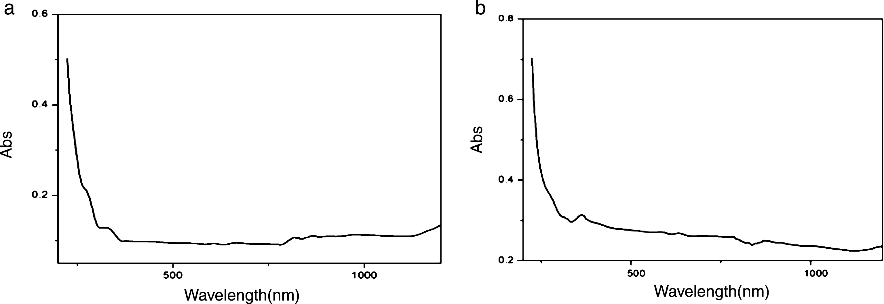
(a) Absorption spectrum of [Ca(dga)(H2O)5].H2O. (b) Absorption spectrum of [Ba(dga)(H2O)]
The thermal stability of the compound 1 and 2 has been studied by thermogravimetry experiments conducted at a heating rate of 10°C/min in nitrogen atmosphere. The compound 1 decomposes in 4 stages. The first stage of decomposition corresponds to the removal of one lattice and two coordinated water molecules with an observed weight loss of 19.0% (19.2% calc.). The second stage of decomposition occurs within a high temperature range of 137 to 397°C with an observed weight loss of 18.3% (19.2% calc.), which cooresponds to the removal of the remaining three strongly coordinated water molecule. The third and fourth stage corresponds to the removal of one diglycollate ligand with a weight loss of 38.5% (40.67% calc.) and finally the residual weight is obtained as CaO with a weight loss of 20.0% (19.9% calc.).
The compound 2 decomposes in three stages. The first stage of decomposition corresponds to the sharp peak at 243°C in DTG curve, with an observed weight loss of 6.8% (6.2% calc.) and corresponds to the removal of one water molecule. The second stage of decomposition at 416°C with an observed weight loss of 24.1% (25.0% calc.) corresponds to the pyrolysis of the ligand moieties and after the third step, the percentage of residual weight obtained from the graph (67.3%) is in well agreement with the calculated value of 68.5%, indicating that the end product obtained is barium carbonate (BaCO3). TG/DTG curves of compounds 1 and 2 are shown in Fig. 8(a, b).
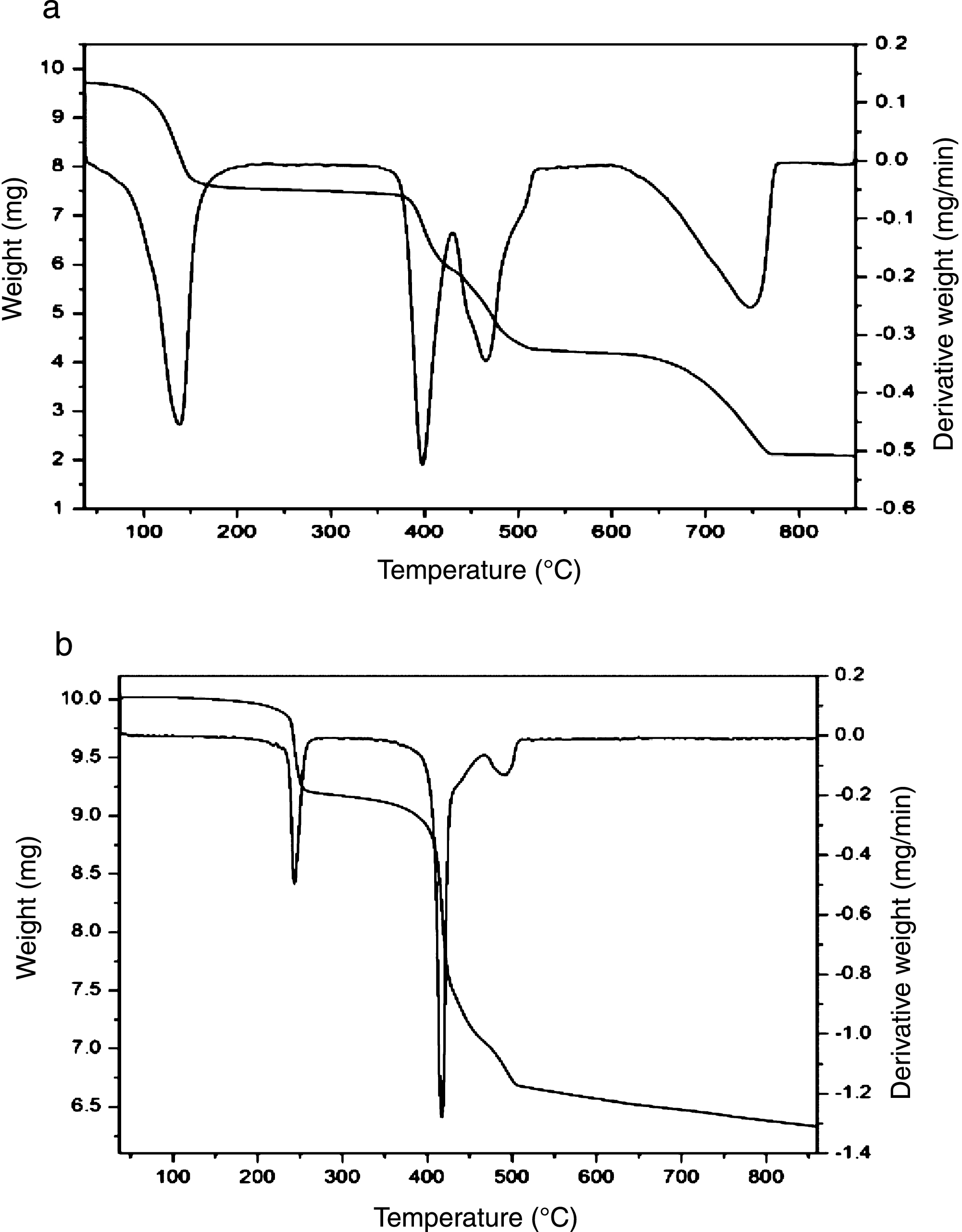
(a) TG/DTG curve of [Ca(dga)(H2O)5].H2O. (b) TG/DTG curve of [Ba(dga)(H2O)].
The solid state photoluminescent spectrum of the complexes
The complex
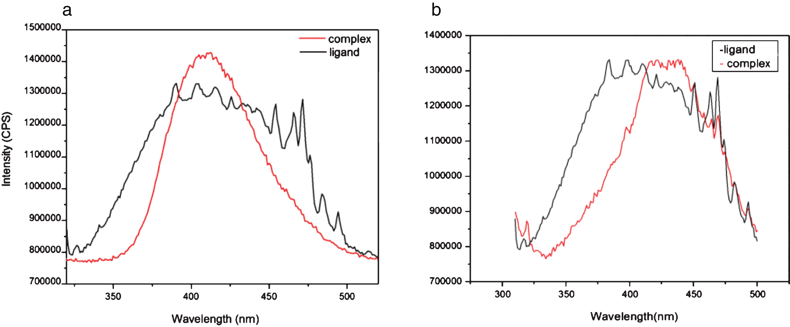
(a) The solid state photoluminescent spectrum of diglycolic acid and [Ca(dga)(H2O)5].H2O. (b) The solid state photoluminescent spectrum of diglycolic acid and [Ba(dga)(H2O)]
Single crystals of two new polymorphs of monomeric complex of 1 and three-dimensional metal-organic framework of complex 2 have been successfully grown by gel diffusion technique at room temperature. Single crystal X-ray diffraction analysis reveals that the complex 1 belongs to monoclinic P21/n, having a monomeric structure and converted in to 2D supramolecular sheets by extensive hydrogen bonding interaction. The complex 2 belongs to orthorhombic space group Pnma, having a 3D framework consists of hydrophobic channels. The composition of the complexes was obtained from the elemental analysis. The FT-IR spectral analysis confirms that the ligand coordinate to the metal atom through carboxylate and etherial oxygen atoms. UV-Visible spectrum shows that that the compounds are highly transparent in the entire visible region. The thermal decomposition behavior of the compounds was provided by TG studies. The crystallinity of the compounds was confirmed by powder X-ray diffraction studies. The luminescent property of the compounds can be used as potential photoactive material.
Footnotes
Acknowledgments
One of the authors, USSM is thankful to the UGC for providing the financial assistance in the form of Junior Research Fellowship. The authors are thankful to SAIF, CUSAT, Kochi, India and Dr. A. Santhosh Kumar, School of Pure and Applied Physics, M. G University, Kottayam, India for providing analytical facilities. We are also thankful XRD Lab, SAIF, IIT-Madras, Chennai, India for single crystal X-ray diffraction measurements and crystal structure refinement.
CCDC no 1501100, 1501101, contains the supplementary crystallographic data for the compounds [Ca(C4H4O5)(H2O)5].H2O, [Ba(C4H4O5)(H2O)] respectively. These data can be obtained free of charge via ![]() or from the Cambridge Crystallographic Data Centre, 12 Union Road, Cambridge CB2, 1EZ, UK; Fax: (+44)1223–336-033; E-mail:
or from the Cambridge Crystallographic Data Centre, 12 Union Road, Cambridge CB2, 1EZ, UK; Fax: (+44)1223–336-033; E-mail: