
Abstract
In this work, we synthesized a simple fluorescent molecule based on quinoline derivative. It displayed excellent sensitivity and selectivity toward copper ions. Upon addition of Cu2+, this molecule exhibited fluorescence quenching due to a fast electron transfer mechanism, the detection limit for Cu2+ of this sensor could be 6.17×10⁻⁷μmol L–1. Simultaneously, the cell imaging experiments were utilized to demonstrate its applicability in biological systems.
Introduction
Fluorescent sensor has become a poweful tool for sensing and monitoring chemical analytes by virtue of its sensitivity, selectivity and fast response [1–4]. In view of many research fields need sensing systems, including chemistry, medical, biology, industrial and environmental science, abundant highly effective fluorescent sensors are designed and synthesized for different requirements, such as detecting important metal ions which are involved in biological processes, or detecting toxic substance in the water or soil, even sensors for explosives and hazardous chemicals are being extensively investigated for the detection of chemical and biological weapons [5]. Moreover, fluorescent sensor allows for the study and control of chemical processes from the laboratory to the industrial scale, and plays an significant role in the food industry for the control of food quality and safety [6]. With the advances in technology, the need for accurate, reliable, real-time biological and chemical sensing is in the spotlight [7, 8]. Cu2+ is one of the most important metal ions in human body, and it is involved many vital physiological processes in biological system [9]. However, excessive Cu2+ in hunman body may cause many problems, such as the damage of protein or nucleic acids [0–12]. And the cellular toxxicity of Cu2+ is related to some serious disease like Alzheimer’s disease or Menkes and Wilson disease [3–15]. Therefore, much attention has been drawn to the development of detecting methods of Cu2+ ion because of its importance in environmental and biological systems. In recent years, the fluorescent sensors for detecting Cu2+ are increasingly concerned and researched [6–18]. Based on previous studies, we found that it is of great importance to select an efficient fluorophore in the preparation of fluorescent sensors. In this study, we design a novel fluorescent sensor which combined quinoline with pyridine moiety (Sensor 1). It displays excellent discernment to Cu2+ among common metal ions,and its favourable affinity with Cu2+ is based on photoinduced electron transfer (PET) mechanism. In addition, the living cells image experiments expand its application prospects in biological field.
Experimental
Materials and methods
All the materials for synthesis were purchased from commercial suppliers and used without further purification. Solutions for spectra detection was HPLC reagent without fluorescent impurity. 1H NMR spectra were taken on a Varian mercury-400 spectrometer with TMS as an internal standard and DMSO-d6 as solvent. HRMS spectra was analysed on an Agilent 1290-micro TOF QII. Fluorescence spectra measurements were performed on a Hitachi F-4500 spectrofluorimeter. The pH measurements were made with Metteler-Toledo Instruments DELTE 320 pH. Cell experiment were applied on an inverted fluorescence microscope (Olympus IX-70) connected with a digital camera (Olympus, c-5050).
UV-vis and fluorometric analysis
The solution of Sensor 1 was prepared in DMF and the metal ion buffer solutions were using various cations dissolved in Tris-HCl buffer solution at pH 7.2. In titration and selectivity experiments, the test samples were prepared by placing appropriate amounts of ions into corresponding solution of sensor 1. For fluorescence measurements, excitation was provided at 325 nm, and emission was collected from 330 to 550 nm, both the excitation and emission slit widths were 5 and 5 nm, respectively.
Synthesis
Preparation of intermediate 2
(1.0 g, 6.89 mmol) 8–hydroxyquinoline and (1.9 g, 13.78 mmol) K2CO3 were dissolved in 25 mL acetonitrile with stirring for 30 min, then added (1.25 g, 7.58 mmol) ethyl bromoacetate in the solution and kept stirring for 6 h at room temperature, distilled the solvent and the generated intermediate was extracted with CH2Cl2 and H2O for 3 times. Kept the organic layer and dried on MgSO4 for 12 h before distilled the solvent. The crude product was purified by column chromatography (EtOAc: petroleum ether = 1:2) on silica gel to obtain compond 3 as red oil (2.18 g, ultimate yield 75%).13 (0.25 g, 1.08 mmol) compond 3 in 2.5 mL ethanol was added dropwise into 0.1 mL hydrazine and kept stirring for 1 h, filtered the generated precipitates and washed them with H2O and CH2Cl2 for 3 times respectively, the precipitates were purified by column chromatography (CH2Cl2: C2H5OH = 1:2) on silica gel to obtain intermediate 2 as white solid (0.23 g, ultimate yield 64%). 1H NMR (300 MHz, DMSO-d6) δ 4.39 (s, 2H, amino-H), 4.76 (s, 2H, methene-H), 7.26 (d, 1H, J 6.0 Hz, Ph-H), 7.53 (d, 2H, J 3.0 Hz, Ph-H), 7.59 (s, 1H, Ph-H), 8.36 (d, 1H, J 3.9 Hz, Ph-H), 8.91 (d, 1H, J 1.6Hz, Ph-H), 9.46 (s, 1H, imino-H). HRMS (FTMS + pESI) m/z, observed: 218.1; C18H16N4O2 [M]+ requires: 217.09.
Synthesis of sensor 1
(0.11 g, 0.5 mmol) intermediate 2 and (0.06 g, 0.5 mmol) acetylpyridine were dissolved in 10 mL DMF and kept stirring at 155°C for 8 h, after removed the solvent,the crude product was purified by column chromatography (CH2Cl2: CH3OH = 2:1) on silica gel to obtain sensor 1 as white solid (0.10 g, ultimate yield 59%). 1H NMR (300 MHz, DMSO-d6) δ 1.07 (d, 3H, J 6.3 Hz, methyl-H), 4.61 (s, 2H, methene-H), 7.07 (d, 2H, J 3.0 Hz, Ph-H), 7.33 (d, 1H, J 6.2 Hz, imino-H), 7.55 (m, 4H, Ph-H), 8.02 (d, 2H, J 7.4 Hz, Ph-H), 8.75 (m, 2H, Ph-H). HRMS (FTMS + pESI) m/z, observed: 321.1; C18H16N4O2 [M]+ requires: 320.13. (Scheme 1).

Synthesis of Sensor 1.
SGC-7901 cells were cultured in Dulbecco’s Modified Eagle Medium (DMEM) with 10% (v/v) Fetal Bovine Serum (FBS) in a humidified atmosphere of 5% CO2 at 37 °C. Before cellular imaging, SGC-7901 cells were seeded in 24-well plates and allowed to grow for 24 h. Next Sensor 1 (1×10⁻⁴ mol L–1) was added to the 24-well plates and the cells were incubated for another 30 min. After most of the fluorescence sensor successfully immersed into cells, the culture medium was washed with phosphate buffered saline (PBS, pH = 7.4) to remove the remaining Sensor 1 and the fluorescence images were carried out by the fluorescent microscope (Olympus IX70 – c-5050).
Results and discussion
Cu2+-titration and spectral responses
In order to investigate the identification characteristics of Sensor 1 toward Cu2+, titration experiments were adopted under Tris-HCl buffer solution (pH = 7.2). Sensor 1 revealed an absorption maximum at 319 nm as shown in Fig. 1 ([Cu2+] = 0-1×10⁻⁵ mol L–1), upon addition of Cu2+ buffer solution, the absorption band at 319 nm decreased and had a redshift to 325 nm until the first stoichiometry, so we take 325 nm as the excitation wavelength in fluorescence titration experiment. First we researched the stoichiometry between sensor and determinand, the job’s plot was used to discuss the issue, as shown in Fig. 2, we were informed that the maximum absorbance appeared at w = 50% of Sensor 1, that is to say, when the ratio between probe and Cu2+ became 1:1, the fluorescence intensity of Sensor 1 was reduced to the greatest extent, this situation indicated that Sensor 1 and Cu2+ formed a 1:1 stoichiometry complex. Moreover, we investigated the association constant of complex of Sensor 1 with Cu2+ from the Benesi-Hildebrand expression, as illustrated in Fig. 3, the measured absorbance [1/(A0–A)] at 319 nm varied with 1/[Cu2+] in a linear relationship (R = 0.99847), and according to the linear equation, the association constant of complex of Sensor 1 with Cu2+ was calculated to be 3.179×10⁵ mol–1 [19, 20]. In fluorescence titration experiment, the fluorescence emission of Sensor 1 toward Cu2+ was illustrated in Fig. 4, we could find that the independent Sensor 1 exhibited strong fluorescence at 399 nm in emission spectrum (water: DMF = 5:5, excited at 325 nm). Upon increasingly added Cu2+ into Sensor 1 solution, the fluorescence intensity at 399 nm gradually weakened until fluorescence quenched, the signal transformation was less than 10 s, and the fluorescent quenching process could be observed under the irradiation of a UV lamp. Furthermore, as shown in Fig. 5, the decreasing fluorescence intensity of Sensor 1 depend on the concentration of Cu2+ was in a linearship (R = 0.99717), so we considered that Sensor 1 could quantitative determine Cu2+, and the detecting limit could reach to 6.17×10⁻⁷ mol L–1 by calculation from the linear relation (based on DL = KSb1/S,K = 3; Sb1 is the standard deviation of the blank solution; S is the slope of the calibration curve).
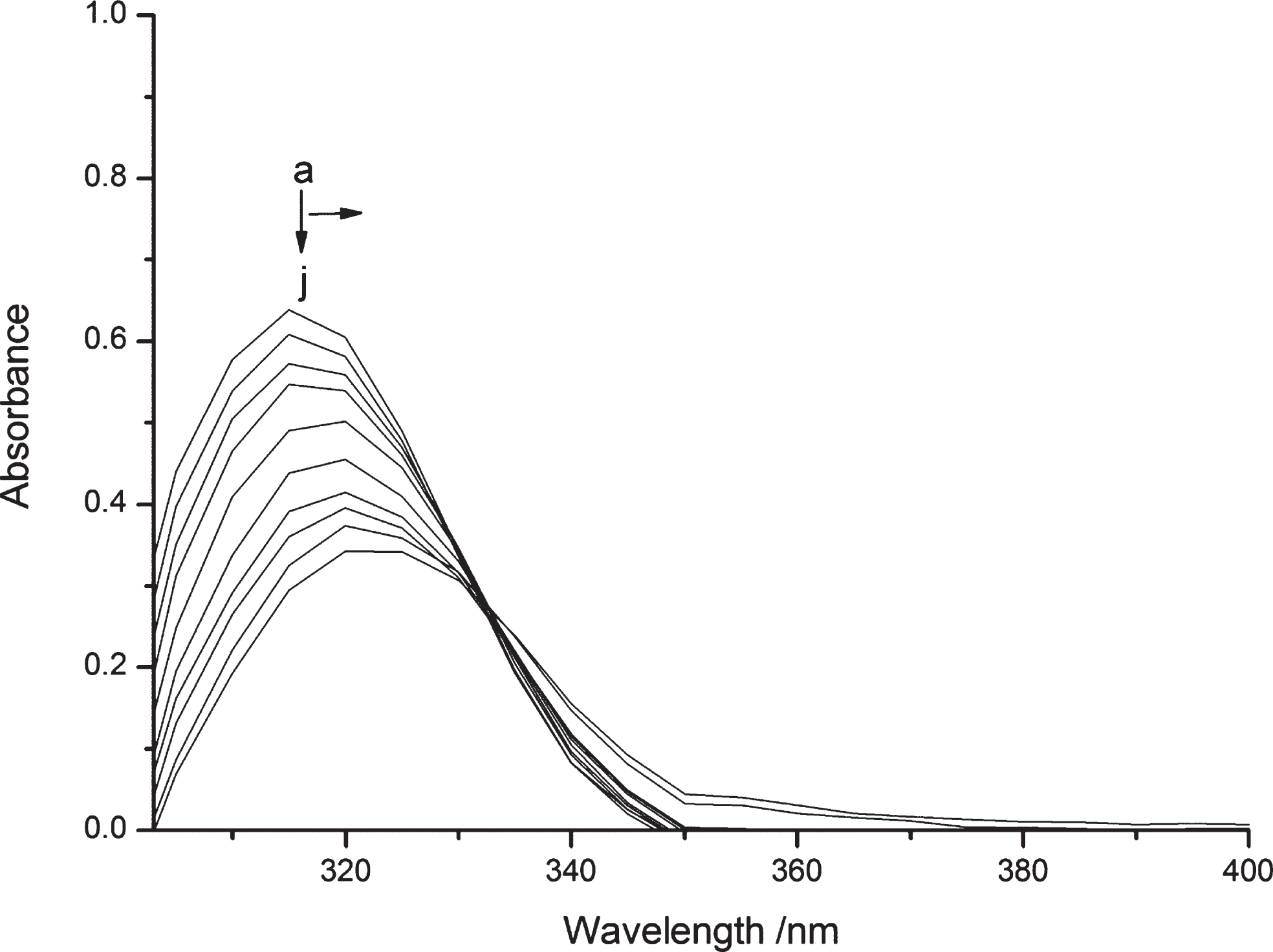
UV-Vis absorption response of Sensor 1(10μmol/L) upon addition of different concentrations of Cu2+ (0-1×10⁻⁵ mol L–1) in Tris-HCl solution (DMF: H2O = 5:5, pH = 7.2).
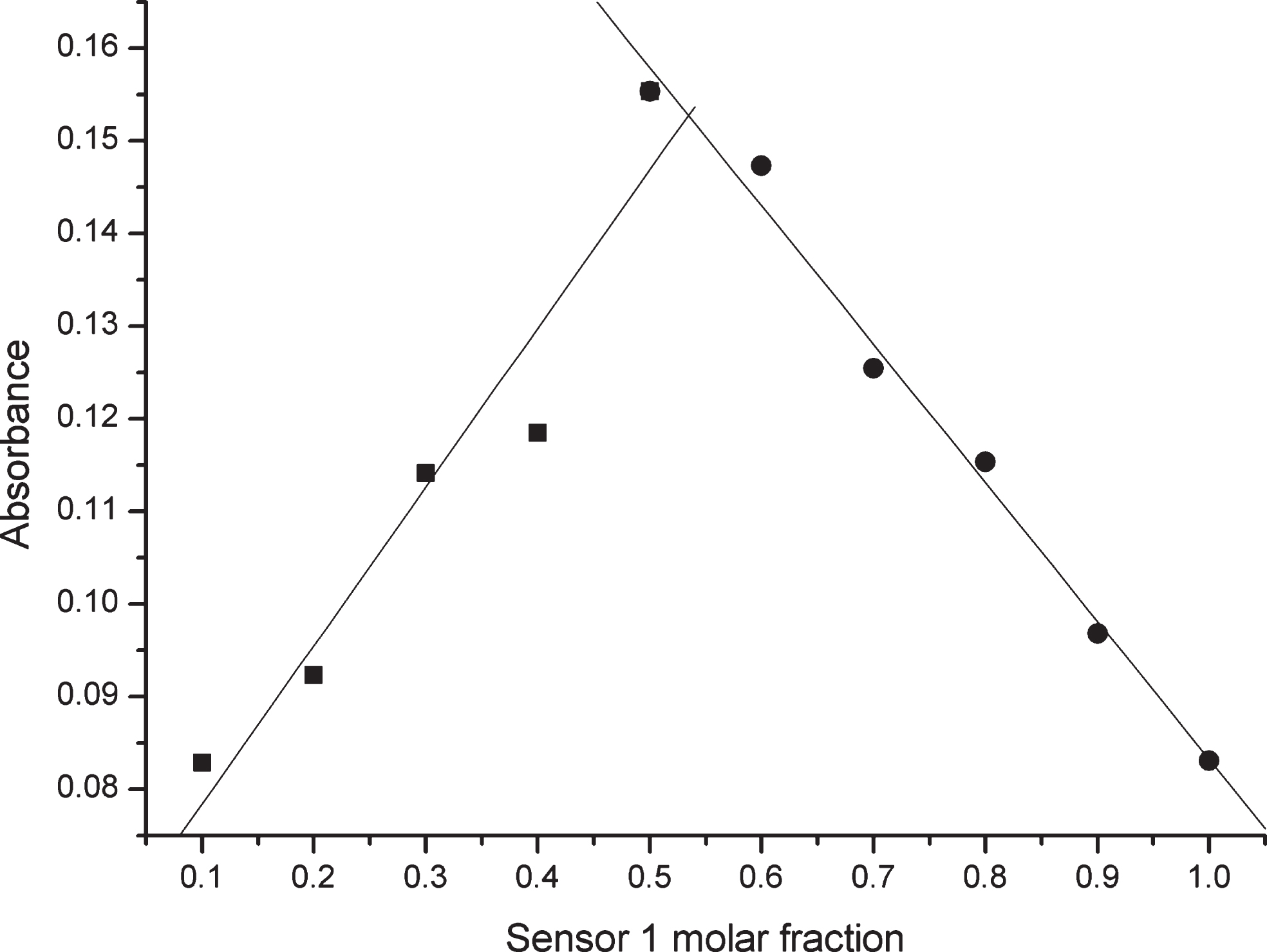
Job’s plot for determining the stoichiometry of Sensor 1 and Cu2+ in Tris-HCl solution (DMF:H2O = 5:5, pH = 7.2) [Cu2+] + [Sensor 1], the total concentration of Sensor 1 and Cu2+ was 1×10⁻⁵ mol L–1.
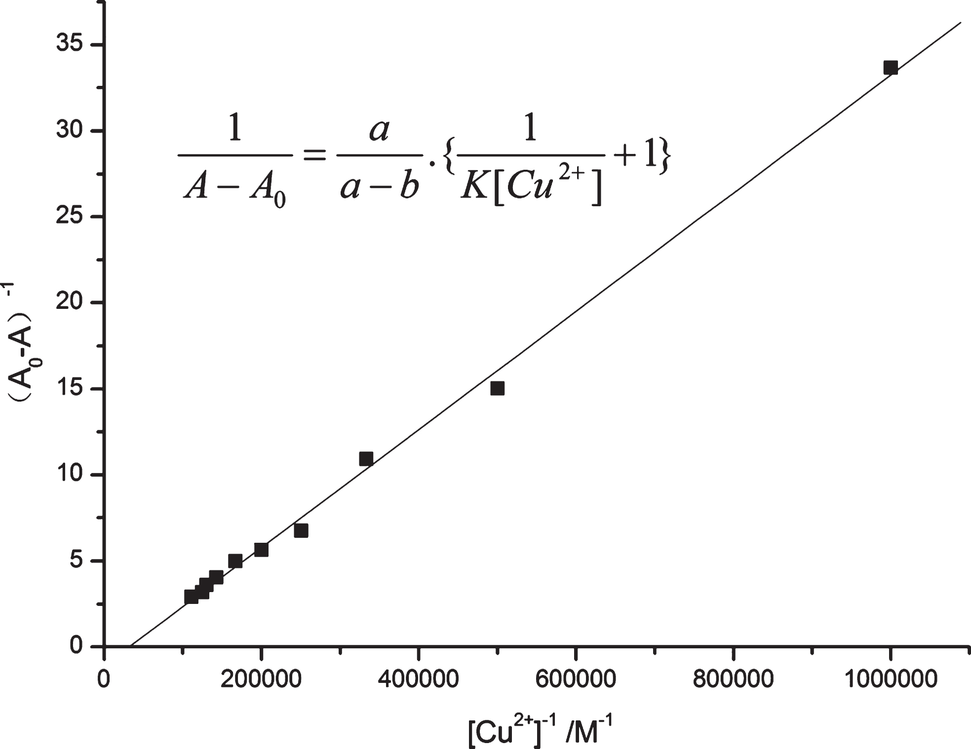
Benesi Hilderbrand plot of Sensor 1 with Cu2+.
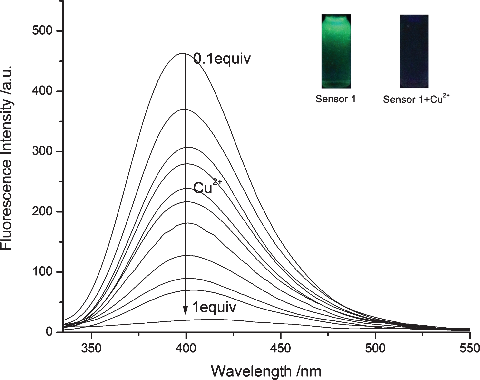
Fluorescence intensities of Sensor 1 (1×10⁻⁵ mol L–1) upon addition of different concentrations of Cu2+ (1×10⁻⁶ mol L–1—1×10⁻⁵ mol L–1) in Tris-HCl solution (DMF:H2O = 5:5, pH = 7.2) (λex = 325 nm).

Normalized response of the fluorescence signal in different Cu2+ concentrations.
In order to explain the observed fluorescence quenching, two factors were taken into account. First, the strong fluorescence intensity of independent Sensor 1 might be attributed to radiational channel from the ΠΠ* state of the emission of the quinoline moiety in the excited state, the excited electrons directly transited to the ground state to emit fluorescence. Because of the special extranuclear structure of Cu2+, when Cu2+ extranuclear electrons coordinated with lone pair of acceptor moiety, the high energy level electrons transfered to the vacated orbit of fluorophore in the excited state, and blocked the electrons transfer to the ground state for emitting fluorescence (PET), this situation brought about a substantial decrease of the fluorescence intensity. Secondary, the coordination between Sensor 1 and Cu2+ caused its conformation restriction, which also quenched the fluorescence.
1H NMR titration experiments were used for further verifying the configuration of Sensor 1-Cu2+. As illustrated in Fig. 6, upon addition of Cu2+ into Sensor 1 in DMSO-d6, remarkable changes occured in the chemical shift of protons in Sensor 1,especially in the acceptor moiety. Concretely, the obvious change between δ 4.75 and δ 4.61 in the spectrum of Sensor 1 revealed that proton O influenced the process of Cu2+ bound with Sensor 1. The proton Hd and Hj,k indicated the downshift from δ 7.07 to δ 7.19 and δ 8.75 to δ 8.85 with Δδ up to 0.12 and 0.1 respectively. Meanwhile, the other aromatic protons demonstrated inappreciable downshift, so the above-mentioned experimental results provenly affirmed the conclusion that the nitrogen in quinoline and pyridine moiety were both related to the coordination between Sensor 1 and Cu2+.
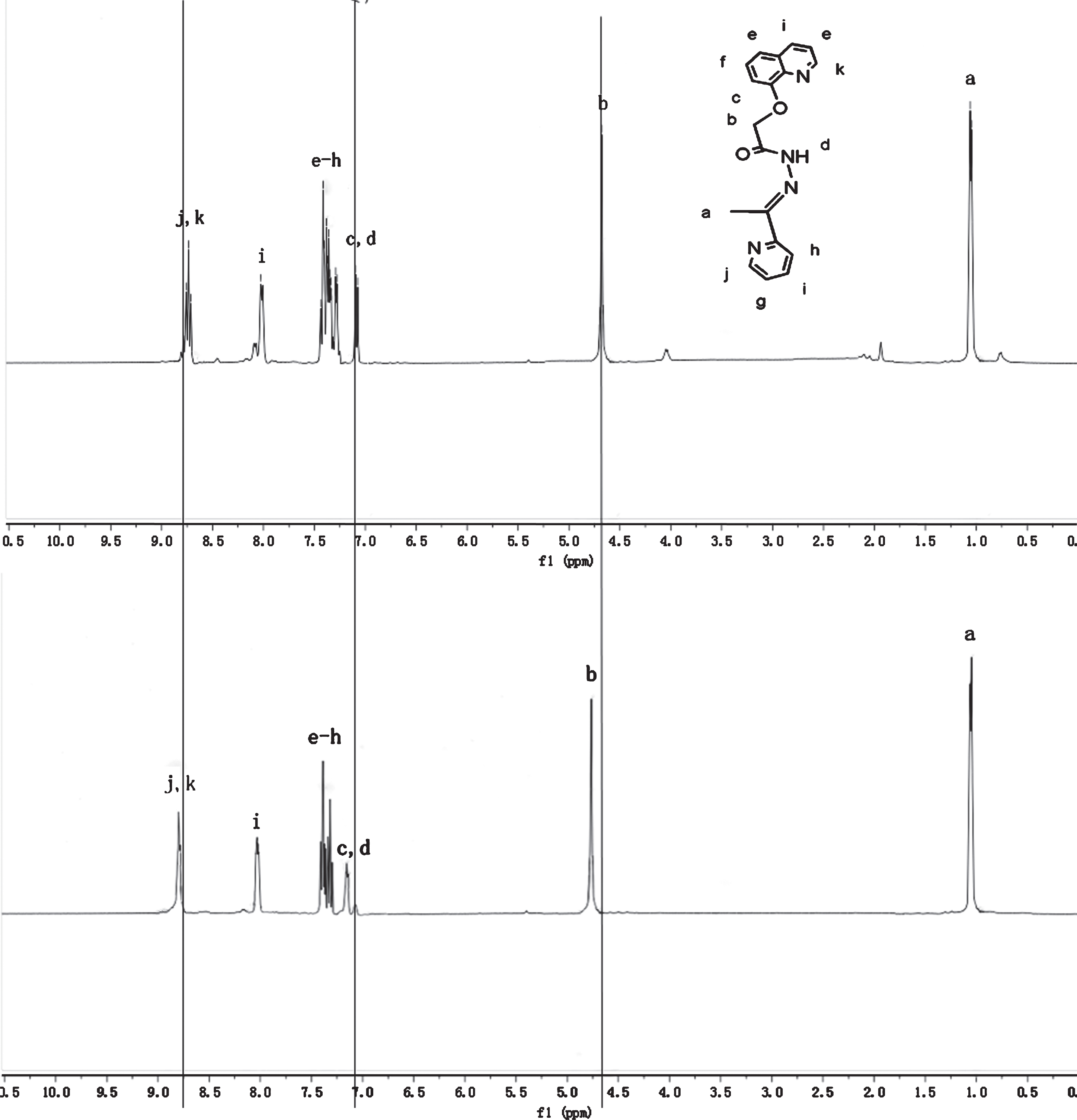
1H NMR (300MHz) spectral changes of Sensor1 in DMSO-d6 upon addition of Cu2+.
In order to further discuss the sensing ability of Sensor 1, selectivity experiments and competition experiments were applied to examine the detectability of Sensor 1 toward Cu2+. In selectivity experiments, the fluorescence response of Sensor 1 (1.0×10⁻⁵ mol L–1) was examined upon addition of various metal ions such as Na+, K+, Ca2+, Cd2+, Hg2+, Mg2+, Cu2+, Ba2+, Mn2+, Fe2+, Pb2+, Zn2+, Fe3 +, Al3 + (1.0×10⁻⁵ mol L–1) in DMF–Tris buffer solution [V(DMF)/V(H2O) = 5:5, pH 7.2]. As demonstrated in the black bar of Fig. 7, only Cu2+ caused a remarkable flourescence quenching, the adjunction of other metal ions such as Na+, K+, Ca2+, Mg2+, Cd2+, Ba2+, Pb2+, Al3 +, Fe2+ and Mn2+ did not affect the fluorescence intensity at 399 nm, besides some metal ions like Fe3 +, Zn2+, Hg2+ had inappreciable effect to the fluorescence. These above results revealed the good selectivity of the Sensor 1 toward Cu2+ preliminarily, to further validate the selectivity, we utilized competition experiments in order to ulteriorly test the disturbance of other metal ions in detecting Cu2+. First, 1.5 equiv. Cu2+ was added in Sensor 1 solution to quench the fluorescence of Sensor 1, then blended 10 equiv. Na+, K+, Mg2+, Ca2+, Cd2+, Fe2+, Ba2+, Mn2+, Hg2+, Pb2+, Zn2+, Fe3 + and Al3 + into the fluorescence-quenched solution, respectively. Next, the fluorescence intensities of the mixed systems were measured once again. As illustrated in the red bar of Fig. 7, all the fluorescence intensities of the mixed systems at 399 nm were still quenching. So we could conclude that these common metal ions would not induce any distinct disturbance of Sensor 1 in Cu2+ detection. In essence, the specific recognition about Cu2+ of this sensor was depended on the special extranuclear structure of Cu2+, compared with these transition metal cations, the extranuclear electrons of Cu2+ exclusively coordinated with lone pair of acceptor moiet of Sensor 1. So the above fluorometric analysis had certified that Sensor 1 could be an excellent fluorescent sensor in Cu2+ detection.
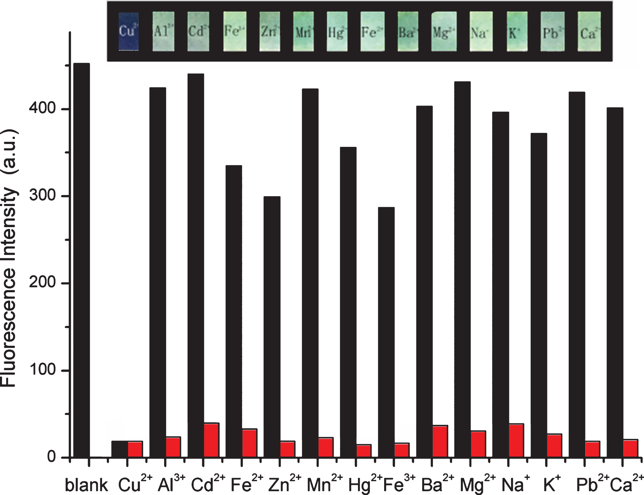
Fluorescence intensities of sensor 1 (1×10⁻⁵ mol L–1) in the presence of various metal ions (black bar) and competition experiment (red bar) in Tris-HCl solution (DMF:H2O = 5:5, pH = 7.2) (λex = 325 nm,λem = 399 nm).
As description above, Sensor 1 exhibit good sensitivity and selectivity towards Cu2+ over common metal ions, in order to expand its biological application, we explored the tracing ability of Sensor 1 for Cu2+ in living Human gastric cancer cells (SGC-7901). The fluorescence microscope images were obtained upon irradiation at 405 nm with a band path from 395 to 425 nm under identical exposure conditions. The living SGC-7901 cells were incubated with 1×10⁻⁴ mol/L Sensor 1 in DMF solution for 30 min at 37 °C in 5% CO2 atmosphere, after that, the phosphate buffered saline (PBS, pH = 7.4) was used to wash the incubated cells for 3 times. As illustrated Fig. 8a, the cumulated Sensor 1 in the living SGC-7901 cells induced obvious intracellular fluorescence, this demonstrated that Sensor 1 had successfully immersed in the cells, then added 2×10⁻⁴ mol L–1 Cu2+ solution into the Sensor 1-immersed SGC-7901 cells, as shown in Fig. 8b, the intracellular fluorescence quenched obviously, a tentative conclusion was drawn that the intracellular Cu2+ induce the change of the fluorescence. In addition, Fig. 8c depicted that the experimental cell profiles were clear and integrated, the introduction of Sensor 1 and Cu2+ did not damage the cell activity. Generally speaking, Fig. 8 demonstrated the fluorescence signal transformation of Sensor 1-supplemented SGC-7901 cells before and after treated with Cu2+, the above cell-permeable experiments had proven that Sensor 1 had potential values in biological application.
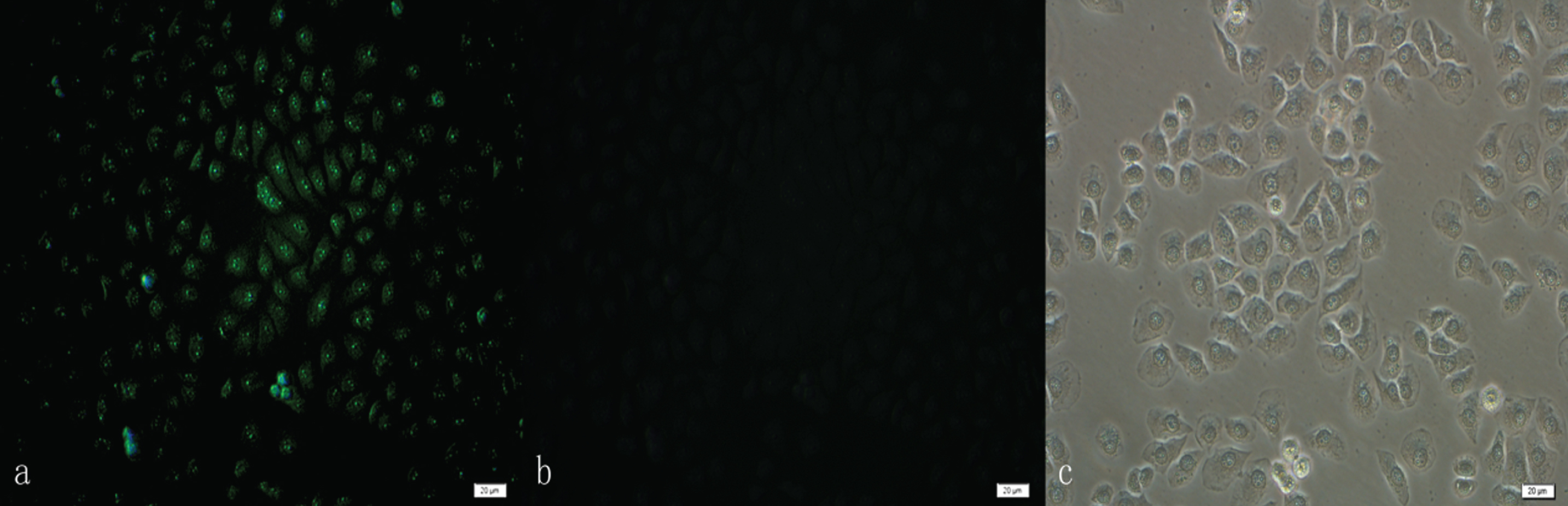
Fluorescence microscope imaging of SGC-7901 cells incubated with 1×10⁻⁴ mol L–1 Sensor 1 before and after treating with 2×10⁻⁴ mol L–1 of Cu2+ (a) SGC-7901 cells incubated with Sensor 1, (b) SGC-7901 cells incubated with Sensor 1 and Cu2+, (c) bright field image of SGC-7901 cells incubated with Sensor 1 and Cu2+.
We have developed a novel fluorescent sensor for recognizing Cu2+ by conjugation of quinoline and pyridine fluorophores. Among common metal ions, only the introduction of Cu2+ induced the fluorescence quenching of Sensor 1. The titration experimental results revealed that Sensor 1 displayed good selectivity and sensitivity to Cu2+ with a 1:1 stoichiometry mode, the association constant of complex of Sensor 1 with Cu2+ was 3.179×10⁵ mol–1. Furthermore, this sensor could be used for quantitative detection of Cu2+, the detecting limit was calculated to be 6.17×10⁻⁷ mol L–1. Then through 1H NMR titration, the recognition mechanism was further confirmed. In addition, the cell imaging experiments demonstrated its good biocompatibility. Finally, we hope this Cu2+ sensor will have broad application in the other field.
Footnotes
Acknowledgments
The authors gratefully acknowledge the support of the Youth Science Foundation of Changchun University of Science and Technology (XQNJJ-2016-11) and the Natural Science Foundation of Inner Mongolia (No. 2015BS0202).