
Abstract
The CH3-(SBA-15) and (SBA-15)-Eu2O3 host-gust nanocomposite materials were synthesized by post-synthesis method, ultrasonic synthesis method (UM) and ultrasonic assisted synthesis methods (UAM). The X-ray diffraction (XRD), infrared spectroscopy (IR), scanning electron microscopy (SEM), transmission electron microscope (TEM) analysis and nitrogen adsorption-desorption method were used to observe the structure, the surface morphology and the chemical composites of the prepared products. The luminescent properties, and the luminescent intensity of the samples were studied by excitation and emission spectra. The results showed that, for the (SBA-15)-Eu2O3 composite materials, the hexagonal pore structure of the SBA-15 molecular sieve was maintained. The luminescent intensity of the UM sample was about 6 times higher than that of the mechanical mixture of SBA-15 and nano-Eu2O3 (5 wt %). The excitation spectrum intensity was 2 times higher than that of the mechanical mixture.
Introduction
The mesoporous materials are widely used as adsorbents, carriers, non-homogeneous catalysts and in other scientific research areas owing to their neat structure and huge surface area, which will enhance their adsorption and catalytic ability [1–4]. In 1992, the scientists of the Mobil corporation used the surface-active agent as a template agent to synthesize the M41S series mesoporous materials, which possessed large surface area, regular arrangement and adjustable pores [5, 6]. In the last years, a variety of mesoporous molecular sieves (including MCM-41, MSU-1 and the SBA-15 series) were synthesized with different materials, such as metal-oxide molecular sieves, all silica molecular sieves, non-oxide skeleton molecular sieves, phosphate-based molecular sieves, heteroatom molecular sieve and non-metallic oxides [7–10]. SBA-15 is one series of mesoporous molecular sieves, since Zhao [11] et al. reported the SBA-15 molecular sieve firstly, it appears to be more attractive than other mesoporous materials because it has the huge surface area, regular pore structure, excellent thermal stability, and easily modified surface, which enhances the scientific value of the SBA-15 molecular sieve highly.
Rare earth luminescent materials have a lot of excellent characteristics and broad applications and have become a researching hotspot. They have particular photic, electric and chemical characteristics and have a broad application in high-powered luminescent apparatus, magnetism materials, catalysts and others functional materials [12, 13]. Eu is an important rare earth element and shows the emission over 550–750 nm, so, it can be used as the activation reagent of red phosphor, and was also used as fluorescent material of the blue phosphor. In recent years, europium oxide was also used as the stimulated emission fluorescent material in the new X-ray medical diagnostic system, in colored lenses, optical filters, magnetic bubble storage devices, control materials of the atomic reactor, screen worn materials and structural materials [14]. In our team, we have investigated the preparation and luminescence properties of SBA-15 doped with Eu2O3, ZnO, Er3+, Zn2+ [15, 16].
In this study, the SBA-15 molecular sieve was prepared by hydrothermal synthesis method. The SBA-15 molecular sieve was modified by trimethylchlorosilane. Eu2O3 was assembled into the SBA-15 molecular sieve by ultrasonic method (UM) and ultrasound-assisted method (UAM). The prepared materials were characterized and analyzed by powder XRD, infrared spectroscopy (IR), low temperature N2 adsorption-desorption, scanning electron microscopy (SEM), transmission electron microscopy (TEM) and fluorescence spectra. And the luminescent properties were studied.
Experimental
Chemical reagents
Tetraethyl orthosilicate (TEOS, 98%, Fluka), poly (ethylene glycol)-block-poly (propylglycol)-block-poly (ethylene glycol) (EG20PG40EG20, 5800, Aldrich), trimethylchlorosilane (A.R., Shanghai Chemical Corporation of Chinese Medicine Group), Nano-Eu2O3 (Shanghai Yu Long Co., Ltd.). 2 mol·L–1 hydrochloric acid solution was prepared from concentrated hydrochloric acid (A.R., Shanghai Chemical Corporation of Chinese Medicine Group) by suitable dilution. The water used in experiments was deionized water (Prepared by ourselves).
Preparation of SBA-15 molecular sieve
In the acidic conditions, the triblock copolymer EG20PG40EG20 was used as template, and TEOS was used as silica source. In a typical synthesis, 2.0 g of the template was dissolved in 60 g of 2 mol·L–1 hydrochloric acid and 15.0 g of deionized water. Then, 4.25 g of TEOS was added, and the mixture was stirred at 40°C for 24 h. The mixture was poured in a teflon-liner autoclaves, and was treated at 100°C for 48 h. The product was filtered, washed with deionized water, and dried at room temperature. The material was calcined at 550°C for 24 h to eliminate the template.
Modification treatment of the SBA-15 molecular sieve surface
Trimethylchlorosilane was used to silanize the SBA-15 molecular sieve in this study. The specific operations were performed as follows: 0.5 g of the SBA-15 molecular sieve was added into 15 mL 1% (v/v) of trimethylchlorosilane of ethyl alcohol solution. The mixture was stirred at the room temperature for 24 h. The product was washed with the pure ethyl alcohol until the Cl- was disappeared. The product was dried at the room temperature. The above product was calcined at 550°C for 24 h. The product was cooled to room temperature, and the white powder was obtained.
The aim of silanization to the SBA-15 molecular sieve surface was to eliminate silanol on the out surface of SBA-15 molecular sieve, but the silicon-hydroxyl on the internal surface could be reserved. It leads to the fact that the Eu2O3 could be mainly introduced into the pores of the SBA-15 molecular sieve.
Preparation of (SBA-15)-Eu2O3 composite materials
Characterization
Powder X-ray diffraction (XRD) patterns were collected on a Siemens D5005 diffractometer using Cu-Kα radiation (λ= 1.5418 Å and operating at 30 kV and 20 mA). Fourier transform infrared (FT-IR) spectra were obtained on a Bruker Vertex 70 FT-IR spectrometer. Powder samples (1% wt) were dispersed in KBr (99% wt) pellets for IR analysis. Scanning electron microscopic (SEM) images were recorded on a JEOL JSM-5600L SEM instrument. Transmission electron microscopic (TEM) images were taken on a JEOL 2010 TEM instrument. Adsorption-desorption study of nitrogen were performed on a micromeritics ASAP2010M volumetric adsorption analyzer at 77 K. The samples were degassed in vacuum at 573 K for 12 h before measurement. Surface areas were calculated based on the BET (Brunner-Emmett-Teller) method, while pore size distribution were computed using the BJH (Barrett-Joyner-Halenda) method. Luminescence spectra were collected on a SPEX-FL-2T2 luminescence spectrometer at room-temperature.
Results and discussion
Analysis of XRD
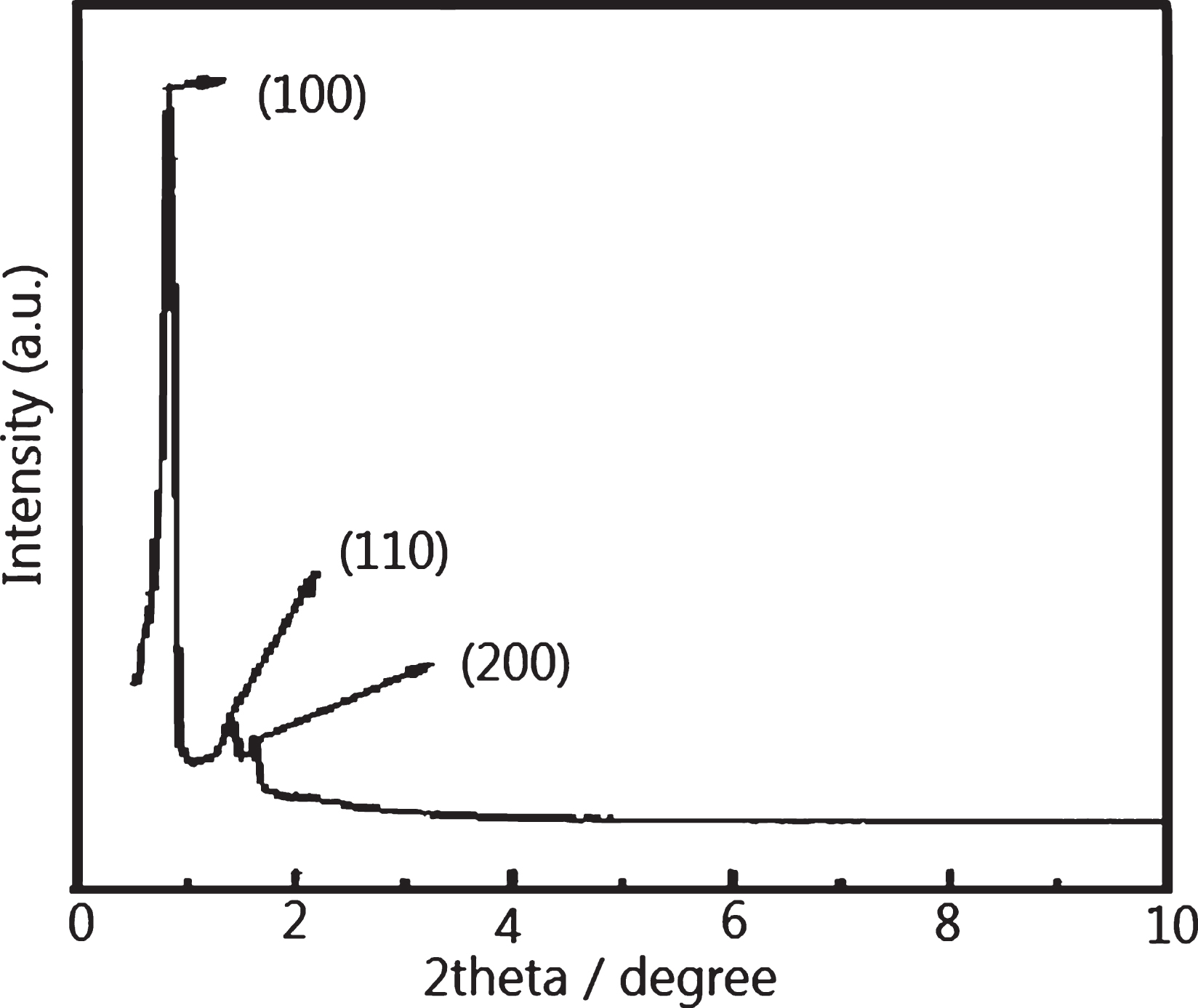
Figures 1 and 2 presented the XRD patterns of the SBA-15 molecular sieve powders and CH3-(SBA-15) sample, respectively. From the Fig. 1, the four reflection diffraction peaks were denoted to (100), (110), (200) and (210), respectively. It indicated that the SBA-15 molecular sieve had a high order and regularity framework structure, two-dimensional hexagonal pores structure. Three characteristic peaks were presented in Fig. 2, which were denoted to (100), (110), and (200), respectively. After silanization modification, the ordered hexagonal pore structure of the SBA-15 was maintained.

The XRD diffraction of the SBA-15.
The XRD diffraction of the silanized SBA-15.
Figure 3 showed the XRD diffraction of the SBA-15 molecular sieve powder, UM sample and UAM sample. From Fig. 3, three characteristic diffraction peaks of (SBA-15)-Eu2O3 samples appeared, which were denoted to (100), (110) and (200), respectively. It indicated that the (SBA-15)-Eu2O3 samples maintained the highly ordered hexagonal pore structure. It showed that the preparation methods had a minor damage to the framework of the SBA-15.
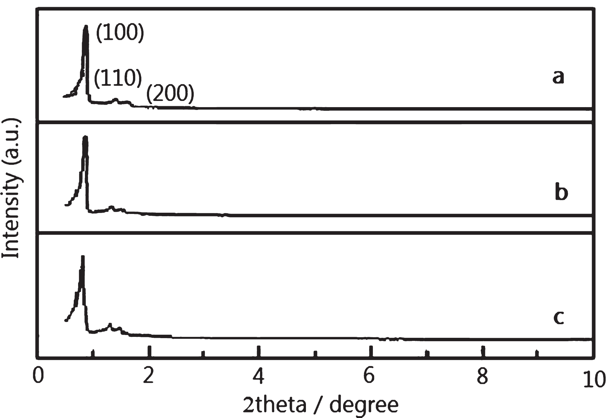
The XRD pattern of(a) SBA-15 (b) UM sample (c) UAM sample.
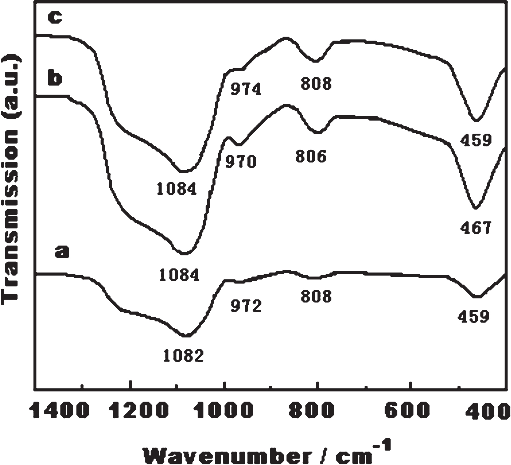
Figure 4 presented the FT-IR spectra of the SBA-15 powder sample (S), SBA-15 sample treated with silane but not calcined (SSN), and SBA-15 sample treated with silane and calcined (SSC). The absorption peaks were listed in Table 1.

Fourier transform infrared spectrums (FT-IR) of a) SBA-15; b) silane treated and calcined SBA-15; c) silane treated but not calcined SBA-15.
Fourier transform infrared absorption peaks
The FT-IR spectra of the SSN samples presented an absorption bands at 2965 cm–1. It suggested that there was the methyl group in the sample. For the SSC sample, the characteristic infrared absorption bands disappeared. It indicated that methyl had separated from the SBA-15 framework after calcination.
Figure 5 showed the FT-IR spectra of the SBA-15, UM sample and UAM samples. From the Fig. 5, the FT-IR spectra of the (SBA-15)-Eu2O3 samples were the same as that of the SBA-15 sample, and displayed four absorption peaks. They corresponded to the characteristic infrared absorption bonds of the SBA-15. It indicated that the structure of (SBA-15) had not been damaged when the Eu2O3 was introduced in.
Fourier transform infrared spectra (FT-IR) of a) SBA-15; b) UM sample; c) UAM sample.
Figures 6–8 showed the SEM images of the SBA-15, UM and UAM samples, respectively. From the SEM images, the samples displayed fibrous structure. The size of the samples were as follow: the diameter of the fiber of the SBA-15 was 396±10 nm, that of the UM samples and UAM samples were 417±10 nm and 430±10 nm, respectively.
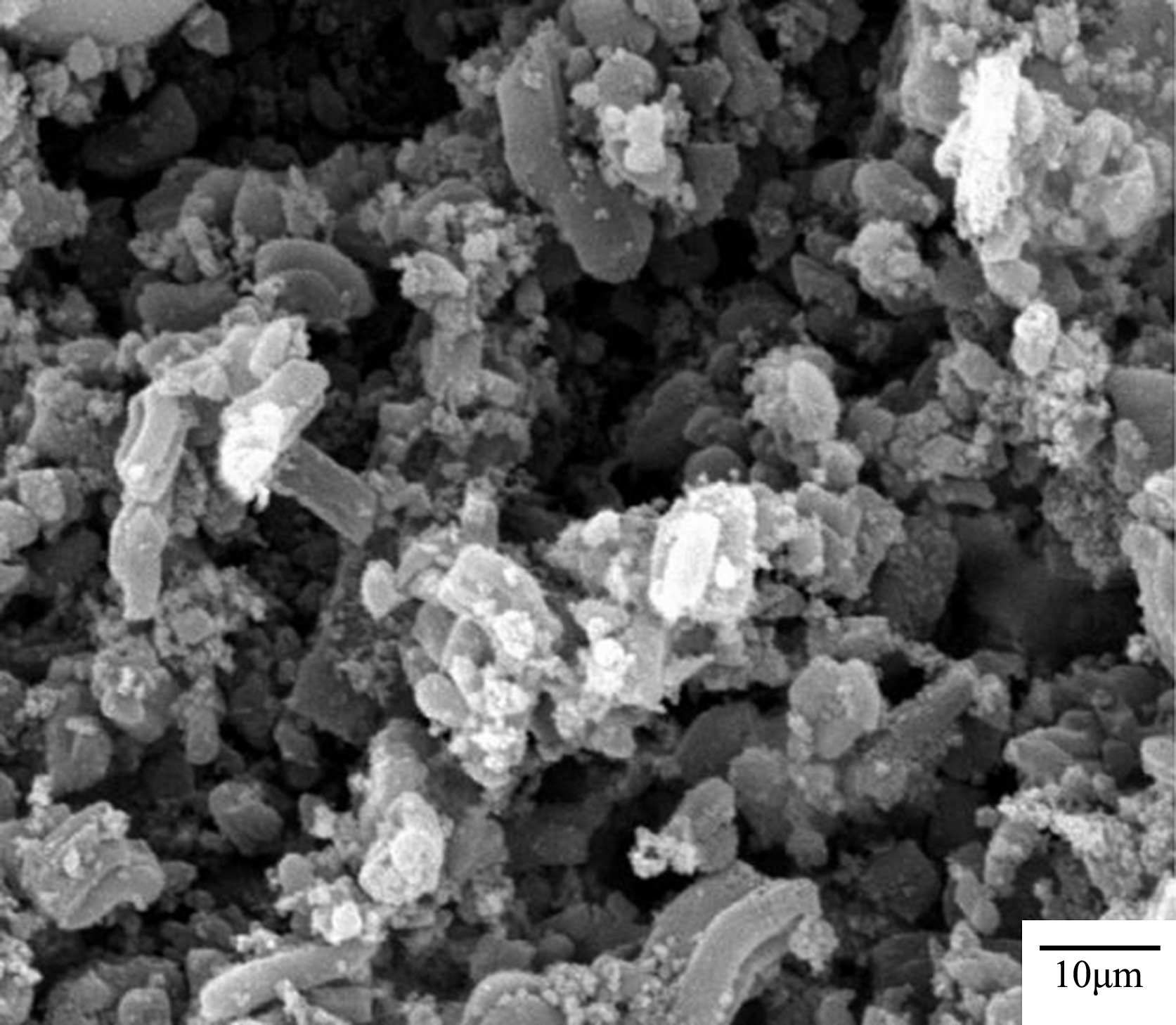
The SEM images of the SBA-15.

The SEM images of UM sample.

The SEM images of UAM sample.
Figures 9–11 showed the TEM images of the SBA-15, UM and UAM samples, respectively. It showed that the (SBA-15)-Eu2O3 samples had two-dimensional hexagonal mesoporous pore structure. It could show that the porous structure of the SBA-15 molecular sieve was maintained after the Eu2O3 was assembled into the SBA-15.
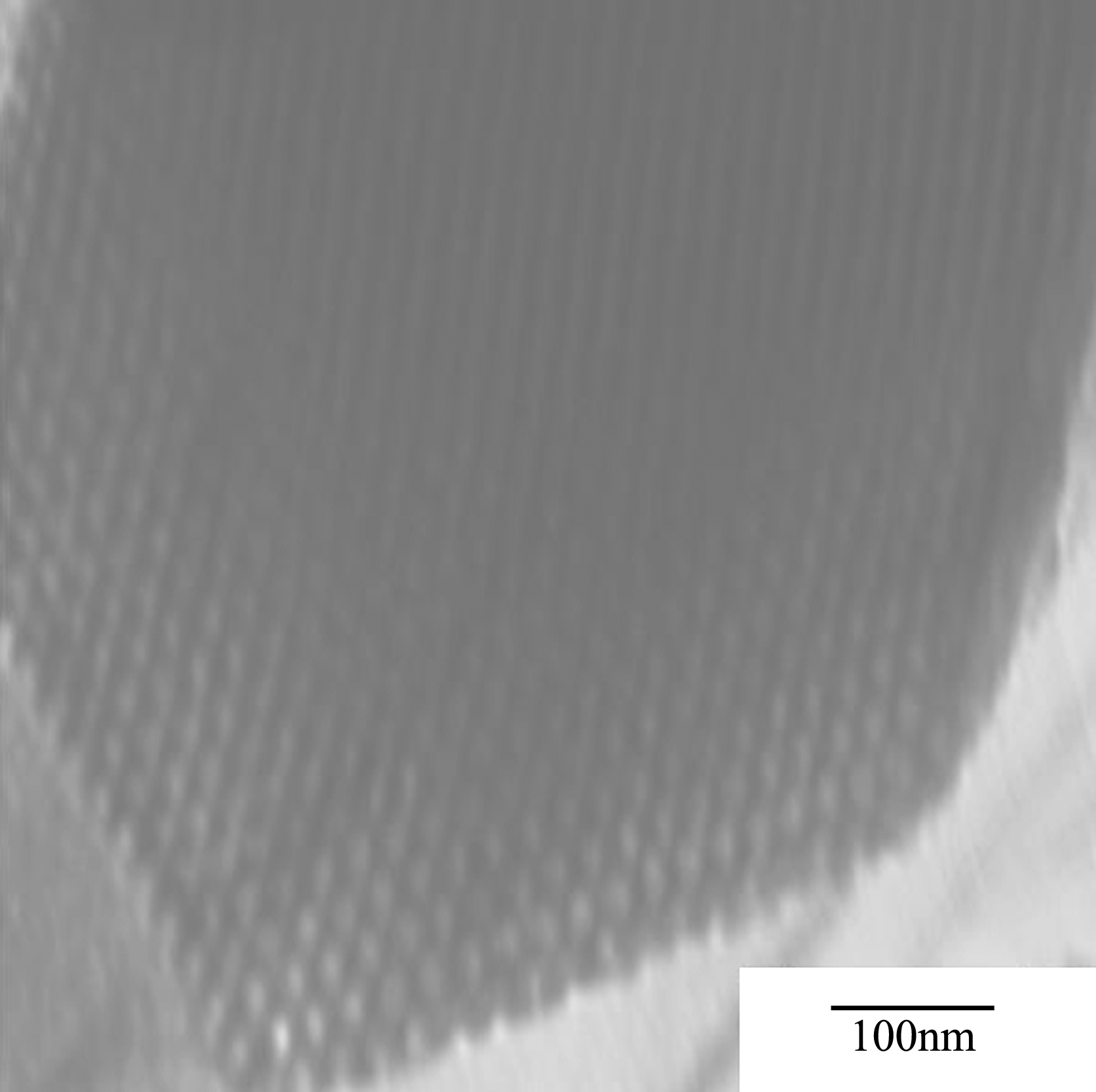
The TEM image of SBA-15.

The TEM image of UM sample.
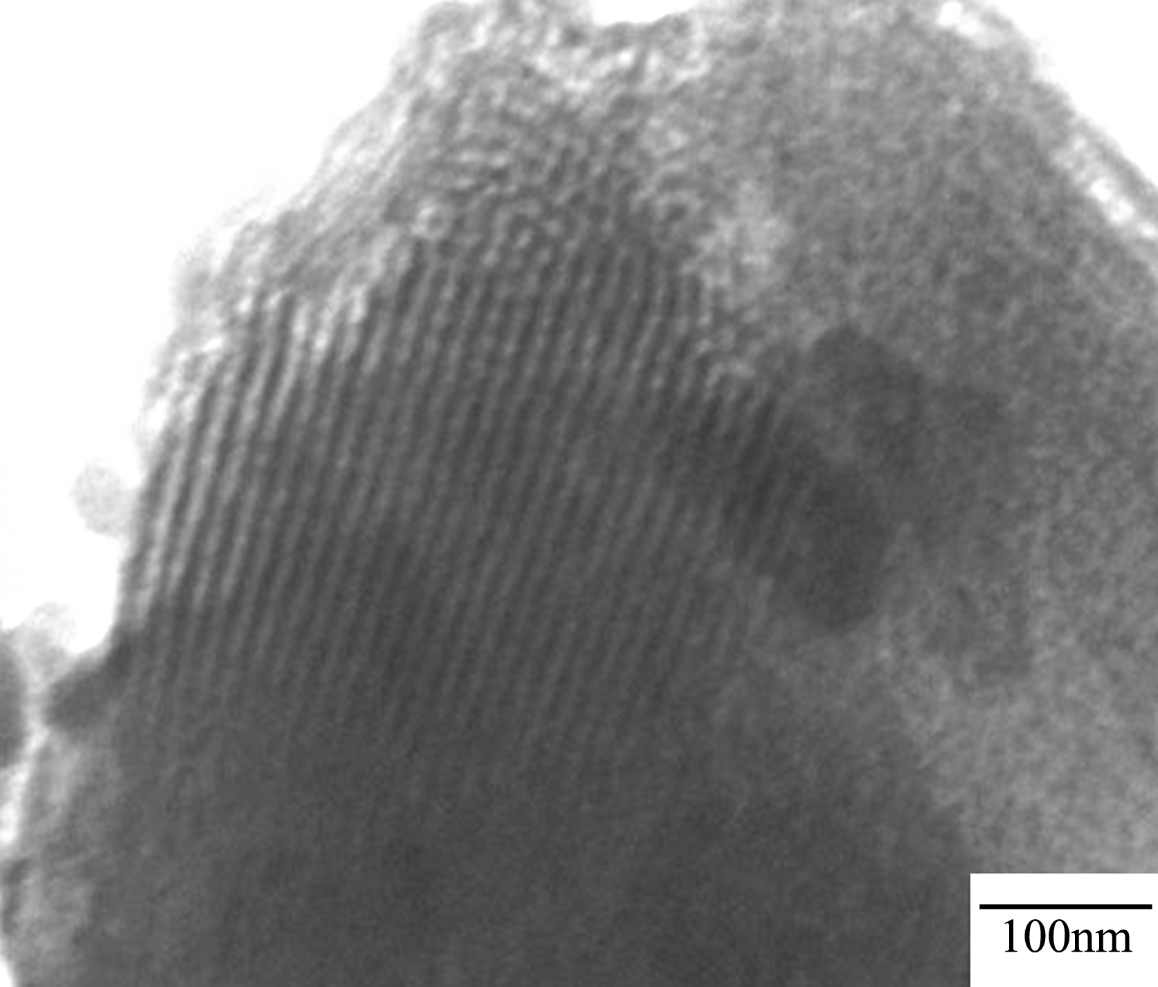
The TEM images of UAM sample.
Figure 12 showed the low temperature nitrogen adsorption-desorption isotherms of the SBA-15 molecular sieve and the (SBA-15)-Eu2O3 materials.
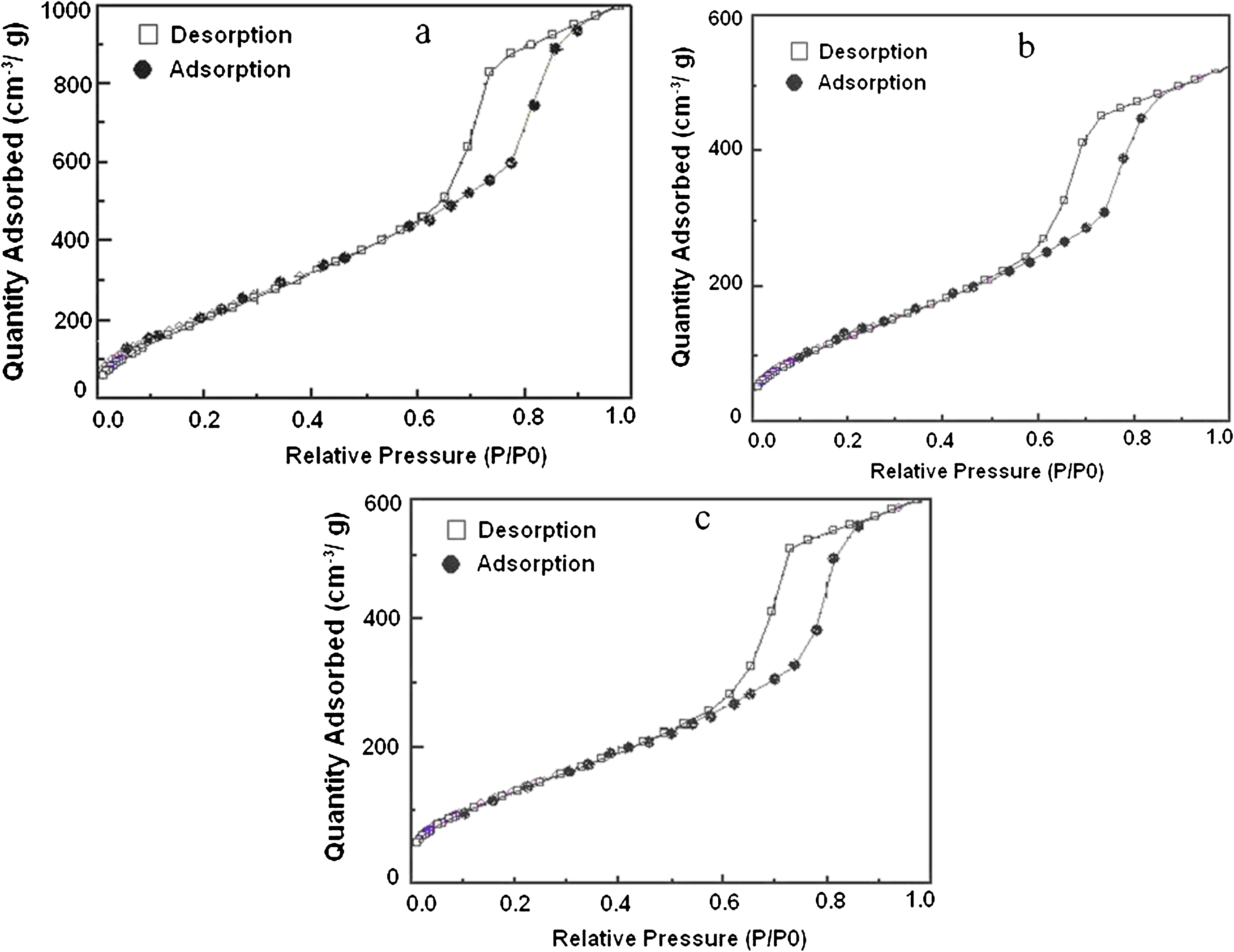
Low-temperature N2 adsorption-desorption isotherms. a) SBA-15; b) UM sample; c) UAM sample.
From the low temperature N2 adsorption-desorption isotherms of the samples, they were the typical irreversible IV adsorption-desorption isotherms, and had a H1-type hysteresis loop. The H1-type hysteresis loop is the main features of the cylindrical porous of the mesoporous materials. There were a clear adsorption branch and a desorption branch in the isotherms. The adsorption branches were very steep. It indicated that the samples had a narrow pore size distribution. It was validated by the porous size distribution curves in Fig. 13.
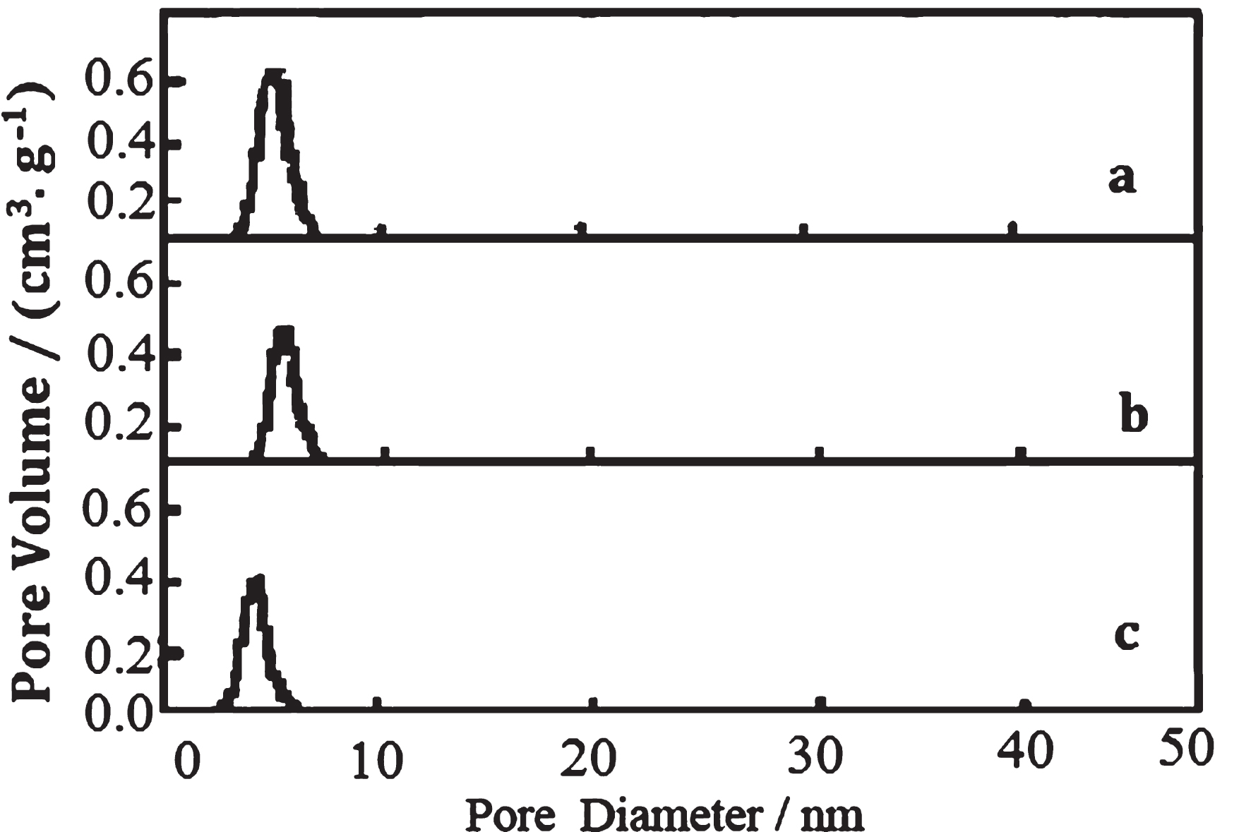
Pore size distributions of samples a) SBA-15; b) UM sample; c) UAM sample.
In the low-temperature N2 adsorption-desorption isotherms, the adsorption was mainly monolayer adsorption at a relatively low pressure. The narrow pore structure could not impede the gas desorption in the desorption process. Therefore, the capillary condensation could not occur at a relatively lower pressure. Thus, desorption would not be delayed, and the adsorption-desorption was reversible. With the gradual increase of the relative pressure, the volume of adsorption gas increases in the pore, and the adsorption was mainly multi-molecular layer adsorption. The adsorption and desorption branches of the adsorption-desorption isotherms inflected sharply when the relative partial pressure (p/p0) was 0.63 for the SBA-15, 0.58 for the UM sample, and 0.53 for the UAM sample. The phenomenon was due to the capillary condensation when the relative pressure was added to a certain degree. It led to the result that the adsorption branch and desorption branch of the adsorption-desorption isotherms inflected sharply. In the isotherms, there existed a H1-type hysteresis loop. It showed that the desorption lagged behind the adsorption process. The adsorption and desorption processes were both irreversible. The larger pore led to the relatively higher partial pressure, and the phenomenon of capillary condensation was occurred. The phenomenon disappeared when the relative partial pressure (p/p0) was 0.9 for the SBA-15, 0.85 for the UM sample and 0.83 for the UAM sample. When the relative pressure was higher, the channels of the materials were filled, and the capillary condensation was finished. The gas was mainly adsorbed on the outer surface of the materials, and this process was reversible.
In this paper, the surface areas of the samples were calculated by BET (Brunner-Emmett-Teller) method, and the pore size distribution was calculated using BJH (Barrett-Joyner-Halenda) method. The surface areas, the pore volume and the pore sizes were calculated from desorption branched of the low temperature nitrogen adsorption-desorption isotherms. The pore wall thickness was obtained from the account, and the parameters are shown in Table 2.
Pore structure parameters of samples
aa0 =
The excitation and emission spectra of the rare earth ions are due to the inner 4f-4f transitions. According to the law of the spectrum, the electric dipole transition is used to be forbidden when ΔI is 0. But transitions can be occurred in reality. The phenomenon is due to the mixture of the 4f configuration and the opposite parity configuration of g or d as well as the symmetry deviation. It makes the originally prohibited f-f transition become possible. The mandatory f-f transitions make the line characters and peak of the spectra lower. It leads to the fact that the excitation energy can not be effectively absorbed. It is one of the impact reasons that the luminescence efficiency of the trivalent rare earth ion is lower. The trivalent rare earth ions have a very small transition probability between the 4f, and the excitation life is far longer than ordinary atoms. The outer electron shells of the trivalent rare earth ions are filled, and 4f orbit is in the inner. Thus, the 4f transitions of the trivalent rare earth ions are protected by the full outer electron shell. It is hardly interfered by external field. Thus, the emission spectrum of the f-f transition is a sharp line characteristic spectrum, and the emission is inherent of the rare earth ion itself. It has not been influenced by the external environment.
In addition to 4f-4f transitions, there are d-f transitions. According to the law of the spectrum, the transition is possible when Δ l is1. The characteristics of the d-f transitions are almost absolutely opposite with 4f-4f transitions. The spectrum of d-f transition is a broadband spectrum, fluorescence lifetime is shorter, and intensity is higher. Compared with the 4f transitions, the 5d transitions are the outer layer transition, and the d-f transitions are greatiy influenced by the outside environment. Luminescence characteristics of the trivalent rare earth ions can be summarized as follows: (1) Fluorescence lifetime is longer; (2) Spectral shape is hardly affected by the external temperature, and temperature quenching. (3) It presents the higher color purity and sharp emission spectra, which is due to the inner electron shell f-f transitions; (4) The transition of the 4f orbital is free from the impact of external field, which is due to the protective effect of the outer shell. So the emission spectra of the luminescent materials have not obviously changed with the matrix.
Figures 14 and 15 showed the emission spectra and excitation spectra of the prepared samples, respectively. The excitation wavelength was 397 nm, the emission wavelength of was 610 nm. The Eu3+ was the center of the host-guest composite materials. The 4f transition of Eu3+ was affected diminutively by the surrounding crystal field. It was due to the shielding protective effect of the 5s26p6 shell. From Fig. 14, the emission peaks at about 623 nm were the intrinsic peaks of the Eu3+, which were produced by the inner 4f transitions. It was compulsory f-f transition. The emission spectra of the UM sample was the strongest among the materials, its intensity was almost 6 times higher than that of the mechanically mixed materials. From Fig. 15, the excitation peak of UM sample was the strongest among the samples, and had the strongest excitation at around 273 nm. Its intensity was about 2 times higher than that of the mechanical mixture samples.
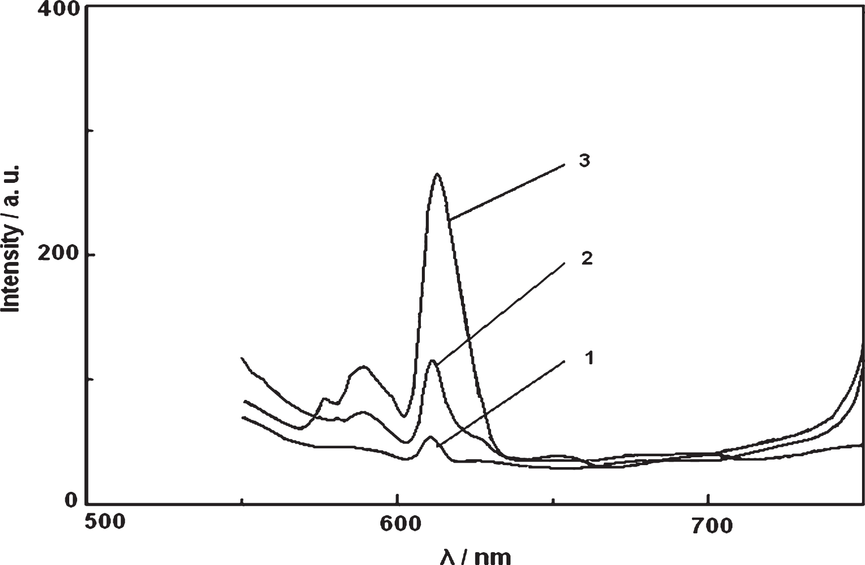
Emission spectra of the samples. 1. SBA-15 and the Eu2O3 mechanical mixture; 2. UM sample; 3. UAM sample.
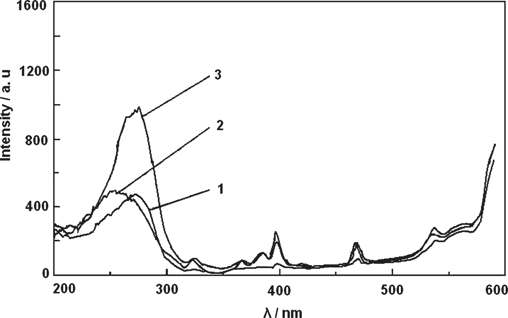
Excitation spectra of the samples. 1. SBA-15 and the Eu2O3 mechanical mixture; 2. UM sample; 3. UAM sample.
The SBA-15 mesoporous molecular sieve was synthesized. It led to the result that the guest material was assembled as much as possible into the pores of the SBA-15 molecular sieves. The (SBA-15)-Eu2O3 host-guest composite materials could be prepared by the ultrasonic method or ultrasonic aid method. The framework of the (SBA-15)-Eu2O3 host-guest composite materials was not damaged. The (SBA-15)-Eu2O3 host-guest composite materials had the luminescent properties, and the luminescent intensity of the UM sample was the best. Its emission peak intensity and the excitation peak intensity were almost 6 and 2 times of the mechanical mixed samples, respectively.
Footnotes
Acknowledgment
Financial support from “Youth Backbone Teacher Training Project of Henan Higher Education Institutions in 2017 (No. 2017GGJS282)”, “The Science and Technology Development Plan Project of Henan Province in 2018 (No. 182102310611”, “Post-doctoral Science Foundation of Henan province in 2018” and “Startup Project of Doctor Scientific Research of Shangqiu Medical College (No. BSJH001)” are gatefully acknowledged.