
Abstract
In this work, the authors developed a method to promote the SERS activities of gold nanoparticles by deposition of thin silver shell on their surfaces. Spherical gold nanoparticles were used as the model, and a chemical reduction process was carried out to deposition silver shells on the surface of gold nanoparticles (in the form of Au@Ag). The shell thickness can be adjusted by tuning the molar ratio of MAg/MAu, and the optimized the SERS activities by using rhodamine 6G as the probing molecules. The relationship between SERS activity of Au@Ag and shell thickness was studied.
Introduction
Surface-enhanced Raman scattering (SERS) is a spectroscopic tool for the detection and analysis of molecules based on the surface and nanotechnology [1]. Raman scattering of molecules adsorbed on or in the place close to the surface of a SERS substrate can be strongly promoted via electromagnetic or chemical enhancement [2–4]. Containing unique spectroscopic fingerprinting information relating to molecular structure and providing intact data acquisition with high sensitivity, SERS is widely applied in the field of materials science, environmental science, psychobiological testing, public food safety, and detection of explosive particles in luggage, etc [5]. The development and application of SERS relies on the SERS active substrate. Great efforts have been made for the investigations on SERS active substrate [6, 7]. Gold nanoparticles are commonly used SERS substrates. Due to the biocompatibility and chemical stability, gold nanoparticles have been widely employed in the biology and clinic medicine related SERS studies [8, 9]. The drawback of gold nanoparticles in the aspect of SERS as compared to that of silver substrates is the relative lower SERS activity, which might reduce the sensitivity on chemical/biochemical sensing. One way to solve this problem is to deposit a silver shell on the surface of a gold nanoparticle [10, 11]. In this work, a chemical reduction process was developed to archive this aim. Silver atoms deriving from the reduction of silver ions in the presence of ascorbic acid as the reducing agent were deposited on the surface of gold nanospheres (AuNS) and formed silver shells. The obtained nanostructures were marked as Au@Ag (core@shell). The shell thickness can be tuned by controlling molar ratio of MAg/MAu, and the relationship between the shell thickness and SERS activity of Au@Ag was investigated.
Experimental
Materials
HAuCl4·4H2O, Na3C6H5O7·2H2O, AgNO3, ascorbic acid, HCl (36%), HNO3 (68%), and H2SO4 (98%) were purchased from Beijing Chemical Plant. Poly(diallyldimethylammonium chloride) solution (PDDA, 20%) was obtained from Sigma-Aldrich. Rhodamine 6G (R6G) was purchased from Waldeck GmbH & Co. KG.. All reagents were used as purchased without further purification. MilliQ water (18.6 MΩ) was used to prepare aqueous solutions.
Instrument
UV-vis extinction spectra were recorded by a UV-vis spectrometer (200–890 nm, Ocean optics USB Chem4000). Transmission electron microscope (TEM) images were measured with a Hitachi H-8100 IV operating at 200 kV. TEM samples were prepared by dripping a drop of nanoparticle colloid onto the carbon-coated copper grids and dried in the air at room temperature. The Raman spectra were collected by a Renishaw 1000 microspectrometer connected to a Leica microscope with an objective lens of 50× (NA = 0.5). The laser line was 532 nm, and the laser power was 2 mW. The accumulation time for the Raman detection was 10 s.
Preparation of Au@Ag
First, gold nanospheres were prepared by a thermal reduction process in the presence of sodium citrate as the reducing agent [12]. Typically, 50 mL of HAuCl4 solution (1×10⁻³ M) was placed in a round bottom flask and was heated to boiling under magnetic stirring. Then, 4.6 mL of sodium citrate (0.0378 M) was injected into the HAuCl4 solution under vigorous stirring. The solution in the flask changed from pale yellow to wine red (Fig. 1a). The as-prepared gold nanospheres were then stored in an Erlenmeyer flask in dark under room temperature.

Optical photos of AuNS (a) and Au@Ags prepared with the ratio of MAg/MAu equaled to 0.5 (b), 1 (c), 1.5 (d), and 2 (e), respectively.
In the following step, to prepare Au@Ag, 5 mL of as-prepared gold colloid was taken and placed in a reagent bottle (12 mL, Agilent), and ascorbic acid (0.1 M) and silver nitrate (10–2 M) with certain volume were then added successively to the gold colloid under vigorous stirring. To tune the shell thickness of Au@Ag, different amount of silver nitrate (with different molar ratio of MAg/MAu) was adopt as listed in Table 1. The photos of as-prepared Au@Ags were shown in Fig. 1b.
The parameters of reagents used for preparing Au@Ags
UV-vis extinction spectra of Au@Ags
Figures 1 and 2 shows the photos and UV-vis extinction spectra of the as-obtained AuNS and Au@Ags. The AuNS is wine red in color. Its extinction spectrum was displayed in Fig. 2a, in which a single band centered at 521 nm can be observed. With the addition of ascorbic acid as the chemical reducing agent and silver nitrate as the silver deposition source, obvious change in the color of colloidal solution can be seen. As the ratio of MAg/MAu equaled to 0.5, the color of the colloidal solution varied from initial wine red to orange red. Further increase in the ratio of MAg/MAu caused the darken in the color of the colloidal solution. These changes were also proved in the corresponding extinction spectra as shown in Fig. 2b.
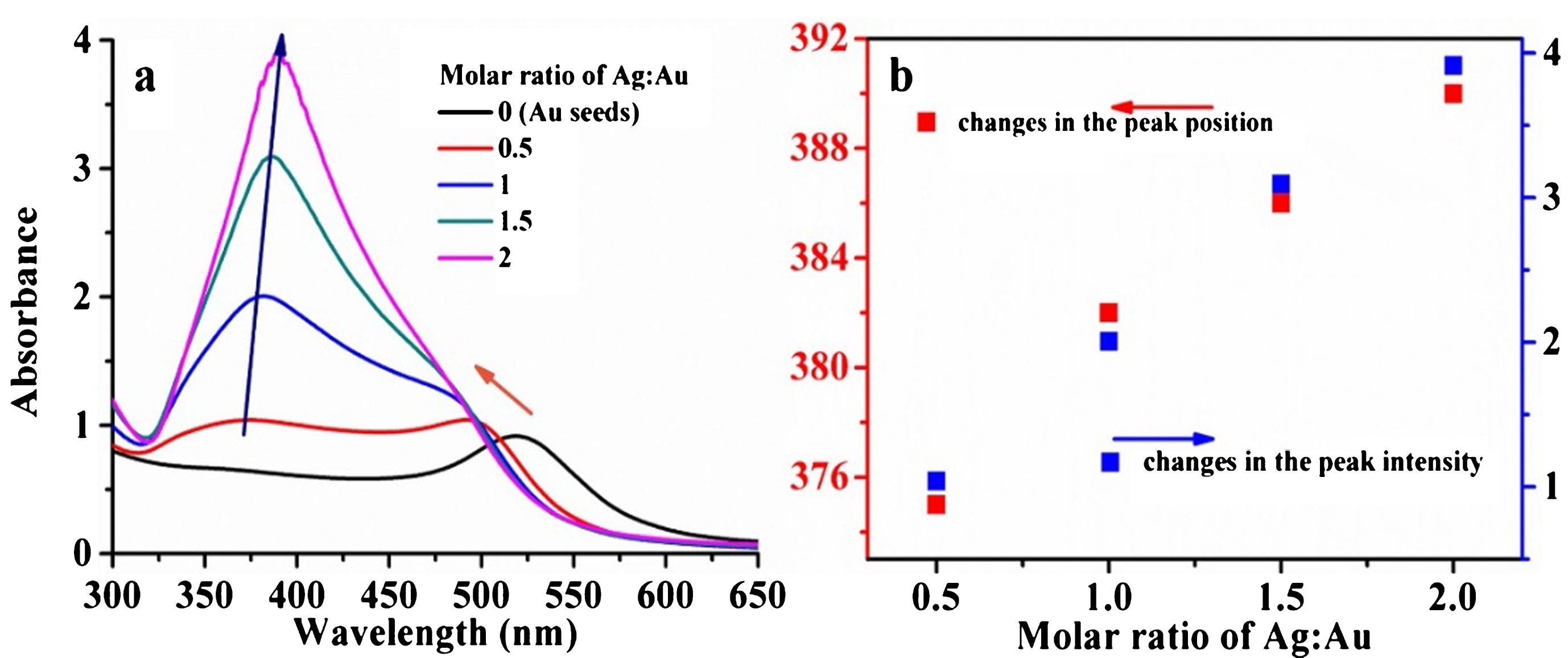
(a) UV-vis extinction spectra of AuNS and Au@Ags prepared with the molar ratio of MAg/MAu changed from 0.5 to 2. (b) Changes in the peak position and intensity of the extinction spectra of Au@Ag as a function of MAg/MAu.
When the ratio of MAg/MAu equaled to 0.5, the addition of silver nitrate caused a blue shift of 521 nm band and the presence of a new band at around 370 nm. These spectral changes indicated the formation of silver shell on the surface of AuNS instead the formation the mixture of silver nanoparticles and AuNS. If the latter one happened, a new band at around 400 nm attributed to silver nanoparticles should be observed, and 512 nm band of AuNS should not shift to shorter waveband. Thus, Au@Ags were formed during the reduction of silver nitrate in the presence of AuNSs as the cores. The movement of the 521 nm band can be attributed to the coupling effect of the gold core and the silver shell. As the amount of silver ions increases, the band attributed to the silver shell continuously moved toward longer waveband, from around 370 nm to 390 nm, and the band intensity increased as well. The band attributed to gold core showed a little blue shift and was gradually evolved as a shoulder [13].
The changes in the band position and band intensity attributing to the silver shell of Au@Ag as a function of MAg/MAu were investigated and shown in Fig. 2b. As can be seen, both the degree of redshift of the band position and the band intensity increased as the function of MAg/MAu, and displayed quasi-linear dependent relationship. However, as more silver nitrate was added, with the ratio of MAg/MAu bigger than 2, the as-prepared Au@Ag was instable, obvious precipitate can be found in the colloidal solution.
To identify the structure of the as-prepared nanoparticle, TEM images of AuNS and Au@Ags were collected and shown in Fig. 3. Uniform nanospheres can be seen in the TEM image (Fig. 3a) of AuNS. The average size was measured to be 12.7 nm. TEM images (Fig. 3b–e) of Au@Ags prepared with different ratio of MAg/MAu proved that the as-prepared nanostructures were indeed core-shell structure. Due to contrast difference in TEM image, the dark area located at the center of the particle corresponded to gold, and the light area located at the periphery corresponded to silver. Thus, the as-prepared nanoparticles shown in Fig. 3b-e are gold core/silver shell structures. The Au@Ags prepared with different ratio of MAg/MAu were uniform nanospheres as well, the average sizes were measure to be 15.9, 17.2, 19.2, and 21.9 nm, as the ratio of MAg/MAu increased from 0.5 to 2, as shown in Fig. f. The thickness (h) of the silver shell was estimated by the function of h = (dAu @ Ag–dAuNS)/2, where dAu @ Ag represented the average diameter of Au@Ag, and dAuNS represented the average diameter of AuNS, considering the fact that gold core kept unchanged during the formation of Au@Ags. The calculated shell thicknesses were 1.6, 2.6, 3.2, and 4.6, as the ratio of MAg/MAu increased from 0.5 to 2. The shell thickness of Au@Ag showed a quasi-linear dependence with the ratio of MAg/MAu. Thus, the shell thickness of Au@Ag can be well controlled by tuning the amount of silver ions added with suitable ratio of MAg/MAu.
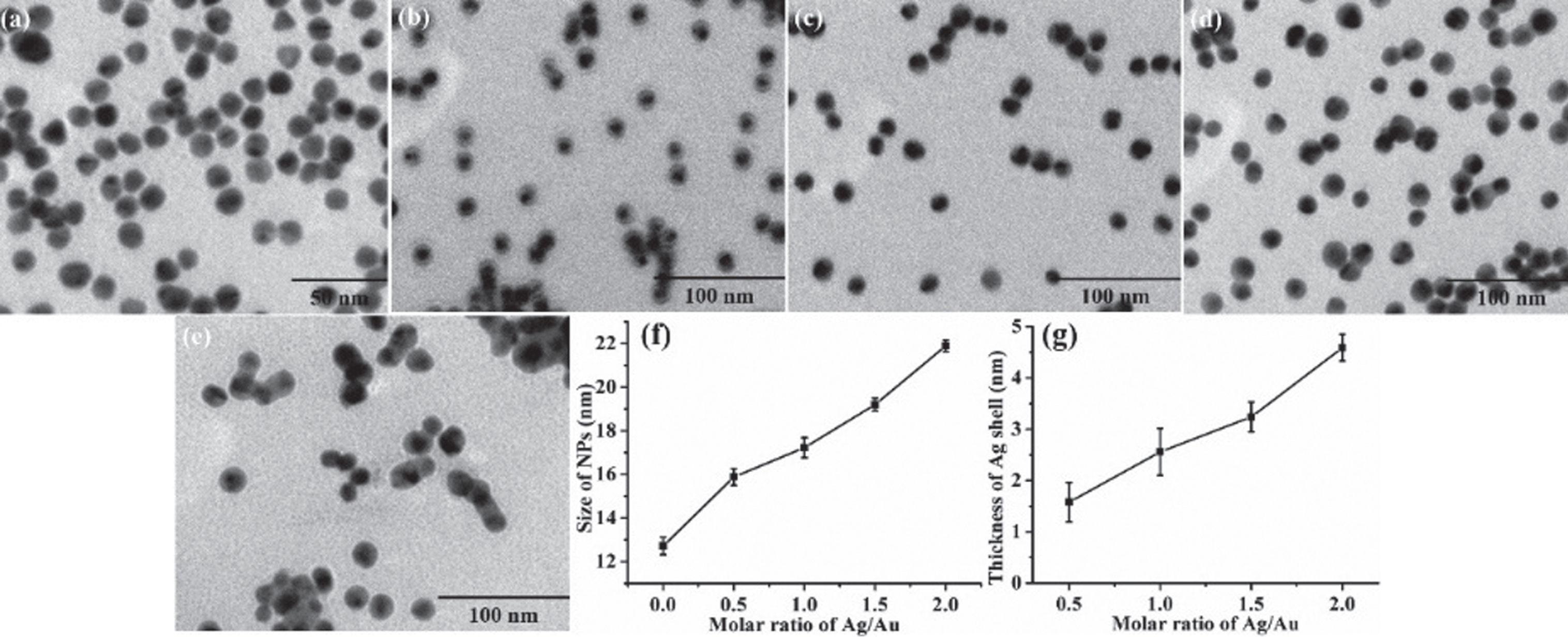
TEM images of AuNS (a) and Au@Ags prepared with MAg/MAu of 0.5(b), 1(c), 1.5 (d), and 2(e), respectively. The changes in size (f) and shell thickness (g) of AuNS and Au@Ag as a function of MAg/MAu.
By using R6G as the probe molecule, the SERS activities of AuNS and Au@Ags with different silver shell thicknesses were studied and compared. In order to ensure the dispersion of the particles and the reproducibility of the SERS activity, nanoparticles were fixed on the surface of glass slide before Raman test. A self-assembly process was introduced to fix nanoparticles on the surface of glass slide. At first, glass slides (5 mm×5 mm) were immersed and heated in a Piranha solution containing H2SO4 and H2O2 with volume ratio of 7:3 for about 1 h. This process can modify the surface of glass slides with hydroxyl groups under acidic conditions, and make them negative charged. A negative charged surface was not suitable for the immobilization of nanoparticles prepared in this work, which were negative charged as well due to the presence of citrate as the stabilizer, via electrostatic assembly. Thus, a positive charged bridge was needed. PDDA, a kind of positive polyelectrolyte, was chosen as the mediate bridge. In the following step, glass slides were soaked in the PDDA solution (1%) for 12 h. After thorough rinse, glass slides were then immersed in the as-prepared colloidal solutions for about 12 h. In this way, AuNS or Au@Ag can be immobilized on the surface of glass slide, and were used as the SERS substrates.
To exam the SERS activities of AuNS and Au@Ags, R6G, a commonly used dye molecule, was chosen as the probing molecule. Before SERS collection, SERS substrates were soaked in solution of R6G (ethanol solution, 10–4 M) for 0.5 h. Then, the substrates were washed with ethanol to remove physical adsorbed molecules, and dried by N2 blow.
SERS spectra collected from AuNS and Au@Ags prepared with different ratio of MAg/MAu were shown in Fig. 4. Characteristic peaks attributed to R6G can be observed in the SERS spectra collected on each substrates. For example, the in-plane bending of the C-C-C ring produced a characteristic peak at 612 cm–1, the out-of-plane bending vibration of the C-H bond generated a characteristic peak at 774 cm–1, and the in-plane bending vibration of the C-H bond located at 1183 cm–1. The peak at 1311 cm–1 was due to the in-plane bending vibration of the N-H bond, and the peaks at 1358, 1362, 1510, 1574 and 1650 cm–1 were assigned to the stretching vibration of the aromatic C-C structure [14]. These results manifested that AuNS and Au@Ags prepared with different ratio of MAg/MAu were SERS active. Their SERS activities were roughly compared by the peak intensities at 612 and 774 cm–1 in the SERS spectra collected on their surfaces, as shown in Fig. 4b. As can be seen, AuNS displayed the lowest SERS activity. The presence of silver shell on the surface of AuNS greatly promoted the SERS activity of AuNS. The thicker of the shell, the higher SERS activity can be achieved as the ratio of MAg/MAu changed in the range of 0∼1. When ratio of MAg/MAu was bigger than 1, further increase in shell thickness did not promote the SERS activity of Au@Ag further. Thus, best SERS activity was achieved by covering the gold core with a 2.6 nm thick silver shell.
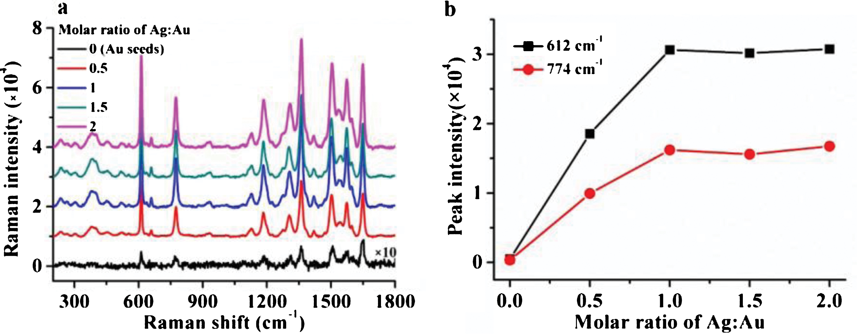
(a) SERS spectra of R6G adsorbed on AuNS and Au@Ags prepared with different MAu/MAg. (b) Changes in the peak intensities at 612 and 774 cm–1 as a function of MAg/MAu.
In this work, SERS activity of AuNS was improved by covering its surface with a silver shell. A chemical reduction process was developed to deposit silver shell on the surface of AuNS, in which ascorbic acid was employed as the reducing agent and silver nitrate was used as deposition source. Thickness of silver shell can be adjusted by tuning the ratio of MAg/MAu. The higher value of MAg/MAu, the thicker shell can be formed on the surface of AuNS. SERS activity of AuNS was promoted in the presence of silver shell, and the thicker of the shell, the higher SERS activity can be obtained. The optimum shell thickness was roughly proved to be 2.6 nm.
Conflicts of interest
The authors declare that they have no conflicts of interest.
Footnotes
Acknowledgments
This work was supported by the Natural Science Foundation of Jilin Province Department of Education (No. JJKH20190857KJ, JJKH20170241KJ).