
Abstract
This study was designed to develop and validate a simple and efficient high performance liquid chromatography (HPLC) method to determine flunixin concentrations in Asian elephant’s (Elephas maximus) plasma. Flunixin was administered orally at a dose of 0.8 mg/kg, and blood samples were collected. Flunixin extraction was performed by adding an equal amount of acetonitrile to plasma and centrifuging at 4500 rpm for 25 minutes. The supernatant was removed, and flunixin was analyzed using HPLC-UV detection.
Two methods were developed and tested utilizing two different mobile phases either with or without adding methanol (ACN: H2O vs. ACN: H2O: MeOH). Both methods showed excellent linearity and reproducibility. The limit of detection was 0.05 ug/ml and limit of quantification was 0.1 ug/ml. the efficiency of flunixin recovery was maximized by the addition of methanol to mobile phase (ACN: H2O: MeOH as 50:30:20) at 95% in comparison to 23% without methanol.
In conclusion, adding methanol to HPLC methods for extraction of flunixin from elephants’ plasma yielded higher recovery rate than without methanol.
Introduction
Flunixin meglumine (Banamine ®) is a nicotinic acid derivative (2 - methyl – 3 – trifluoromethyl – phenyl - amino – nicotinic acid) (Fig. 1) in the form of meglumine (n-methyl glucamine) salt with a pKa = 5.82 (weak acid) [1]. Flunixin is a potent non-steroidal anti-inflammatory agent (NSAID) that inhibits cyclooxygenase enzymes (COX). Flunixin is an approved as antipyretic and analgesic agent for horses. It also may reduce blood dynamic effects caused by E. coli in horses [2–5].
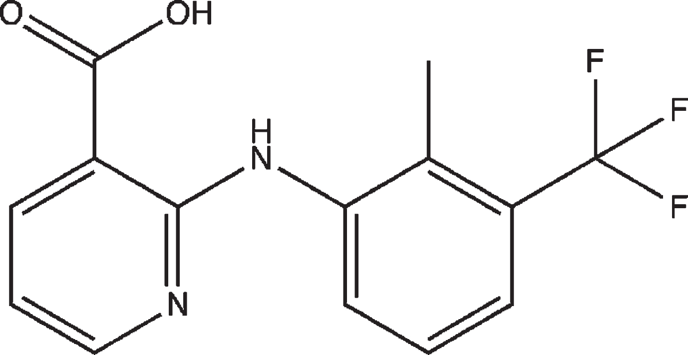
Chemical structure of flunixin.
Foot pathology is a real problem in captive elephants. Osteoarthritis is the major concern among musculoskeletal disorders and foot health problems for elephants in North America zoos with one-half of Elephants in captivity showing foot health care problems in zoos. A higher ratio of captive Asian elephants (Elephas maximus) is affected by osteoarthritis than African Elephants according to Association of Zoos and Aquarium (AZA) [5–7]. The incidence of arthritis increases as the age of the elephant’s increases that often requires therapeutic drug monitoring. Flunixin is the one of most commonly prescribed NSAID agent in elephants [8–10]. The available published pharmacokinetic studies (trimithoprime-sulfamethoxazole, metronidazole, succinylcholine, and ampicillin) involve small numbers of elephants due to geographical dispersion of captive elephants populations in North America [11–15].
Flunixin analytical assays in various veterinary animals like cows, horses, rabbits, camels, and llamas have been performed [16–22]. The analytical techniques used to determine flunixin drug concentrations in these animals included techniques such as High Performance Liquid Chromatography (HPLC), Thin Layer Chromatography (TLC), and Gas Chromatography-Mass Spectroscopy (GC-MS) [16, 23]. A HPLC technique was previously used to detect flunixin in cows and horses since the 80 s until to-date [18, 19]. GC-MS, on the other hand, was used to evaluate flunixin in horses and camels plasma [17–21].
However, HPLC is the most common and convenient analytical technique that determines flunixin in animal’s plasma. Reliable flunixin plasma concentrations are achieved by developing an optimized HPLC assay. For example, flunixin is a weakly acidic drug and favorably analyzed using acidic pH (3.1– 3.5) of the mobile phase to attain the best chromatographic resolution [16]. Although extraction by evaporation was the most commonly used technique to extract flunixin, it provided moderate flunixin recovery from an animal’s plasma [16, 23]. Flunixin Ultra-Violet (UV) absorbance was found to be the highest between 274– 290 nm wavelengths [24].
Elephant’s plasma provides problems that other animals’ plasma do not. In previous studies in our laboratory with Elephant plasma, it was often observed to congeal at room or refrigerated temperature making the analysis of the drug extremely difficult to perform by chromatography [25, 26]. Extraction of the drug, (solid or liquid) from the congealed plasma was also difficult as not all substances can be fully removed. Analysts with the latest chromatographic equipment are reluctant to measure drugs from Elephant plasma due to this problem.
The main aim of this study is to develop and validate a rapid and efficient bioanalytical assay using high performance liquid chromatography (HPLC) in order to detect and determine flunixin concentrations in Asian elephant plasma according to U.S. Food and Drug Administration (FDA) recommendations [27].
Animals
Captive Asian elephants that successfully completing trials were maintained in facilities located in the USA and Canada. This study was approved by the Animal Care and Use Committee at Auburn University (PRN 2015/2609) as well as Animal Care and Use Committees at each participating facility. Routine hemograms and serum chemistry panels were conducted on elephants prior to and immediately following each dose trial. Flunixin meglumine (flunixin) paste (Banamine® Paste; Merck Animal Health, Madison, New Jersey 07940, USA) was given orally with food treats (e.g., peanut butter sandwiches, Gatorade), and blood samples were collected from ear veins using 21-gauge butterfly catheters.
Reagents and Chemicals
All reagents and solvents were HPLC grade. Pure Flunixin meglumine powder was purchased from Spectrum (CA, USA) and Diclofenac sodium salt (internal standard) was obtained from Sigma-Aldrich (Germany). Methanol and acetonitrile were purchased from Fisher Scientific (NJ, USA). Simple acetic acid, which was used to adjust the pH of the mobile phase, was obtained from VWR international (PA, USA).
Instrumentation
Chromatographic analysis was conducted using a Shimadzu Prominence High Performance Liquid Chromatographic system (Kyoto, Japan model LC-2010A HT) equipped with UV Detector. Sample centrifugation was carried out using Eppendorf centrifuge 5415 C (Brinkmann Instruments, NY, USA). Serum samples were filtered via 0.45 um nylon filter (VWR International, USA) before injection into HPLC.
Method development and designs
Two chromatographic methods were evaluated. One was with and the other without the addition of methanol to the mobile phase. The first method consisted of acetonitrile and water at a ratio of 50:50. The second method was with a mobile phase consisting of acetonitrile, water, and methanol in a ratio of 50:30:20. The pH of both studied mobile phases was adjusted to 3.1, and the UV absorbance of flunixin was measured at 278 and 284 nm.
Chromatographic conditions
HPLC analysis of flunixin was performed with a 4.6×150 mm column (Kinetex, Phenomenex, CA, USA) packed with 5-um C-18 chromatographic media. Also, it is connected to a pre-column (Security guard 2.1 – 4.6 mm, Kinetex, Phenomenex, CA, USA) and samples flow through an in-line filter (KrudKatcher ULTRA HPLC, Kinetex, Phenomenex, CA, USA) to further insure removal of impurities. Two mobile phases were studied. The mobile phase of the first method consisted of acetonitrile, water (pH 3.1, pre-adjusted with acetic acid) in the ratio of 50:50 (method I). Methanol was added to the mobile phase of the second method, which consisted of acetonitrile, water (pH 3.1 pre-adjusted with acetic acid), and methanol in the ratio of 50:30:20 (method II). The mobile phases were sonicated for 60 min before use. The flow rate was 0.8 ml/min for the method I, and 0.6 ml/min for method II.
The UV-Detector wavelength was set at 278, and 284 nm and the column temperature was set at 25°C. The injection volume was 20 ul for the method I, and 15 ul for method II respectively. The run time was 12 minutes for the method I, and 8 minutes for method II.
Standard solutions and calibration curves preparation
A stock solution of Flunixin (1 mg/ml) was prepared in methanol. Standard solutions of flunixin in elephant serum were prepared by adding appropriate volumes of the flunixin (1 mg/ml) stock solution to drug-free elephant serum to produce concentrations of 0.1, 0.5, 1, 5, 10, 20, 40 ug/ml. Each concentration was injected into the HPLC in triplicate utilizing both methods’ conditions for flunixin analysis. Three quality control levels were also included at low, middle and high concentrations (1, 5, and 20 ug/ml) and three replicates for each level were also assayed.
For calibration curve and quality control analyses, an appropriate amount of elephant’s plasma was spiked with the appropriate flunixin stock solution volume in order to achieve the calibration curve range (0.1– 40 ug/ml). The average ratios of peak area under the absorbance curve (AUC) of flunixin and diclofenac Na+ (Internal standard) from assayed elephant serum were measured and plotted against the corresponding concentration of flunixin in ug/ml to prepare calibration curves for flunixin plasma concentration determination. The slope, intercept, and correlation coefficient (R2) were determined by linear regression analysis for each method. Linearity was checked with F-test lack-of-fit analysis.
Sample preparation
Blood samples were collected from an Asian Elephant via an ear vein. For the HPLC analysis of flunixin concentrations in the collected elephant plasma samples, 0.5 ml of elephant plasma was pipetted into 2 ml Eppendorf centrifuge tube. For all series of samples (calibrators, quality control levels, and test samples), an equal amount of acetonitrile, and 200 ul of internal standard solution (100ug/ml Diclofenac sodium) were added. Then, the contents of the tube were mixed for 1 min on a vortex mixer and then allowed to sit for 15 min to denature. After that, the mixture was centrifuged at 4500 rpm×25 min.
The supernatant was filtered and transferred to a new HPLC glass tube. The previously determined volume for each method was injected into the HPLC.
Statistical analysis
Mean, standard deviation (SD) and Analysis on variance (ANOVA) tests were used to analyze the data.
Results
Appropriateness of chromatographic conditions
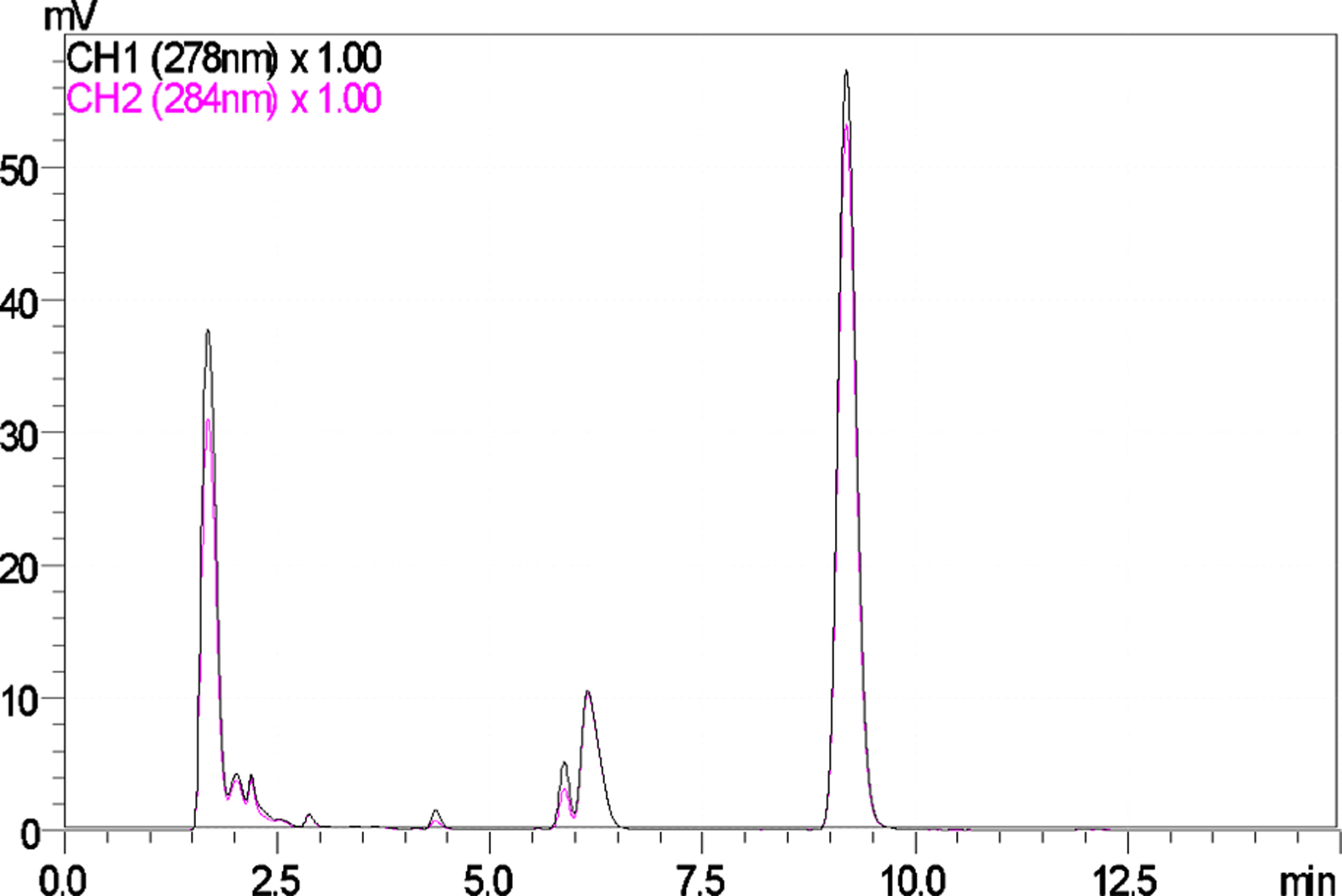
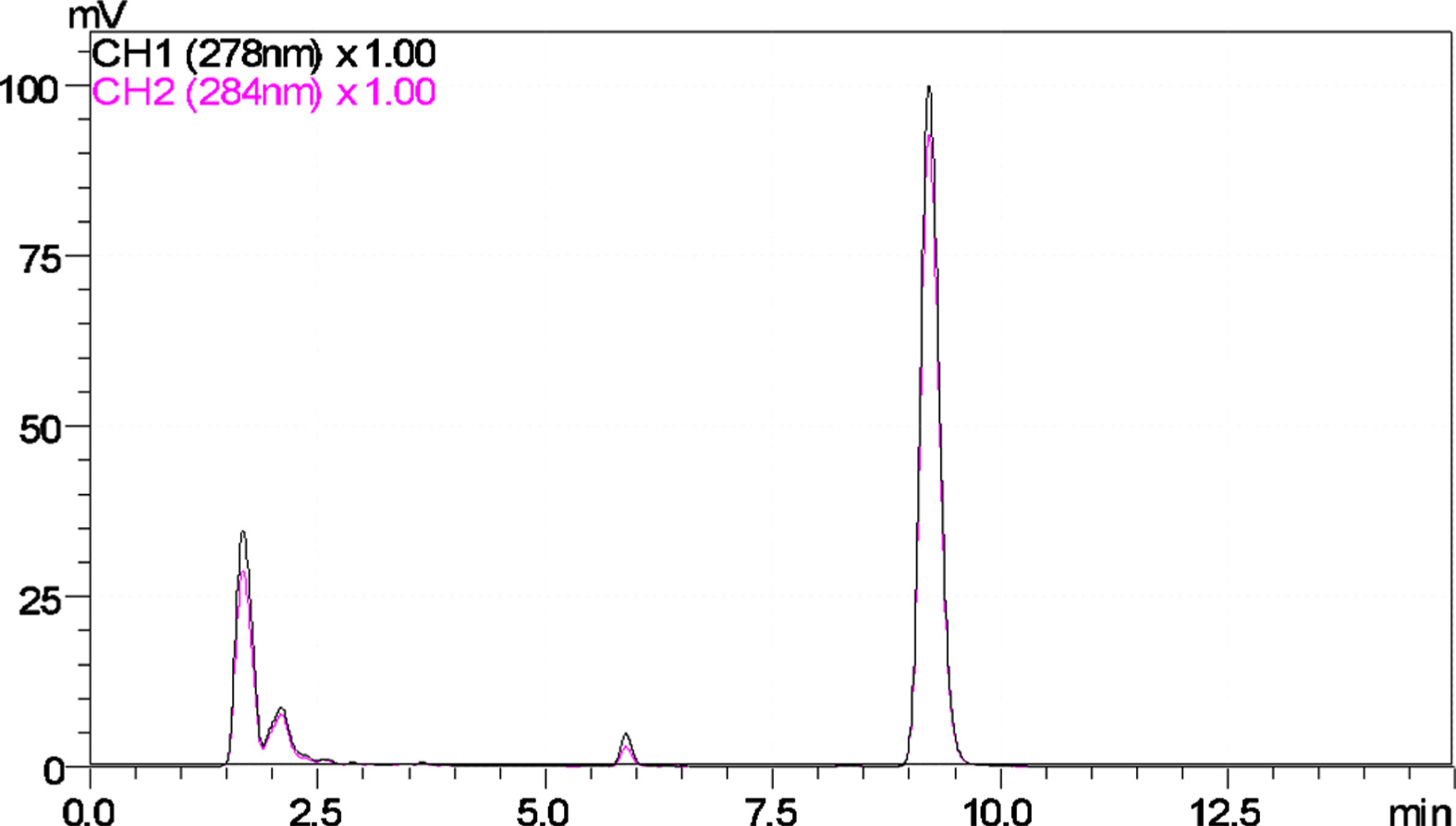
The addition of methanol to the method II (ACN: H2O: MeOH) gave better separation between flunixin and internal standard than the method I (ACN: H2O). Retention times of flunixin and diclofenac were about 6.2 and 9 minutes with mobile phase in the method I, and 6.5 and 7.5 minutes with mobile phase in method II (Fig. 2, 3). Flunixin optimal UV absorbance was at 278 nm. Figures 4 and 5 report the chromatogram of elephant serum without flunixin but with diclofenac. Complete separation was observed between plasma components, flunixin, and diclofenac for method II. A small shoulder, apparently from a substance in plasma, overlapped with flunixin in method I (at 5.8 min).
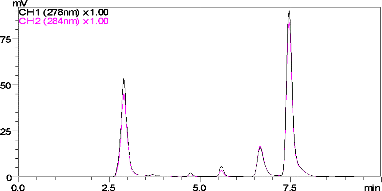
Method II (50:20:30) elephant plasma spiked with 10 ug/ml Flunixin and internal standard (Diclofenac Na+) 15uL injection volume.
Method I (50:50) elephant plasma spiked with 10 ug/ml Flunixin and internal standard (Diclofenac Na+) 15uL injection volume.
Method I (50:50) Blank elephant plasma with internal standard (Diclofenac Na+) 15uL injection volume.
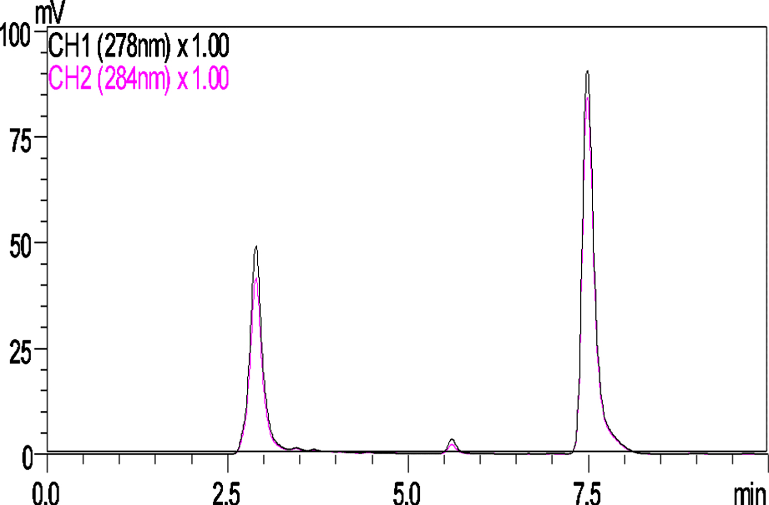
Method II (20:30:50) Blank elephant plasma with internal standard (Diclofenac Na+) 15uL injection volume.
The validity of the HPLC assay method was performed according to FDA recommendations for assessment. The validity of proposed HPLC assay methods included the assay’s linearity, LOD, LOQ, accuracy, precision, and selectivity.
Linearity
For the previous two described HPLC methods, an excellent linear relationship was found. Plotting the average chromatograms ratio of peak areas (AUC) of flunixin to the internal standard’s AUC versus concentration range (0.1 – 40 ug/ml) a straight line was observed (Fig. 6, 7).
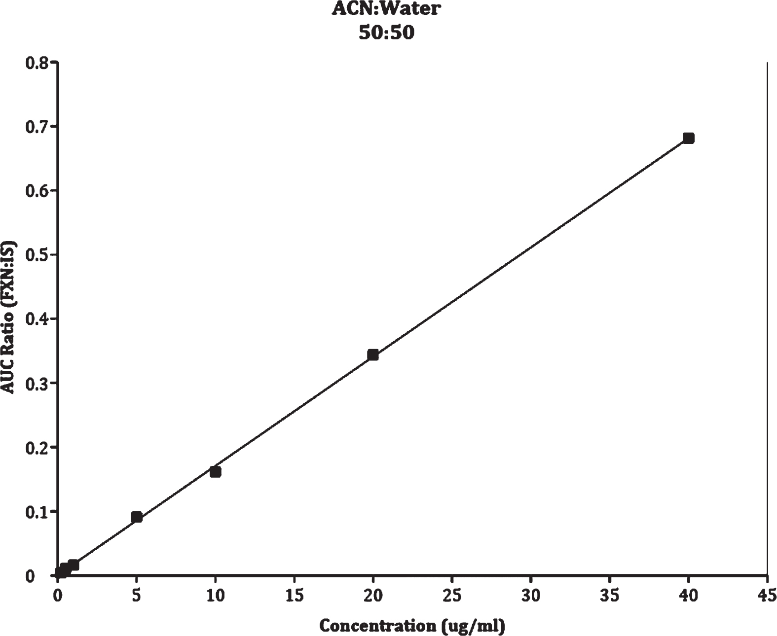
Calibration curve of Method I ACN: Water at ratio of 50 : 50.
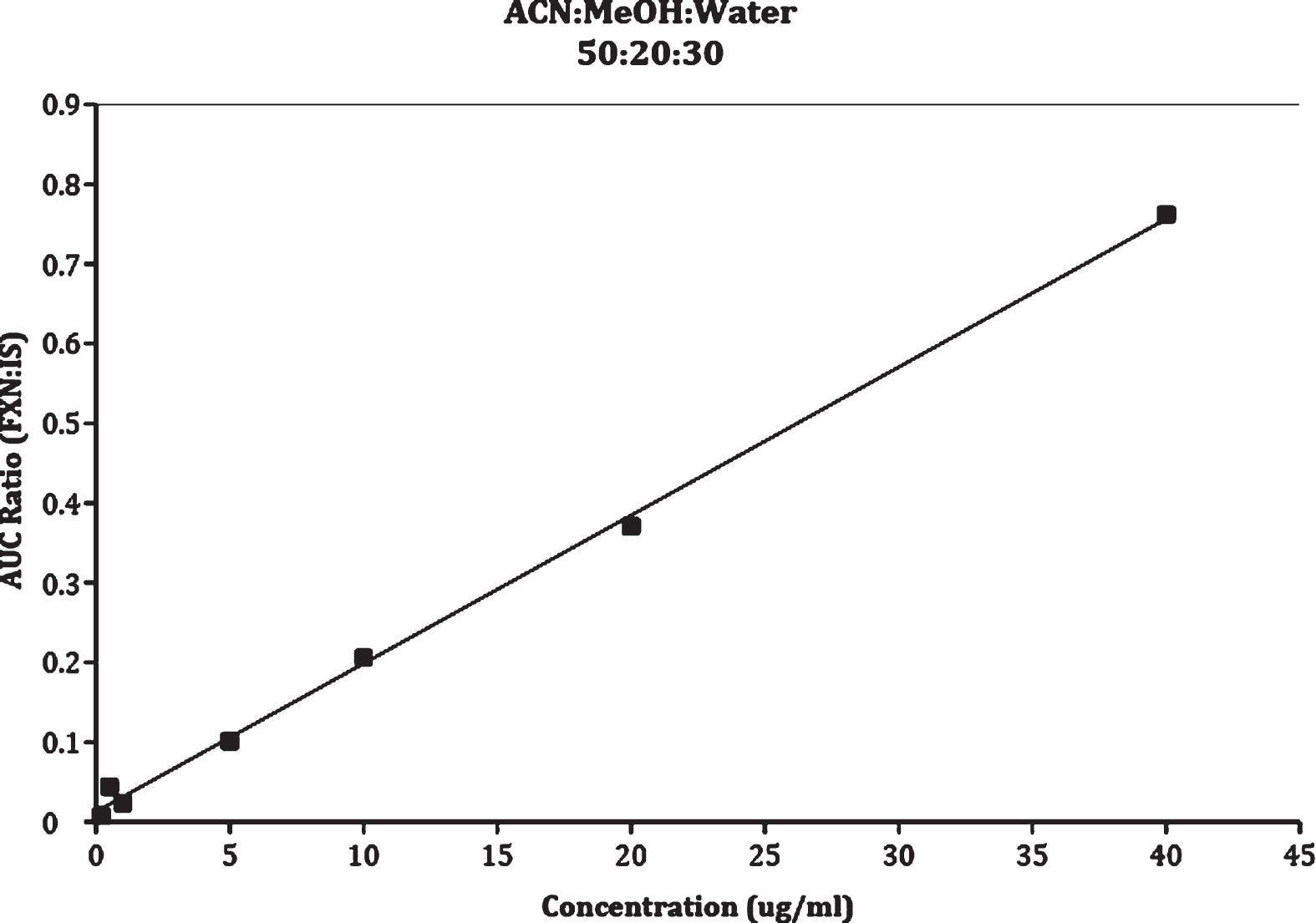
Calibration curve of Method II ACN: Methanol: Water at ratio of 50 : 20 : 30.
The concentration range contains 7 points for both methods. Linear regression on the calibration curves were performed, and one-way ANOVA test was used to calculate the slope and intercept for method I (Y = 0.017 X + 0.0007, and R2 = 0.9996), and for method II (Y = 0.0186 X + 0.0128, and R2 = 0.997); Where Y is absorption peak area ratio (flunixin AUC: Internal standard AUC), C is the corresponding concentration (ug/mL), and R2 is correlation coefficient (Fig. 7). The high R-square value (R2) indicates the goodness of fit of the calibration curves’ linearity for both methods.
The lower limit that can be reliably detected by both HPLC methods’ conditions was 0.05 ug/mL (LOD). The limit of quantification below that the flunixin concentration cannot be measured is 0.1 ug/ml. The calibration curve shows a non-linear pattern below 0.1 ug/mL (LOQ).
Accuracy
Performing three replicate analyses at three concentration levels (quality controls) 1, 5, 20 ug/ml on three consecutive days were done to assess intra-day and inter-day recovery of flunixin from the proposed two methods. Method I, showed very low recovery rate (inter-day, and intra-day) of flunixin from elephant’s plasma compared to method II. The flunixin inter-day recovery percentage with the addition of methanol (method II) was between 94.5 – 96.4% in comparison to 22.8 – 26.94% from method I (without methanol). The intra-day recovery rate of flunixin from the method I (without methanol) was between 23.2 – 31.4 % in comparison to 93.4 – 94.7 % with method II (with an addition of 20% methanol) Tables 1, 2.
Method I Accuracy and Precision
Method I Accuracy and Precision
Method II Accuracy and Precision
The coefficient of variation (% CV) was calculated for the averages of peak areas ratio (FXN: IS) for three flunixin elephant plasma concentrations 1, 5, 20 ug/mL. The intra-day % CV was between 0.7 – 1.3 for both methods, and the inter-day % CV was between 0.16 – 3.7 for both methods (Table 1, 2).
Selectivity
The selectivity of the proposed HPLC method was established by measuring its ability to detect and determine pure flunixin by HPLC-UV absorbance. To ascertain the specificity of each method’s conditions for the drug, a blank plasma was tested under the same chromatographic conditions that were used for the tested plasma samples.
There were no interfering peaks at flunixin retention’s time using method II while with method I, an unknown substance from the plasma produced a peak that was interfering with the flunixin peak from tested plasma samples. The internal standard (Diclofenac sodium) was separated completely from flunixin peak and plasma peaks in the chromatogram for both methods (Figs. 2– 5).
Robustness
Flunixin determination is affected by extreme changes in pH and temperature during HPLC analysis. Column temperatures above 30° C gave inconsistent peak behavior and readings. Also, a pH that lies between two points higher or lower from flunixin meglumine pka = 5.82 results in significantly different peak areas. Under the chromatographic conditions in this study, both methods were robust enough to any small change in either pH and column temperature which was set as a constant at room temperature (∼25° C).
Application
Method II was applied successfully for assaying flunixin in serum from one Asian elephant (Elephas maximus). An oral dose (0.8 mg/kg body weight) of flunixin paste was administered to the animal. Blood samples were collected at 15 different time points after dose administration. Plasma samples were assayed by Method II and the concentration versus time curve of flunixin was plotted on a semi-logarithm graph (Fig. 8), and pharmacokinetic analysis was performed (Table 3).
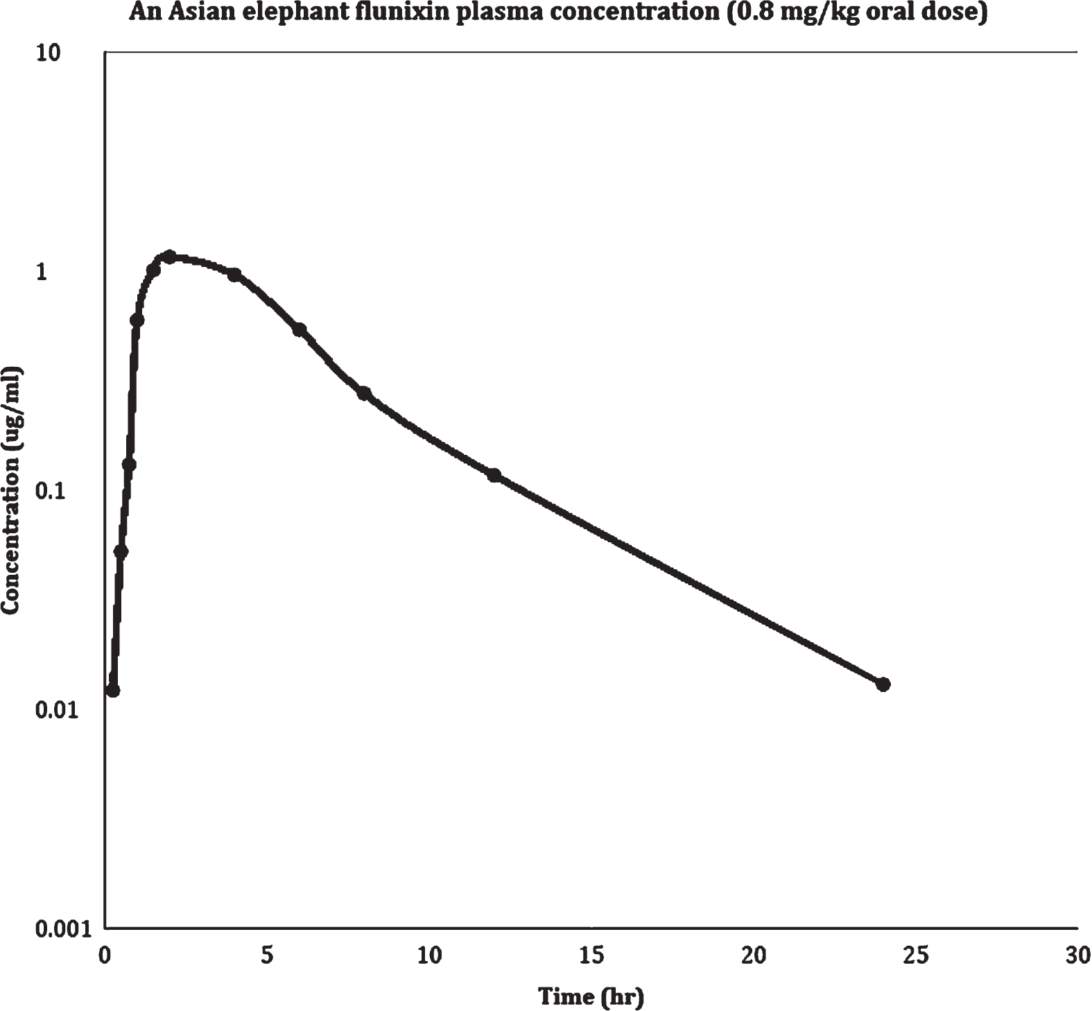
Flunixin plasma concentration versus Time curve in Asian elephant after 0.8 mg/kg oral dose.
Pharmacokinetic parameters for an Asian elephant using the proposed HPLC-UV method (Method II) after administration of flunixin as 0.8 mg/kg (of body weight) orally
This study was able to assay and determine flunixin in Asian elephant plasma after oral administration (0.8 mg/kg). Flunixin was detected by utilizing HPLC with UV detection using two different mobile phases. The recovery rate of flunixin from elephant serum was significantly improved when methanol was added to the mobile phase in method II (95% vs. 23%). Methanol also provided full separation between flunixin and unknown elephant plasma peaks (Fig. 2, 3). Unlike other studies, the addition of methanol to mobile phase provided higher flunixin recovery rate and better flunixin peak’s resolution than phosphate, acetate, and ACN alone [16, 19]. Many studies suggest different types of buffers be used in the mobile phase. In this study, deionized nano-particle free HPLC grade water was used to create the buffer utilizing glacial acetic acid to adjust the pH to 3.1. Using purified water in the mobile phase (with pH adjusted at 3.1 with acetic acid) was an excellent choice without increasing extra cost to the analyses and without introducing possible error of utilizing a buffer formation or error in preparation.
Elephant’s plasma may thicken and become almost milky-gel-like in texture at room temperature. This property makes it very hard for HPLC analysis of drug in plasma by techniques like LC-MS or GC-MS. The extraction of drugs from elephant’s plasma is tedious and time-consuming. Two pharmacokinetic studies were performed on NSAIDs in elephants. Ibuprofen and phenylbutazone were determined by HPLC methodology [25, 26]. In both studies, elephant’s plasma was treated with acetic acid and double extracted by organic solvents.
In this study, using an equal volume of acetonitrile added to elephant plasma followed by centrifugation provided high flunixin extraction from the elephant’s plasma for HPLC analysis. Treatment of plasma samples containing flunixin by strong acids before extraction resulted in extreme changes in flunixin HPLC analysis [19]. Therefore, the extraction procedure was performed with acetonitrile alone.
The developed Method II is a simple, inexpensive and efficient process to detect and determine flunixin from elephant’s plasma. It has a high extraction recovery rate, and the limit of detection was as low as 0.05 ug/ml. Besides, the method showed excellent linearity and precision. The injection volume in this study was 15 ul that is much less than previous NSAIDs studies on elephants [25, 26]. The very small injection volume allows for possible repetitions to assay the elephant’s plasma. With this study, flow rate of 0.6 ml/min was optimal to provide a reasonable run-time and increase the column’s shelf life by producing lower pressure.
Conclusion
The proposed HPLC method (Method II) is reliable and provides a useful process for detecting flunixin from Asian elephant’s plasma. It has the advantage of high sensitivity, efficacy and time-saving efforts. Validated Method II (with addition of methanol) provided a higher rate of Flunixin recovery than Method I (without methanol). Therefore, Method II is more favorable for use in future flunixin analyses in elephants.