New benzoselenadiazole based BODIPY probe was synthesized and characterized with spectroscopic techniques like 1H, 13C, 77Se and mass spectrometry. The fluorescence of the probe quenched in the presence of superoxide ion, showing “Turn-off” response without interfering with other ROS even at higher concentrations. The detection limit was found to be 21.28μM. Kinetic study reveal the fast reaction between the probe and superoxide with good photostability. Also, the probe was studied for biological activity and found to be good agent for anti-inflammatory and antioxidant activities.
Organoselenium compounds possess trustworthy detection for biologically active species such as reactive oxygen species (ROS), reactive nitrogen species (RNS), biothiols, metal ions and so forth [1–5]. The excess generation of ROS (O2·–, H2O2, tBuOOH, –OCl, ·OH and tBuO·) in human body is linked with various kinds of disorders for instance Alzheimer’s, Parkinson’s, and cancers diseases [6–9]. One of the ROS, superoxide (O2·–), has very short half-life; hence its real time recognition is essential. Fluorescence microscopy is used to detect certain analytes demands new molecular design and synthesis which make this field more challenging [10]. Fluorescence technique is significant for detecting biologically important species because of its high sensitivity, fast response, ease of execution and high capability for real time detection [11].
Cancer chemo-preventive activities have received greater attention in recent years. Many naturally occurring as well as synthetic compounds have been reported for cancer chemoprevention [12–15]. The role of selenium as a cancer chemo-preventive agent was supported by many preclinical and clinical studies [16–18]. The supplement of selenium was observed to be beneficial for the treatment of various types of cancers [19–23]. Numerous effective organoselenium compounds have been developed to achieve superior chemo-preventive potency and selectively reduced side effects by structural modification. So, the applications of organoselenium compounds in cancer chemotherapy is an interesting field for research [24–27].
Selenium containing heterocyclic compounds has received more attention due to their pharmacological properties [28]. Ebselen and their analogue containing Se–N bond own in-vivo anti-inflammatory and in-vitro glutathione peroxidase-like (GPx) activities [29–31]. It catalytically reduces harmful hydrogen peroxide into less toxic compounds [32]. Also, it prevents oxidative damage of lipid membrane and other cellular components [33–35]. The pyridine fused selenium containing compounds possesses antibacterial and anti-inflammatory property also, the selenamides exhibit GPx mimic activity [36–42]. Apart from ebselen, various arrangements over Se-N bond reported in recent years include selenodiazole, selenocyanates, isoselenocyanates and charged selenazolinium salts [43].
In comparison with ebselen, selenadiazoles are more active due to presence of second nitrogen atom in the structure rather than positive charge. This may lower electron density and can encourage electrophilic reactivity which required for biological processes [21]. The first series of 4-Methyl-1,2,3-selenadiazole-5-carboxylic acid amides was synthesized for their in-vitro and in-vivo antitumor property against tumour cell lines [5]. 1,2,5-Selenadiazolo[3,4-d, 3,4-d]pyrimidine-5,7(4H,6H)-dione was appeared to be cytotoxic against various human cancer cells [44].
This research article describes the synthesis and selectivity of benzoselenadiazole based BODIPY probe for the detection of superoxide ion by photophysical experiments and some important pharmacological properties.
Experimental section
Materials
All the reactive oxygen species (ROS) were purchased from Sigma Aldrich. All the chemicals and solvents used for experiments were of analytical grade and used without further purifications. Fetal bovine serum (FBS), Trypsin-EDTA and Phosphate buffered saline (PBS) were obtained from Thermo scientific (Gibco). Four pathogens viz. two gram-negative bacteria [Escherichia coli (ATCC 25922) and Salmonella typhi (ATCC 23564)] and two-gram positive bacteria [Staphylococcus aureus (ATCC 25923) and Bacillus subtilis (ATCC 6633)] were purchased from National Chemical Technology, Pune, India. Double distilled water was used for the entire analysis. Silica gel (60–120) was used to perform Column chromatography. The 1H and 13C NMR spectra of all the synthesized compounds were recorded in deuterated chloroform (CDCl3) as a solvent and Tetramethylsilane (TMS) as internal standard. The NMR spectra were recorded using Bruker avance II (300 MHz) NMR spectrometer. The 77Se NMR spectra were acquired on JEOL JAPAN, ECZR Series 600 MHz spectrometer in deuterated dimethyl sulfoxide (DMSO-d6) using diphenyl diselenide as an external standard. Mass spectra of compounds were recorded using AB SCIEX 3200 Q TRAP LC/MS/MS spectrometer. UV-vis spectra were recorded on Shimadzu UV2450 PC recording spectrophotometer. The fluorescent spectra were obtained from Shimadzu RF 5301PC. 5-methyl-2,1,3-benzoselenadiazole (2) and 2,1,3-benzoselenadiazol-5-carbaldehyde (3) were synthesized by previously reported procedure [45].
Synthesis of compounds
Synthesis of compound 4
2,1,3-Benzoselenadiazol-5-carbaldehyde (3) (1.00 g, 4.73 mmol) and pyrrole (3.17 g, 47.34 mmol) were stirred in round bottom flask at room temperature under inert atmosphere. A catalytic amount of Trifluoroacetic acid (TFA) was added to the reaction mixture. The reaction was continuously stirred for overnight. Progress of the reaction was monitored by thin layer chromatography (TLC). After completion of the reaction, excess of pyrrole was removed under reduced pressure. Further, the crude product was purified by column chromatography using dichloromethane (DCM) (Yield: 32%). Spectroscopic data:1;H NMR (300 MHz, CDCl3): δ (ppm) 10.09 (bs, 2H), 7.67–7.64 (m, 1H), 7.44–7.40 (m, 2H), 6.70 (s, 2H), 5.88 (s, 2H), 5.55 (s, 1H). 13C NMR (100 MHz, CDCl3): δ (ppm) 160.7, 159.7, 145.1, 131.8, 131.3, 122.3, 120.9, 117.6, 107.7, 107.1, 43.6. MS (ESI): Calculated for C15H12N4Se: 328.02, found m/z 327.50 (M-H)+.
Synthesis of compound 5
Compound 4 (0.50 g, 1.50 mmol) was dissolved in 10 mL of dry tetrahydrofuran (THF) at room temperature. 2,3-Dichloro-5,6-dicyano-1,4-benzoquinone (DDQ) (0.35 g, 1.50 mmol) was added to the reaction mixture and the reaction was stirred for 1 hour. The consumption of dipyrromethane was monitored by TLC. Further triethylamine (1.40 mL, 15.20 mmol) and boron trifluoride etherate (BF3.OEt2) (1.90 mL, 15.20 mmol) were added to the reaction mixture. The reaction was continued to stir for another 3 hours. The crude product was purified by column chromatography (Ethyl acetate:Pet ether, 50 : 50) to yield compound 5 in 26%. Spectroscopic data:1H NMR (300 MHz, CDCl3): δ (ppm) 8.10 (s, 1H), 8.01 (m, 3H), 7.69–7.66 (d, 1H), 7.02–7.01 (d, 2H), 6.60 (bs, 2H). 13C NMR (100 MHz, CDCl3): δ (ppm) 160.4, 159.5, 145.1, 144.8, 134.9, 134.6, 131.3, 131.0, 125.4, 123.4, 119.1. 77Se NMR (114.46 MHz, CDCl3): δ (ppm) 1555; MS (ESI): Calculated. For C15H9BF2N4Se: 374.01, found m/z 374.30 (M)+.
Selectivity of probe 5
Due to poor solubility of probe 5 in aqueous solvent, the probe was dissolved in DMSO solvent to form 5μM stock solution. Different ROS (O2·–, H2O2, tBuOOH, –OCl, ·OH and tBuO·) were generated as per documented in the literature [46–49]. Hydroxyl radical and t-butoxide radical were produced by Fenton reaction. The stock solutions of ROS (0.1 M) were prepared in distilled water. After addition of ROS to probe solution (5), all solutions were incubated for 5 min. The UV-vis and fluorescence spectra have been recorded in DMSO at room temperature. The excitation wavelength was taken as 508 nm and slit width was 5.0/5.0 nm for all spectral measurements. For screening experiment, the probe solution (5) (3.0 mL, 5μM) and ROS (10μL, 66 equiv) were taken.
Sensitivity and Limit of detection
In order to determine LOD of the probe (5), increasing concentration of superoxide from 0 to 10μL (0 to 66 equiv) were added to the probe (5) (3.0 mL, 5μM) and emission spectra were recorded. LOD was find out according to the literature method (Eqs. 1 and 2) [50]. The fluorescence emission spectra of the probe (5) were recorded three times and standard deviation was calculated. To calculate the slope, the emission intensity at 547 nm plotted against concentration of superoxide.
For Limit of Detection
The calculation and result from the analysis is given below:
Where, σ is the standard deviation of blank measurement, K is the slop between the fluorescence versus KO2 concentration [50].
Time dependent experiment
The response time experiment was carried out with 5μM probe solution (5) and 10μL (333μM, 66 equiv) superoxide. The emission spectrum was recorded for 1 hour. The excitation wavelength used for experiment was 508 nm and slit width was 5.0/5.0 nm.
Interference study
Superoxide (10μL, 66 equiv) was added to 3 mL of the probe solution (5) (5μM) followed by addition of other ROS (10μL, 66 equiv) takes place in every vial. The emission spectra were recorded at room temperature after 5 min incubation.
Antibacterial investigations
Staphylococcus aureus (ATCC 25923), Bacillus subtilis (ATCC 6633), Salmonella typhi (ATCC 23564) and Escherichia coli (ATCC 25922) were used for antibacterial analysis. These bacterial cultures were procured from NCT (National chemical technology), Pune, Maharashtra. These bacterial cultures were maintained at 4°C on Nutrient agar slants and used for analysis after 16 hours.
DPPH scavenging activity
To determine the radical scavenging activity of probe (5), 2,2-diphenyl-1-picrylhydrazyl (DPPH) (0.80 mL, 0.1 mM in ethanol) was added to the different concentrations of the probe solution (15, 30, 60, 120, and 240μM). The resulting mixtures were incubated for 30 min at 37°C. The absorbance of test solutions was measured at 517 nm. The ascorbic acid was used as standard. The value of IC50 was calculated by plotting a graph of percentage scavenging activity vs. concentration (Equation 3).
The formula for DPPH radical scavenging ability is given below:
(ii) For Cell Cytotoxicity
Where, Ac = Absorbance of control
As = Absorbance of the probe
In-vitro anti-inflammatory activity: Protein denaturation inhibition bioassay
All solutions were prepared in dimethyl sulphoxide (DMSO) and 0.2 M phosphate buffer solution (PBS, pH 6.3). All the solutions of concentration (15, 30, 60, 120 and 240μM/mL) were incubated at 37°C for 20 min. The test solution was heated up to 57°C for 3 min for protein denaturation. After cooling, the absorbance was measured at 660 nm.
Results and Discussions
Synthesis
The 5-methyl-2,1,3-benzoselenadiazole (2), 2,1,3-benzoselenadiazol-5-carbaldehyde (3) were prepared by reported procedures [45]. The purified products were confirmed by 1H NMR spectroscopy before use (Fig. S1, S2). Compound 3 was converted into BODIPY derivative 5 by conventional method reported in literature [51]. Compound 3 was reacted with excess of pyrrole in the presence of catalytic amount of TFA at room temperature. The reaction yielded dipyrromethane derivative 4 (32%).
Synthesis of benzoselenadiazol based BODIPY probe 5.
After characterization, corresponding dipyrromethane derivative 4 was oxidized with DDQ and then reacted with triethylamine followed by BF3 . OEt2 to afford BODIPY derivative 5 in the yield of 26% (Scheme 1). Both the compounds were characterized by common spectroscopic techniques (multinuclear NMR) and mass spectrometry (Fig. S3–S9).
In the 1H NMR spectrum of compound 4, a singlet was appeared at 5.55 ppm for Sp3 –CH. A broad singlet was observed at 10.09 ppm for two –NH protons of pyrroles (Fig. S3). In the 1H NMR spectrum of compound 5, the peak at 5.55 ppm was disappeared, which confirmed the oxidation of Sp3–CH to Sp2 –C= (Fig. S6). The mass spectrum of 4 exhibited molecular ion peak with selenium isotopic pattern at m/z Calculated for C15H12N4Se [M]+: 327.25; found 327.50 (Fig. S5). The 77Se NMR spectrum of compound 5 showed a signal at δ 1555 ppm and mass spectrum showed peak with selenium isotopic pattern at m/z calculated for C15H9BF2N4Se [M]+: 374.01, found m/z 374.30 (Fig. S8, S9). These characterization data confirmed the formation of selenium compounds 4 and 5.
Photophysical studies
UV-vis and fluorescence studies of compound 5
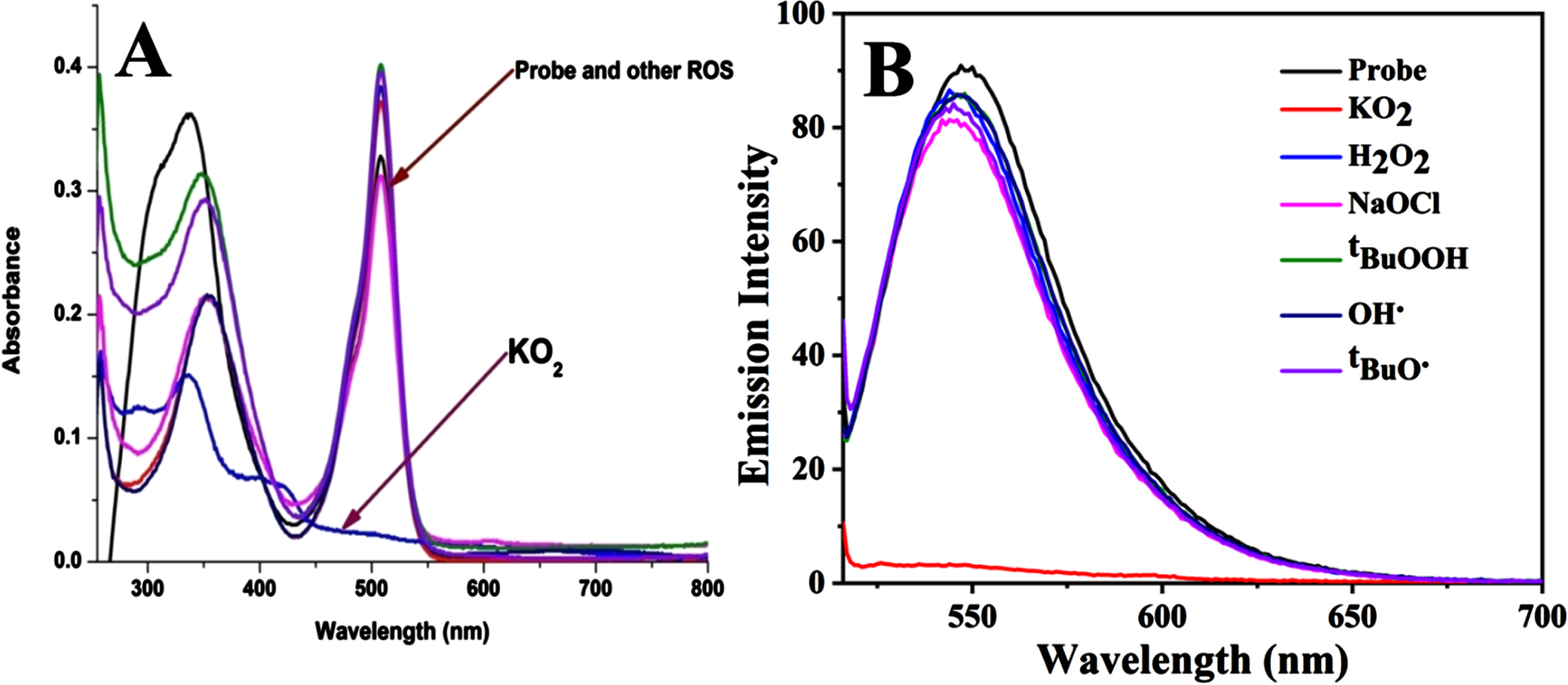
After thorough characterization, in order to understand the chemosensing behaviour of probe 5, the probe was screened with different ROS (O2·–, H2O2, tBuOOH, –OCl, ·OH and tBuO·) in DMSO solvent. The probe solution was light orange in colour and showed absorbance in the visible range of 450 to 550 nm. Upon addition of various ROS to the probe solution, it was observed that the probe with superoxide displayed absorbance band between 300 to 400 nm. However, no noticeable change was observed with other ROS in absorption band (Fig. 1). The significant colour changes of the probe solution from orange to colourless was observed. Probe 5 exhibited fluorescence emission band at 547 nm upon excitation at 508 nm. The addition of various ROS to the probe did not affect the emission intensity of the probe, however, in the presence of superoxide, the fluorescence emission intensity of the probe quenched effectively. It exhibited almost 29-fold decrease in fluorescence emission intensity (Fig. 1, S11).
A) UV-vis spectra of probe 5 (5μM) in DMSO with various ROS (O2·–, H2O2, tBuOOH, –OCl, ·OH and tBuO·; 330μM); B) Emission spectra of probe 5 (5μM) in DMSO with ROS (330μM), incubated for 5 min, at rt (λex = 508 nm andλem = 547 nm); Slit width 5/5 nm respectively.
To find out the sensitivity of probe 5 towards superoxide, the fluorescence titration experiment was performed with increasing concentration of superoxide at excitation wavelength of 508 nm (Fig. 2). The fluorescence emission intensity of the probe at 547 nm gradually decreased with successive addition of superoxide from 0 to 333μM. The probe exhibited a good linear relationship between the fluorescence emission intensity at 547 nm and concentration of superoxide. The detection limit of probe 5 for superoxide was calculated as 21.28μM (Fig. S12).
Emission spectra of probe 5 (5μM) in DMSO with increasing concentration of superoxide (0-333μM) incubated for 5 min, at rt (λex = 508 nm and λem = 547 nm); Slit width 5/5 nm respectively.
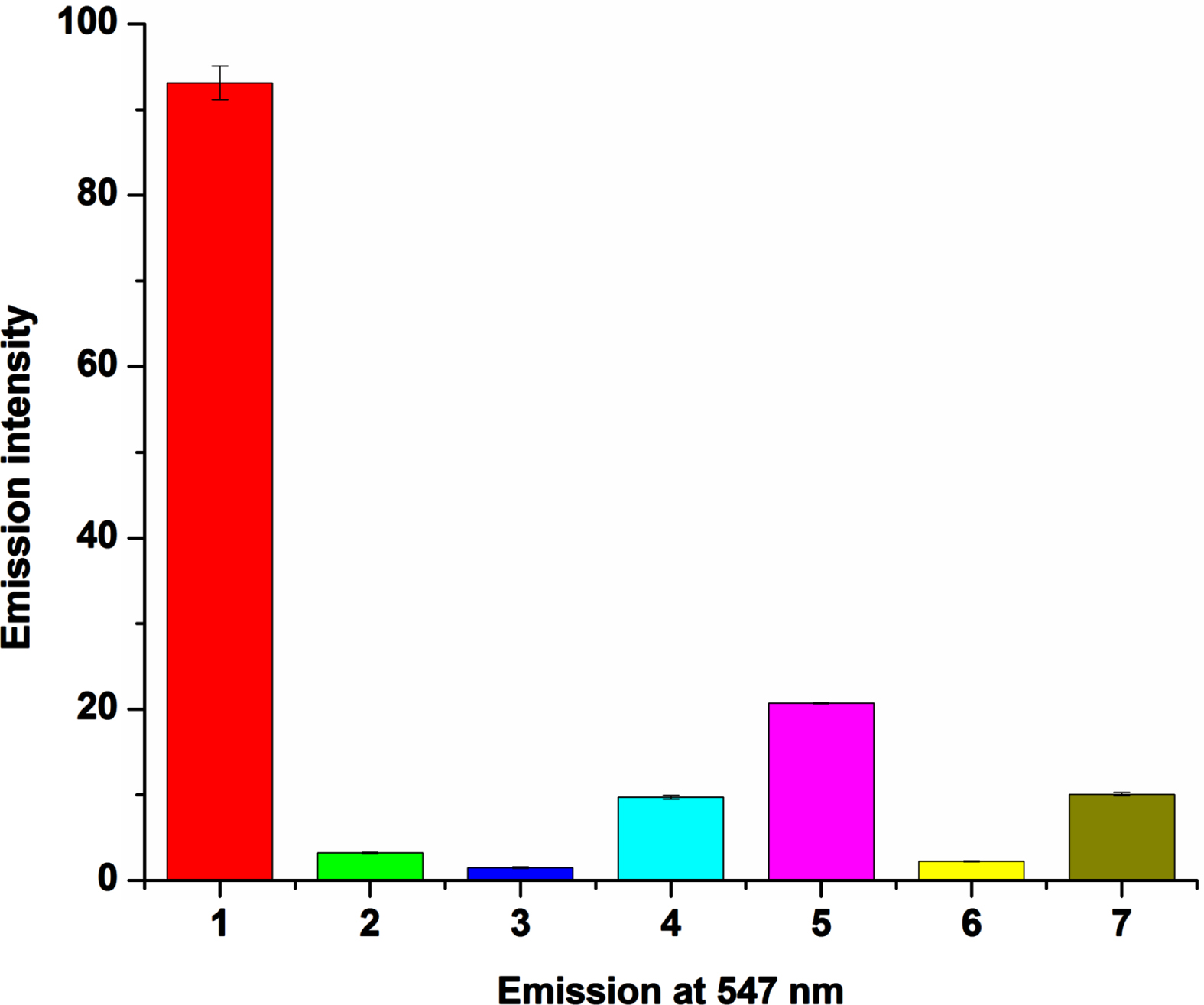
Further, to explore the sensitivity of probe 5 towards superoxide, competitive experiments were performed in DMSO solvent. The fluorescence spectra of probe 5 with superoxide in the presence of various ROS (H2O2, –OCl, tBuHO2, ·OH, tBuO·) were investigated. The change in fluorescence intensity of probe 5 caused by superoxide was unperturbed in the presence of other ROS. These experimental results of the probe with superoxide implied that the fluorescence quenching does not affect with other ROS (Fig. 3).
Fluorescence spectra of probe 5 (5μM) and superoxide with various ROS (330μM); incubated for 5 min. at rt (λex = 508 nm and λem = 547 nm); Slit width 5/5 nm respectively. (1 = probe, 2 = probe+O2·–, 3 = probe + O2·–+ H2O2, 4 = probe + O2·–+–OCl, 5 = probe + O2·–+tBuHO2, 6 = probe + O2·–+·OH, 7 = probe + O2·–+tBuO·).
Kinetic study experiment is very important for practical application of the probe. The reaction of probe 5 with superoxide was completed quickly and it could be observed that fluorescence intensity was quenched rapidly and reached a plateau within fraction of second. The fluorescence intensity of the probe (5) remained unchanged under continuous irradiation for 60 min, which signified that the probe was sufficiently stable for superoxide detection (Fig. 4).
Time dependent emission spectrum of probe 5 with superoxide (330μM) at rt, (λex = 508 nm and λem = 547 nm); Slit width 5/5 nm respectively.
Mechanism study
To investigate the mechanism involved in the detection of superoxide, probe 5 was reacted with superoxide (330μM) in DMSO (Scheme 2). The product formed was analysed by mass spectrometry. In the mass spectrum, peak observed at 389.20 m/z refers to an oxidized product (C15H9BF2N4SeO) of 5 (Fig. S10). The peak in the spectrum showed selenium isotopic pattern and confirmed the proposed product. This result suggested that the selenium from the probe oxidised to selenoxide.
Reaction mechanism for detection of superoxide by compound 5.
Biological Activities
Antioxidant activity of compound
Screening of probe 5 for in-vitro antioxidant activity was carried out by using DPPH free radical scavenging assay. From the study it was found that the probe possessed slightly better antioxidant activity (IC50 = 104.4μM/mL) as that of shown by ascorbic acid (IC50 = 107.38μM/mL). Table 1 shows IC50 values and free radical scavenging ability along with standard deviation for the probe (5) and standard (Vitamin-C) (Fig. S13). This study suggested that the probe acts as an antioxidant.
Calculated IC50 values of antioxidant activities of probe 5 and Vitamin-C
Compound
(% I±S.D.) at various concentrations
IC50μM/mL
15μM
30μM
60μM
120μM
240μM
Probe 5
(16.22±0.02)
(26.31±0.06)
(36.85±0.05)
(64.23±0.04)
(88.38±0.07)
104.4
Vitamin-C
(15.62±0.04)
(21.15±0.04)
(38.97±0.08)
(66.01±0.02)
(85.63±0.04)
107.38
Anti-inflammatory assay of probe
In order to determine the protective nature of protein towards denaturation, probe 5 was screened for anti-inflammatory assay (Fig. S14). The study suggested that the probe (5) (IC50 = 117.65μM/mL) possessed comparable anti-inflammatory activity with standard drug Diclofenac sodium (IC50 = 113.64μM/mL) (Table 2).
Anti-inflammatory assay of probe 5 and standard drug Diclofenac sodium
Compound
(% I±S.D.) at various concentrations
IC50
μM/mL
15μM
30μM
60μM
120μM
240μM
Probe 5
24.47±1.88
38.17±1.06
56.14±0.80
70.93±0.62
80.24±0.80
117.65
Diclofenac sodium
24.84±1.64
45.83±2.01
60.08±0.47
71.45±0.31
82.70±0.37
113.64
Conclusions
In summary, a benzoselenadiazole based BODIPY probe (5) was portrayed for the recognition of superoxide. The probe was characterized by common NMR spectroscopy and mass spectrometry. Treatment of the probe with superoxide results in colour change indicating the selectivity of the probe towards superoxide. The probe with superoxide exhibited fluorescence “turn-off” response even in the presence of various reactive oxygen species. The detection limit of the probe for superoxide was found to be 21.28μM. The time dependent analysis explored that the superoxide could quench the fluorescence of the probe within fraction of second and it last for longer time. As we know that when ROS levels are too high, they cause harm and directly oxidise biological macromolecules including lipids, proteins, and nucleic acids, which exacerbates the inflammatory response and leads to a variety of inflammatory diseases. Therefore, the probe was screened for different biological activities like anti-inflammatory, anti-oxidant, and anti-bacterial activity (Table S1; Fig. S15, S16). Antioxidant assay showed better activity when compared with standard and equivalent anti-inflammatory agent like standard. Hence, the probe is showing multiple functionalities like superoxide sensor, anti-oxidant and anti-inflammatory activities.
Footnotes
Acknowledgments
S.T.M. and P.P.D. acknowledge the Science and Engineering Research Board (SERB), Govt. of India, New Delhi, for financial support (YSS/2014/000726). D.S.S. thanks to University Grant Commission in the terms of UGC-NET Fellowship for SRF. We acknowledge SAIF, IIT Bombay for core instrumentation facility.
The supplementary material is available in the electronic version of this article: .
ChaudièreJ.YadanJ.C.ErdelmeierI.Tailhan-LomontC.MoutetM. In Oxidative processes and Antioxidants, ed. by R. Paoletti (Ravan Press, New York, 1994) pp. 165.
43.
JainV.K.PriyadarsiniK.I. Organoselenium Compounds in Biology and Medicine Synthesis, Biological and Therapeutic Treatments; India, 2017.