
Abstract
The increase of antibiotic-resistant strains has necessitated the generation of antibacterial agents that do not induce microbial resistance. The present study was conducted to evaluate the antibacterial effect of copper-coated carbon nanotubes (Cu/CNTs) synthesized by plasma-enhanced chemical vapor deposition (PECVD) on two strains of gram positive (Staphylococcus aureus) and gram negative (Escherichia coli) bacteria. First, the PECVD method was used to deposit carbon nanotubes (CNTs) on high-resistivity silicon wafers previously decorated with nickel catalyst by an electron beam gun. These nanotubes were then coated with copper thin films (Cu, 0– 60 nm) in a vacuum evaporator using the Direct Current (DC) Magnetron Sputtering method. The morphology of PECVD-grown Cu/CNTs was investigated by scanning electron microscope (SEM) and transmission electron microscope (TEM). The antibacterial properties of as-synthesized Cu/CNTs against Escherichia coli (E. coli) and Staphylococcus aureus (S. aureus) were determined using Standard Plate Count (SPC). The results showed that increasing the coating thickness of Cu/CNTs had intensified their antibacterial activity. The SEM and TEM images confirmed the morphological modification of the samples after coating with copper.
Keywords
Introduction
Antimicrobial resistance (AMR) is one of the most important threats to public health and global development. As a result of the global expansion of drug resistance in recent decades, serious problems have arisen in the treatment of infections, including the increased risk of disease outbreaks, complications and mortality [1]. Bacterial resistance to common antibiotics used in the treatment of infectious diseases in humans and animals has been reported frequently [2]. The increasing emergence of antibiotic-resistant pathogenic bacteria has prompted researchers to search for antibacterial agents that are less expensive to eliminate bacteria and do not cause microbial resistance [3, 4]. Thus, outstanding attention has recently been directed towards the production of antibacterial nanoscale materials [5, 6]. As reported, the combination of nano-oxides with some metals, such as copper, silver and zinc, has shown antibacterial potential [7, 8]. Metal nanoparticles (MNPs) are synthesized by suitable metal ions through chemical, physical, or biological methods [9].
Nanotechnology holds great promise for achieving a reasonable, rapid, and durable method of producing antibacterial agents [10]. Numerous applications have been reported for inorganic nanoparticles (iNPs) and related composites as antimicrobial agents [11]. Nanotubes (NTs) belong to a promising group of nanomaterials with various new and different choices of magnetic, optical and electronic properties [12]. Among all the techniques used in this science, Plasma-enhanced chemical vapor deposition (PECVD) is a thin film deposition process from chemical vapor state to solid state on a substrate. Compared to other chemical vapor deposition (CVD) methods, this technique is performed at a relatively lower temperature and has a higher deposition rate [13]. In this method, the NTs are aligned vertically and thus it is suitable for the deposition of metals, composites and alloys [14]. It has been found that the implementation of this method leads to the formation of a film that is able to manipulate and disrupt the behavior of bacteria [15]. The addition of some materials to mixtures and composites has been reported to create antimicrobial properties [16]. For example, copper and its derivatives have long been known to have antibacterial activities [17]. Researchers have achieved a wide range of applications for copper and its compounds according to chemical composition and size [18]. Copper surfaces can efficiently and naturally inactivate a broad spectrum of microorganisms such as bacteria, fungi and viruses [19]. The antibacterial effect of MNPs is due to both their fine size and high surface-to-volume ratio, which cause nanoparticles (NPs) to be in close contact with the bacterial membrane. Copper coating on carbon nanotubes (CNTs) has been able to enhance the antibacterial potential [20].
Recent years have witnessed a special attention towards the use of functionalized CNTs [21]. Mohan et al. (2010) investigated the efficient growth of silver (Ag) and copper (Cu) nanoparticles on antibacterial multi-walled carbon nanotubes (MWCNTs). The findings showed that Ag-MWCNT and Cu-MWCNT could be used as antimicrobial agents in antibacterial monitoring systems and biomedical equipment [22]. Copper-coated carbon nanotubes (Cu/CNTs) synthesized by ion beam-assisted deposition (IBAD) showed much stronger bactericidal activity compared to the bare type on Escherichia coli and Staphylococcus aureus [23]. In a research, carbon nanostructures have shown antibacterial, antifungal and antiviral properties [24]. Nasehnia et al. (2004) studied the role of catalyst, temperature and time on the growth of CNTs by PECVD method. According to their findings, the catalyst was necessary for the process and the temperature should be higher than 800 °C [14]. Abdolghaderi and Shafiekhani (2019) evaluated the effect of deposition time on changing the structure of AgNPs fabricated by radio-frequency plasma enhanced chemical vapor deposition (RF-PECVD). They found that the increase in time caused an increase in the density of nanoparticles on the surface and thus a decrease in the resistance of the films [25]. A research used a low temperature method to grow vertically aligned carbon nanotubes (VACNT) by combining PECVD and active screen plasma (ASP) methods for the in-situ low-cost high-efficient preparation of catalyst films. In this method, they were able to grow VACNT films from the catalyst films subsequently at a temperature of ≤500°C, which was a lower and more affordable temperature [26].
A team studied phytochemically functionalized AgNPs and CuNPs embedded in MWCNTs to improve antimicrobial and anticancer properties. In this study, Cu(NO3)2 and AgNO3 precursors were reduced to CuNPs and AgNPs using Terminalia arjuna bark extracts under microwave irradiation in the presence of MWCNTs in aqueous medium, and the results showed biocide activities against microbial and cancer cells [27]. Sivaraj et al. (2020) investigated the Cu substituted hydroxyapatite/functionalized multi-walled carbon nanotube (Cu-hydroxyapatite/f-MWCNT) composite coating on 316 L SS implant to increase corrosion resistance, antibacterial and bioactive potentials. Compared with other NPs, Cu-hydroxyapatite/f-MWCNT nanocomposites are effective against Escherichia coli and other microorganisms [28]. Copper coated on nanotubes shows good antibacterial properties due to the large surface area that copper has with CNTs, its seems in other researches [29].
The current research has tried to combine copper with carbon nanotubes (Cu/CNTs) using PECVD in order to achieve an agent with enhanced antimicrobial properties against two strains of gram positive (S. aureus) and gram negative Escherichia coli (E. coli) bacteria. First, the PECVD method was used to deposit carbon nanotubes on high-resistivity silicon wafers previously decorated with nickel catalyst by an electron beam gun. These nanotubes were then coated with copper thin films in a vacuum evaporator using the Direct Current (DC) Magnetron Sputtering method. Next, the PECVD-grown Cu/CNTs was characterized by X-ray diffraction (XRD), scanning electron microscope (SEM), transmission electron microscope (TEM), and Field Emission Scanning Microscopy/Energy Dispersive X-ray Analysis (FESEM/EDX). Finally, the antibacterial properties were determined using Standard Plate Count (SPC).
Experimental section
Preparation of the substrate
One-inch slices of silicon wafer, <100>, type P, were used to prepare the substrate. It is resistant to high temperatures. The slices were completely cleaned of any contamination. In this way, they were washed first with water and then with acetone-alcohol solution for 10 minutes with an ultrasonic device. Finally, the substrates were dried under nitrogen gas.
Preparation of the catalysts
Transition metal catalysts were needed for the growth of CNTs in the PECVD method, because the absence of such metals led to the creation of amorphous carbon in the process. The size and diameter of the catalyst particles are related to the produced nanotubes [30]. In this research, nickel was used as a catalyst. There would be no growth in the absence of this catalyst. Nickel is added as an intermediate metal that causes Releases carbon atoms and converts them to nanotube. For each sample, a thin nickel film (9 nm), as a catalyst, was deposited on the silicon wafer substrate using an electron beam gun at a temperature of 120°C and a deposition rate of 0.01 nm/s.
Then, the samples were placed in the quartz tube of the DC-PECVD system (Model: SensIran PE-802). The step of nanotube growth was carried out in this system; First, the substrate sample prepared in the reactor was placed on the negative pole of the plasma and the system was brought to vacuum conditions to remove excess gases. In this case, the system vacuum was under pressure. The temperature of the sample was brought to 650°C by the device heater and kept constant for 15 minutes along with hydrogen gas blowing at a flow rate of 20 sccm. Optimum temperature setting prevents the production of undesirable Si-OH bonds [31] Until nanometer nickel islands form, the temperature was adjusted [14]. By turning on the DC plasma, this step was continued by applying hydrogen plasma (with a current of 30 mA and a voltage of 500V) for 5 minutes at the same temperature of 650°C. until that the nickel atoms have the necessary kinetic energy to merge with each other, And it was crystallised because of nickel ions that have entered the amorphous layers of nickel. Thus, nanometer islands were formed under suitable temperature and plasma conditions. Acetylene gas (C2H2) was introduced into the system at a flow rate of 5.4 sccm as a source of carbonization. Current and voltage in this case were 32 mA and 600 V, respectively, and the optimal contact time of CNTs was 15–25 minutes. These conditions appeared to have provided the energy necessary to break the carbon-carbon bond. The carbonation of the system was tested at different times and it was found that increasing the contact time caused the size of the nanotubes to change, and thus the contact time was limited to 15 to 25 minutes to achieve the desired growth of CNTs. After the contact time was over, the introduction of acetylene gas into the system was stopped, and the temperature was lowered to 150°C without stopping the flow of hydrogen gas. At this stage, the growth process ended and the sample was slowly harvested from the system.
Copper coating on the carbon nanotubes
At this stage, the copper thin films were deposited on the grown CNT substrates at different times using a DC magnetron sputtering system (Model EDS-160). Thus, the device was first cleaned of any contamination. Copper, as the target material, was positioned in the system and then the substrates on which CNTs had grown were attached to the sample holders and placed inside the system. The system pressure was reached to 6.2 × 10⁻⁵ mbar by the vacuum pump, and then the vacuum pressure was reached to 10–2 mbar by the rotary pump. In the next step, argon gas was introduced into the chamber, applying voltage to the cathode (the target material) caused the formation of an argon gas plasma environment. The introduction of argon gas caused the existing vacuum to be broken to a pressure of 10–2 mbar. To deposit different thicknesses of copper, each of the samples (substrates) was placed in the anode position for different periods of time by rotating the holders. The thickness was measured by a quartz crystal system and displayed on a monitor [14]. The general conditions of deposition are shown in Table 1. The morphology and structure of the samples were characterized by SEM and FE-SEM (Model: Sigma, ZEISS company, Germany; DES detector: Oxford Instruments, UK) and TEM (Model: EM10C-100 kV, ZEISS company, Germany; Tecnai 20: FBI company).
Physical specifications of copper film deposition on carbon nanotubes using a DC magnetron sputtering system (Model EDS-160)

Physical specifications of copper film deposition on carbon nanotubes using a DC magnetron sputtering system (Model EDS-160)
According to the conditions of Table 1, 12 samples (2 series) with six different thicknesses (0– 60 nm) were considered for copper coats. The characteristics of the samples are listed in Table 2. The production and growth of nanotubes was carried out in the Thin Film Laboratory of the Institute of Electrical Engineering, Faculty of Engineering, University of Tehran, Iran.
Specifications of copper-coated carbon nanotubes (Cu/CNTs)

The two bacterial strains of Staphylococcus aureus (ATCC 6538, PTCC 1112) and Escherichia coli (ATCC 25922, PTCC 1399) were prepared from Pasteur Institute of Iran and then tested for the antibacterial effects of as-synthesized Cu/CNTs and bare CNTs (as a control sample) by Standard Plate Count (SPC) method according to standard protocol [32].
Results and discussion
The results of bacterial culture and counting are shown in Table 3. The findings showed that the antibacterial activity was completely dependent on the thickness of the copper films deposited on the nanotubes, and the thickness was also dependent on the copper deposition time. As it is evident in the table, as the thickness of the copper film increased, the antimicrobial activity increased. The results of bacterial count indicated the antibacterial potential of the as-produced samples. The sample with a copper coating thickness of 60 nm showed the highest antimicrobial activity. The control sample, that is bare CNTs or sample without copper coating, showed no antimicrobial properties. Copper ions form an organic complex with the biomolecules of microorganisms that carry sulfur, nitrogen, and oxygen groups, thus disrupting nucleic acid and protein structures, phosphorylation reactions, and osmotic balance. Damage to the cell wall protein results in the destruction of the microorganism [33].
Number of bacterial colonies exposed to different as-produced samples

Number of bacterial colonies exposed to different as-produced samples
According to our results, the bactericidal rate was dependent on the thickness of the product. The antibacterial effect of copper was quite clear because the highest bacterial density was formed in the control sample, that is, carbon nanotubes without copper coating. This issue could be explained by the fact that the surface area of CNTs was expanded by copper coating, hence the copper atoms were able to make better contact with bacteria and destroy them effectively [23]. Copper ions can destroy bacteria by penetrating the cell wall and membrane; by attaching to the free electrons, they can disrupt the cytoplasm and oxidize the nucleus [34]. As the concentration of copper increases on the surfaces (that is, creating a thicker coating), more copper molecules are separated from the surface and damage the bacteria. The bacterial membrane is a structural component that is affected by biocidal challenges such as antibacterial agents and antibiotics [35]. One of the roles of the membrane is to protect the intracellular components. During membrane damage, small ions such as potassium and phosphate tend to leave the cell body, followed by the release of DNA and RNA molecules [36]. The mechanism by which copper destroys bacteria is a smart one. According to a hypothesis, it can be said that copper reacts with cellular endogenous hydrogen peroxide (H2O2) and generates hydroxide radicals, which react with oxygen or hydroxyl radicals similar to the Fenton process [37]. The generated reactive oxygen species (ROS, also called oxygen free radicals) can destroy cell wall proteins [38]. The biocidal activity of Cu NPs is based on the reaction of copper ions with thiols, as Ag NPs also have this property. Amino acids in bacterial cells tend to chelate copper particles [39].
Figure 1 shows the antibacterial effect of copper-coated carbon nanotubes on Escherichia coli in terms of copper film thickness (nm). As can be seen, as the thickness of the copper layer increased, the number of E. coli colonies decreased. The lowest antibacterial rate was in the copper film thickness between 30 and 60 nm.

Antibacterial effect of copper-coated carbon nanotubes on Escherichia coli in terms of copper film thickness (nm).
Figure 2 shows the antibacterial effect of copper-coated carbon nanotubes on S. aureus in terms of copper film thickness (nm). The results showed that increasing the copper film thickness up to 10 nm caused a significant decrease in bacterial growth, but the antibacterial rate became slower from 10 to 30 nm thickness and accelerated again from 30 to 60 nm thickness.
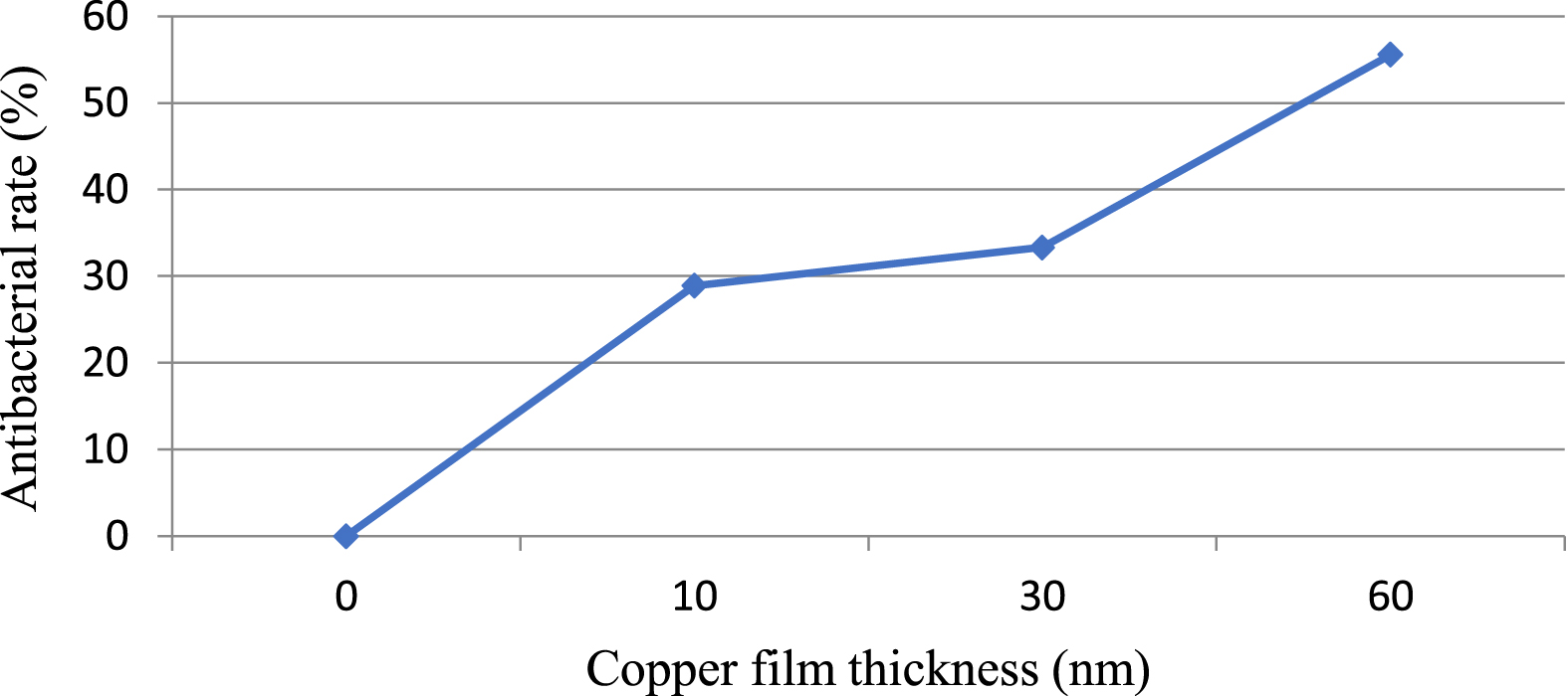
Antibacterial effect of copper-coated carbon nanotubes on Staphylococcus aureus in terms of copper film thickness (nm).
The results showed that sample A1 had more antibacterial properties than sample A0, and it was also observed that the antibacterial rate increased with increasing copper film thickness. The large area-to-volume ratio of the nanotubes caused the interface between the copper coating and the carbon nanotube to become larger, which provided more opportunities for the copper atoms to come in contact with the bacteria for degradation. The results indicated that the copper coating on CNTs enabled the decomposed copper ions to communicate with bacteria, so that the carbon surface coating had a significant effect on the copper contact surface.
The copper-coated samples had more antimicrobial activity against Escherichia coli compared to Staphylococcus aureus, which could be attributed to the cell wall structure of these bacteria. Staphylococcus aureus, like other Gram-positive bacteria, has a thick homogenous cell wall (20–40 nm) containing an extensive peptidoglycan layer [40]. Meanwhile, Gram-negative bacteria such as Escherichia coli have a much thinner cell wall (15 nm) and consist of an inner membrane, a thin layer of peptidoglycan and an outer membrane [41]. In a research on AgNPs, the Ag+ ions destroyed bacterial DNA by colliding with the cell membrane [42]. In gram-negative bacteria such as Escherichia coli, antibiotics, which are among the smallest water-soluble molecules, enter the bacterial body through transport across the cell membrane [43]. In the investigation of the antibacterial properties of single-walled carbon nanotubes (SWCNTs), the results showed that if the metal agent was not present, they could cause damage to the cell membrane of E. coli, but the functionalized ones were more effective on both Gram-negative and Gram-positive bacteria [44]. Cao. (2021) found that the CNT alone had no antibacterial activity [45].
Figure 3 shows the SEM images of CNTs grown by PECVD before copper coating. The analysis of MWCNTs showed a forest of vertically aligned nanotubes, meaning they are oriented on the substrate. The analysis of SEM images showed that the CNTs were aligned parallel to each other and perpendicular to the surface, in line with a study on copper azide confined inside templated CNTs [45]. Comparison of SEM images before and after copper coating showed that the CNTs increased in diameter and the distance between them increased also, in agreement with other studies [46]. The SEM images revealed the favorable growth of CNTs with contact time between 15– 25 minutes. In the first case, it can be said that because the nanotubes have had more time to grow, they have gained more height as a result, but as can be seen, this increase in height has caused the ends of the nanotubes to stick to each other in different places. In addition, the density of nanotubes has increased under these conditions. In growth conditions with shorter contact time, the nanotubes were observed independently of each other with lower density and lower height.
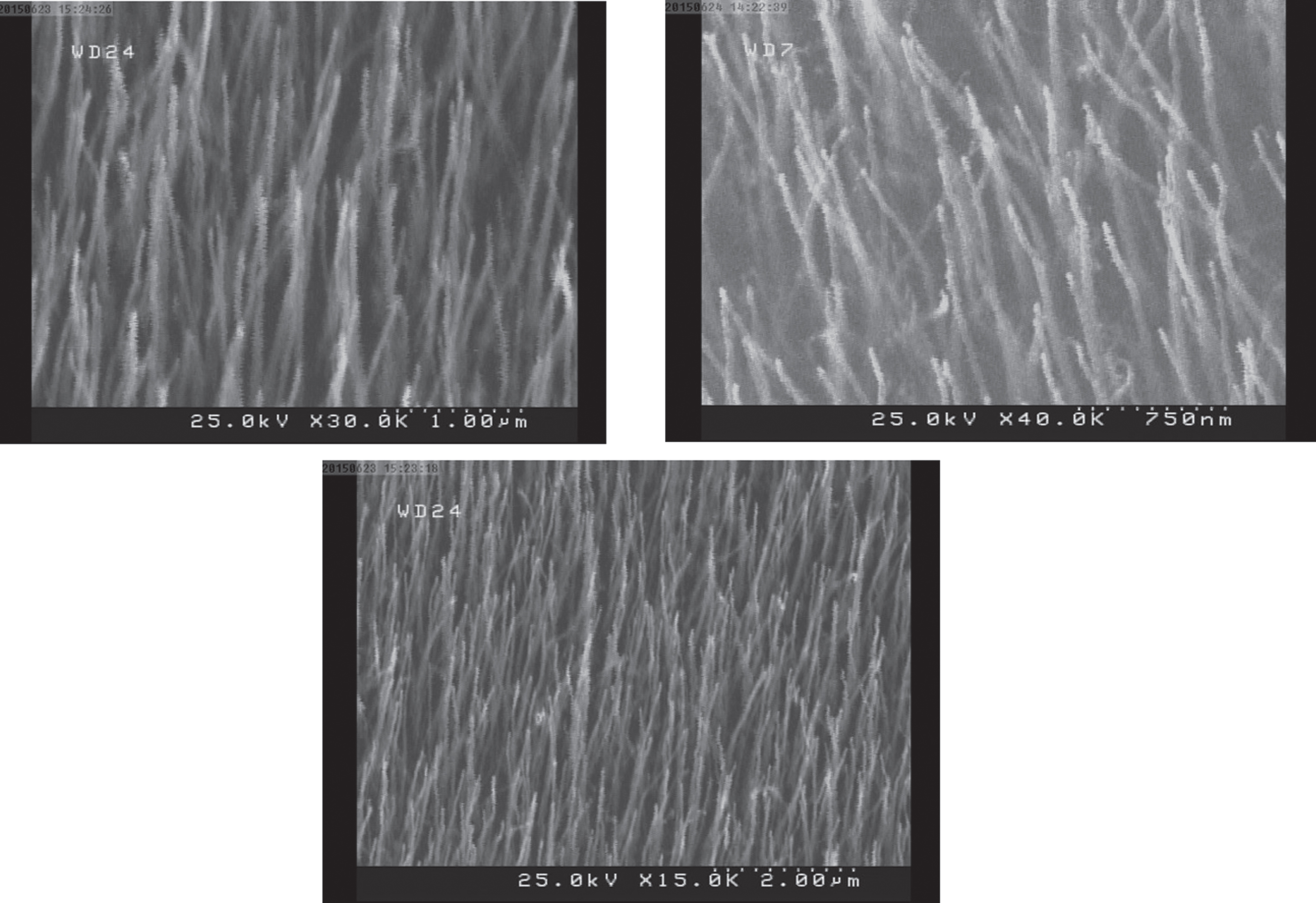
SEM images of carbon nanotube growth before copper coating.
Figure 4 shows the TEM images of the samples before and after coating with CuNPs. The analysis of TEM images showed the deposition of CuNPs on CNTs. The sample a had CNTS with average size of 68 to 94 nm. In the CuNPs-coated samples, the average CNT size was 42 to 55 nm. In the coated samples, the size reduction could be found to some extent. Comparison of FE-SEM images of six samples revealed that increasing the copper film thickness, especially from 30 to 60 nm, had a great effect on the diameter and deposition rate of CuNPs on CNTs. The TEM images also showed that increasing copper film thickness caused CNTs to form denser layers, which can create stronger structures. The copper deposition rate should be slow to align uniformly on the nanotubes. A higher deposition rate caused the nanotubes to connect to each other. If copper was placed on the CNTs in such a way as to cover them in bunches, the interface with CNTs would decrease and therefore the antibacterial potential would decrease. In this study, the coating rate was estimated to be 0.15– 0.16. In accordance with the findings of previous studies, oxygen-containing functional groups, such as carboxyl and hydroxyl, were formed both inside and outside of CNTs [47].
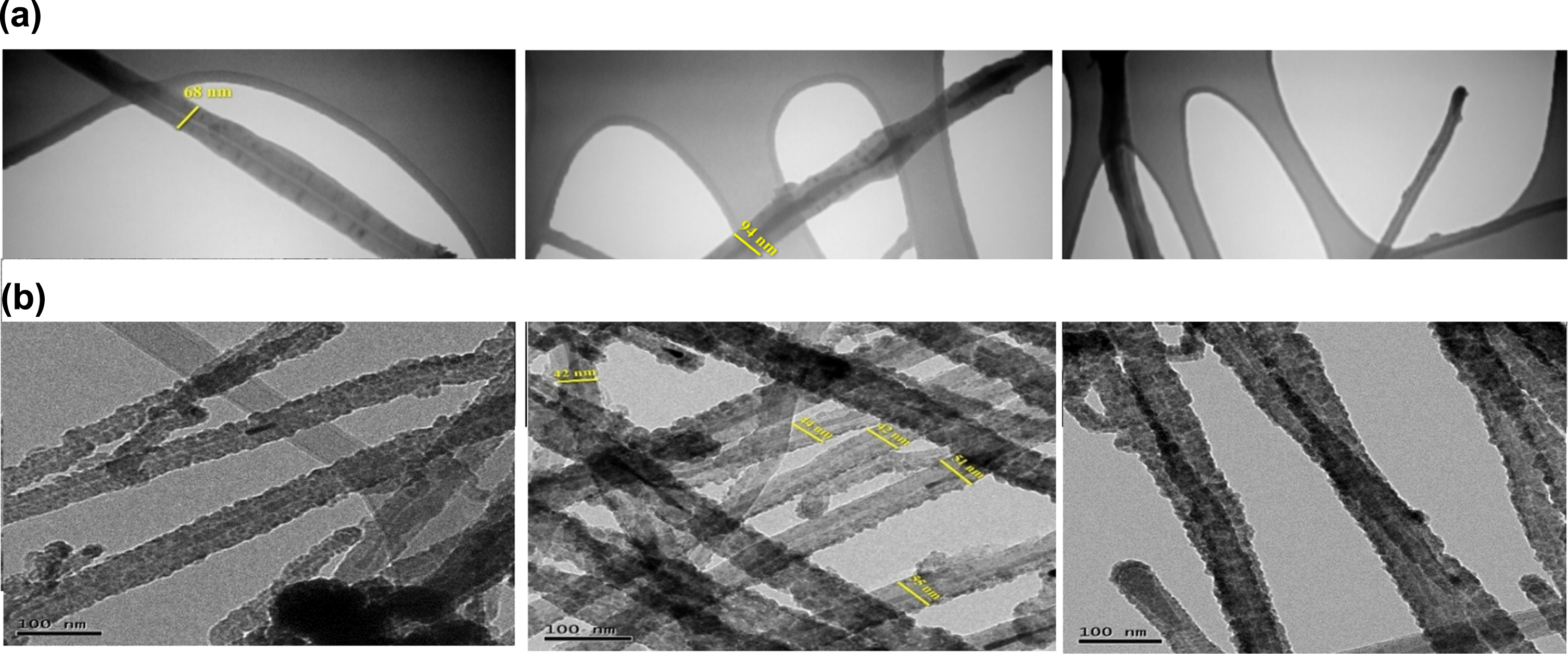
TEM images captured from (a) bare carbon nanotubes and (b) copper-coated carbon nanotubes.
Figure 5 shows FESEM images taken from MWCNTs with different copper film thicknesses. Dc Magnetron Sputtering was used to coat CNTs because this method made it possible to easily and accurately control the rate of copper deposition on CNTs, as one of the main parameters in this study. The FE-SEM and TEM analyzes revealed that the copper film had coated single nanotubes, thus resulting in an increase in bactericidal activity. Observations of FE-SEM and TEM images before and after coating were consistent with the antibacterial test results. FE-SEM images confirmed the uniform distribution of copper surface on CNTs.
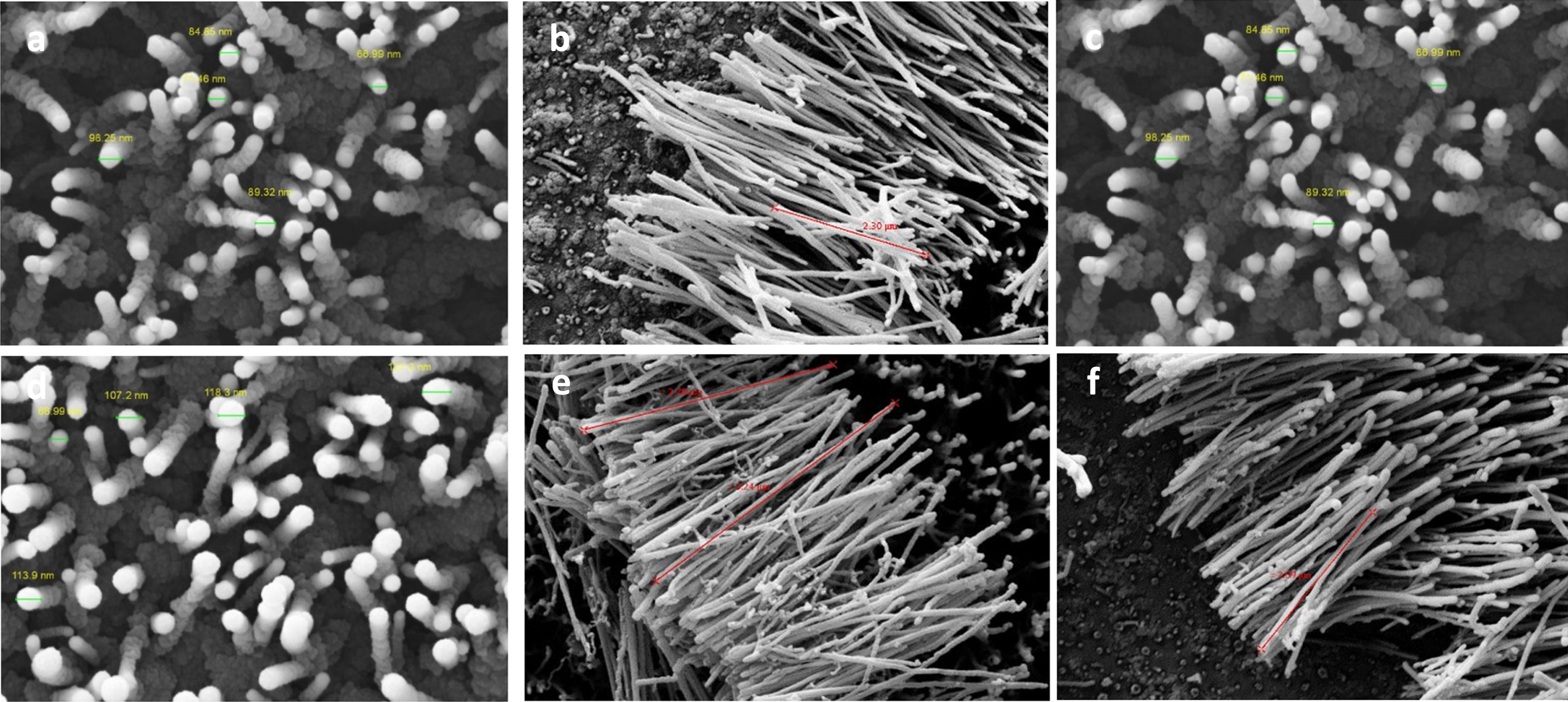
FE-SEM images captured from carbon nanotubes coated with different copper film thicknesses: a) 9 nm, b) 10 nm, c) 15 nm, d) 21 nm, e) 30 nm and f) 60 nm.
Figure 6 shows EDS analyzes for carbon nanotubes coated with different copper film thicknesses. According to the findings, carbon had the highest level before deposition, followed by silica and nickel respectively. Silica and nickel in the sample were related to the bottom layer. In the sample without copper coating, there were peaks of gold, silicon, carbon and nickel. The presence of silicone compounds was due to the type of substrate. Due to the gold coating in the device, the Au ion peak was seen in the spectrum. After copper coating, the EDS spectrum showed peaks related to CuNPs. Bonds were formed between carbon and copper particles [48]. Following the increase in the thickness of the copper film, the amount of carbon has decreased and as a result, the amount of copper increased. At first, the copper content was small, but as the carbon content decreased, the copper content increased. The findings showed that the deposition process and the thickness of the catalyst layer had a great effect on the growth and distribution of carbon nanotubes on the surface. It was also found that as the copper film thickness increased, carbon nanotubes with the density of copper metal spread more on the surface. In this case, the tubular structure was preserved and their structure and morphology were more appropriate. The findings of EDS proved the copper coating. With increasing amount of copper, less dark spots were seen, indicating that copper electrons were attached to the surface of nanotubes. This phenomenon has also been seen in the investigation of nanotube coating with other heavy elements [49]. Copper coating created a relatively homogenous structure but in a fragmented manner. With the increase in the amount of copper, which can be seen in Figure F, carbon and copper are constituent elements, But in lesser amounts of copper coating, other elements are seen.

EDS spectra for carbon nanotubes coated with different copper film thicknesses: (a) 9 nm, (b) 10 nm, (c) 15 nm, (d) 21 nm, (e) 30 nm, (f) 60 nm and (g) before copper coating.
Figure 7 shows the distribution of copper particles on CNTs. Copper nanocrystals were deposited on CNTs. As can be seen, as the thickness of the copper film on CNTs increased, the diffusion and distribution of copper became more and more homogeneous and more structural stability was achieved, in agreement with other researches [50]. The images showed that the nanoparticles were better deposited on the carbon matrix with increasing thickness [51].

Distribution of copper film on carbon nanotubes with different thicknesses: (a) 10 nm, (b) 30 nm, (c) 60 nm.
Figure 8 shows the XRD spectrum for Cu/CNTs. The results of EDS analysis showed no other chemical compounds. The materials were not completely crystalline, so a mixture of crystalline and amorphous materials was found. The XRD spectrum showed the amorphous domains as broad peaks and the crystalline domains as sharp peaks, which could determine the crystallinity of the material. The XRD spectra were related to copper, nickel and carbon peaks. The copper peaks with lower intensity but crystalline structure were seen in the range of 74.9°, 66.5° and 33°. According to XRD analysis, there was no peak higher than that of copper. Copper had a cubic crystal structure. The presence of nickel peak showed that this metal also had a cubic structure.
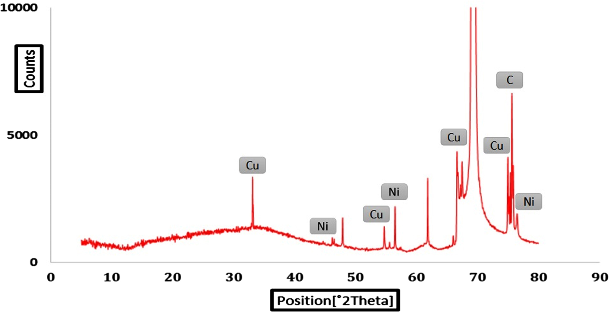
XRD spectrum for synthesized samples,
In the EDS curves, the formation of carbon nanotubes with CuNPs was evident, in consistent with the XRD results. Copper particles were separate particles in the composite of CNTs with copper, in line with previous findings [52]. The spatial structure and high surface area of carbon nanotubes allowed for the deposition of more copper particles, and copper had more space for deposition. The strong Cu peaks were related to Cu 2p3, Cu 2p1, Cu LMM-2 and O 1 s. The C1 s carbon peak was weak, which was due to the deposition of copper on the CNTs substrate, thus leading to the formation of a copper-rich thin layer with some oxygen.
The research findings showed strong antibacterial properties for as-synthesized Cu/CNTs. The Cu film coated on the CNTs enhanced the antibacterial activity due to providing a large surface area. The main mechanism of bacterial removal was due to the copper nanoparticle coating, and the resistance of Escherichia coli to nanoparticles was lower compared to S. aureus. The reason for the removal of bacteria was the mechanism of ROS production, in which highly reactive oxygen compounds, such as hydroxyl radicals (•OH), superoxide anion radicals (O2•–), hydrogen peroxide (H2O2) and NO radicals, are produced as a result of contact with copper nanoparticles; subsequently, these compounds react with and degrade lipids, proteins and enzymes. The formed radicals are responsible for the destruction and removal of the bacterial cell wall. As the copper film thickness increased, the tubes with more copper metal density were distributed on the surface. The findings of FESEM, EDS, TEM, and XRD techniques showed that copper was well coated on multi-walled carbon nanotubes, which played an essential role in the elimination of studied bacteria. The results showed that copper layers can be well coated on individual nanotubes and therefore play an essential role in controlling bacteria; this structure may have the potential to be used in medical devices and biomaterials. It is suggested to use other elements that have antibacterial properties in future studies. Also, other bacteria should be used to test the antibacterial properties.
Footnotes
Acknowledgments
This research has been extracted from the PhD dissertation in Physics written by Mr.