
Abstract
BACKGROUND:
Prolonged high fat diet consumption was reported to cause metabolic disorders including obesity, NAFLD and insulin resistance. NAFLD is one of the common causes of liver failure with lipid accumulation and inflammation as the major driving forces for its progression.
OBJECTIVE:
The study was aimed at evaluating the benefits of Aloe vera supplementation on lipid profiles, antioxidant properties, liver function as well as the histology of liver, heart and brain on high fat diet induced toxicity in BALB/c mice.
METHODS:
Eighteen mice were divided into three groups (n = 6). Group 1 received normal diet (Vital feed), group 2 received high fat diet (HFD) i.e. 70 g of normal diet plus 30 g of margarine, while group 3 received high fat diet plus Aloe vera (HFD+AV) i.e. 100 g of HFD plus 20 g of Aloe vera gel. The mice were fed for 10 weeks and euthanized thereafter. The liver function, lipid profiles, antioxidant properties as well as liver, brain and heart histology were evaluated.
RESULTS:
The levels of cholesterol, triglycerides and low density lipoprotein were significantly increased (P < 0.05) in the HFD treated mice compared to the control. Liver catalase and superoxide dismutase activities were significantly increased (P < 0.05) in HFD+AV treated mice compared to the control and HFD treated mice. The liver of HFD+AV treated mice showed normal architecture while those of HFD treated mice showed numerous hepatic vacuoles indicative of fat droplets.
CONCLUSIONS:
Aloe vera supplementation regulated liver function and prevents hyperlipidemia. The resultant effect increased antioxidant activities thereby preventing liver injury and brain damage.
Introduction
The incidence of Non-alcoholic fatty liver disease (NAFLD) has increased drastically in recent years. NAFLD is one of the common causes of liver failure usually progressing from steatosis to fibrosis and cirrhosis [1]. Previous studies have identified lipid accumulation and inflammation as the major drivers for the progression of NAFLD. Consequently, most preventive strategies focused on controlling high fat consumption and improving metabolic pathways to reduce accumulation of fat [2]. While a number of compounds with promising potentials for the treatment of NAFLD are being evaluated, getting an effective animal model for testing these compounds still constitute a major challenge [3]. An effective animal model of NAFLD is expected to develop obesity, hepatic inflammation, insulin resistance and dyslipidemia. Although animal models have not sufficiently depicted human NAFLD, prolonged high fat diet consumption was reported to cause metabolic disorders including cardiovascular disease, NAFLD, insulin resistance and obesity in rodents [4]. Previous studies reported that high fat diet induced obesity and subsequently led to fat accumulation in the liver and muscles tissues coupled with oxidative stress and dyslipidemia [5–7]. Other diseases may include NAFLD, Non-alcoholic steatohepatitis (NASH), neuropathy, cardiovascular diseases and type 2 diabetes [4].
Due to the efficacy and reduced effect of natural products, they are preferred in the treatment and prevention of obesity and its related diseases such as type 2 diabetes, dyslipidemia and NAFLD [8]. The World Health Organization (WHO) reported that traditional medicines played a vital role in the prevention and treatment of chronic diseases [3]. Aloe vera (Aloe barbadensis Miller) belongs to the family Liliaceae. It is a succulent plant adapted to growing in the tropics [9]. Aloe vera is used as an anticancer, anti-diabetic, anti-inflammatory and antiseptic agent [10]. It was reported to improve glucose metabolism, reduce fat accumulation and prevent obesity [11]. Aloe vera is also used in the production of many cosmetics and drugs. In the United States, it is used as a nutritional supplement in food and drinks while in Chinese traditional medicine, it is called Elixir of Youth because of its wide range of medicinal uses [12]. Aloe vera leaf contains gymnemic acid which binds to receptors that prevent glucose intake in tongue and suppress glucose absorption in the intestine [13]. The present study was aimed at evaluating the benefits of Aloe vera supplementation on lipid profiles, antioxidant properties, liver function as well as the histology of liver, heart and brain in high fat diet induced toxicity in BALB/c mice.
Materials and methods
Plant material
Aloe vera were collected from a farm in Maiduguri, Nigeria and deposited in the herbarium (UMM/FPH/ASH/001) Faculty of Pharmacy, University of Maiduguri. The plant consists of a colourless gel enclosed in a spiny leaf. The leaf of Aloe vera were gently removed to extract the gel. The process of gel extraction was performed each day for diet formulation.
Animal treatment and Ethics approval
Eighteen (18) male BALB/c mice were purchased from the National Veterinary Research Institute (NVRI) Vom, Nigeria. The mice were kept in the Department of Biochemistry animal house for one week to acclimatize in conditions as follows: 20°C–29°C and 45% humidity. The study was approved by the Department of Human Anatomy Ethical committee, University of Maiduguri (UM/HA/UGP20.21-023) and was carried out following the ARRIVE guidelines and the United States guide for the care and use of laboratory animals.
Experimental design
Six weeks old mice were randomly divided into three groups (n = 6). Simple randomization was used for the distribution. Group 1 (Control), received normal diet (15% crude protein, 4% fat, 70% carbohydrate and 6% fibre). Group 2, the high fat diet group (HFD), received a mixture of 70 g normal diet and 30 g margarine while group 3 received 100 g HFD plus 20 g Aloe vera group (HFD + AV). The ratio of HFD and Aloe vera was selected based on the local Aloe vera supplementation in Northern Nigeria for obesity and liver disease treatment. The administration lasted for 10 weeks (Fig. 1). All the mice had free access to drinking water and their body weights were monitored weekly. The mice were euthanized at the end of the 10th week and the blood of each mouse was collected and centrifuged at 5000 rpm for 10 minutes.
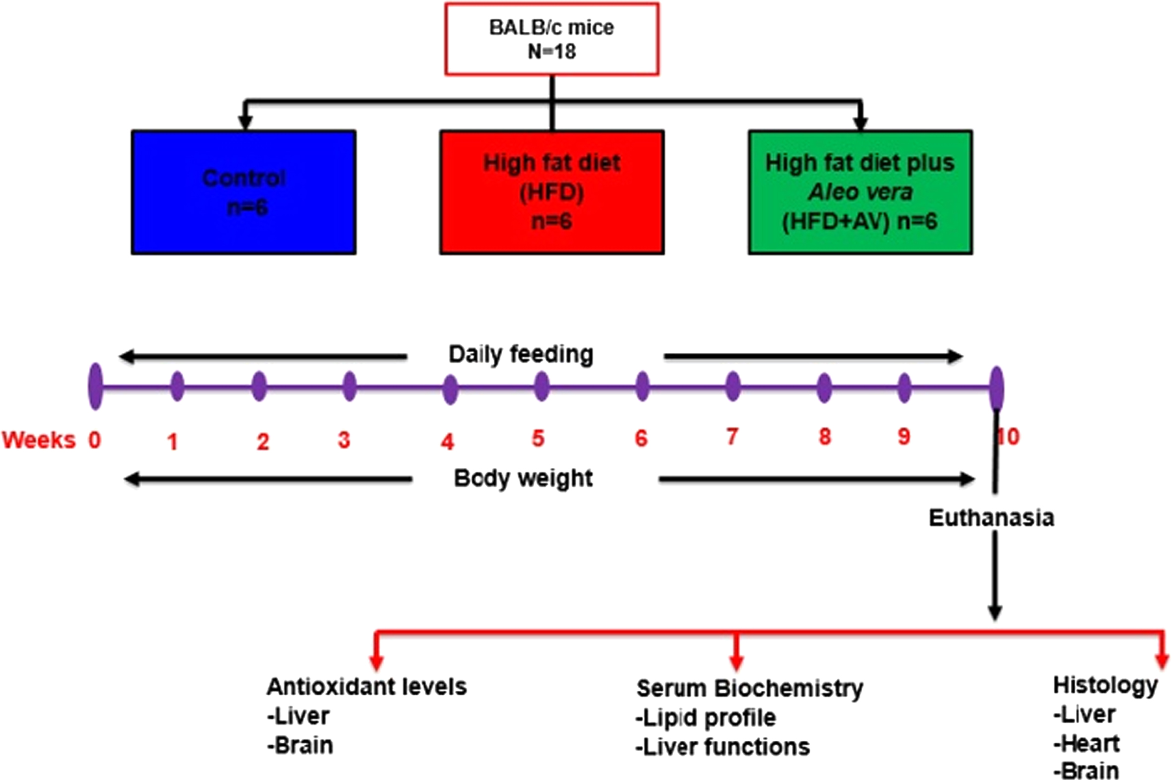
Summary of experimental design
The serum levels of liver function markers [albumin, total protein concentration, aspartate aminotransferase, alanine aminotransferase, alkaline phosphatase) and lipid profile (cholesterol, triglycerides, high density lipoprotein and low density lipoprotein) were evaluated using enzyme linked immune-sorbet assay (ELISA) kit (NeoScientific, USA) according to manufacturers’ instruction.
Antioxidant activity
The liver, heart and brain were dissected. Part of the liver and one half of the brain were homogenized in phosphate buffer (pH 7.2) and the supernatant was used to determine the activity of catalase, superoxide dismutase and reduced glutathione (GSH). Catalase activity was evaluated by the method described by Aebi [14]. Superoxide dismutase was determined by the method of Fridovich, [15] while GSH concentration was conducted as described by Rajagopalan et al. [16].
Histological study
The liver, heart and dentate gyrus were fixed in 10% formalin, dehydrated in graded alcohol, cleared in xylene and embedded in paraffin wax. Tissue sections were sectioned at 5μm and stained with heamatoxylin and eosin (H&E). Micrographs were taken at x40 and x200 magnifications using microscope digital camera (AmScope, UK). The mice groups were concealed from all researchers except one while preparing the samples for analysis and during interpretation to avoid bias.
Statistical analysis
One-way analysis of variance and Sidak post-hoc test was conducted using GraphPad prism 7 (GraphPad, USA) and the results were expressed as Mean±SEM. P < 0.05 was considered statistically significant.
Results
Body weight changes
The body weight changes of control group was similar to that of HFD and HFD +AV treated groups of mice (Fig. 2), as there was gradual increase in body weight from weeks 1–10.
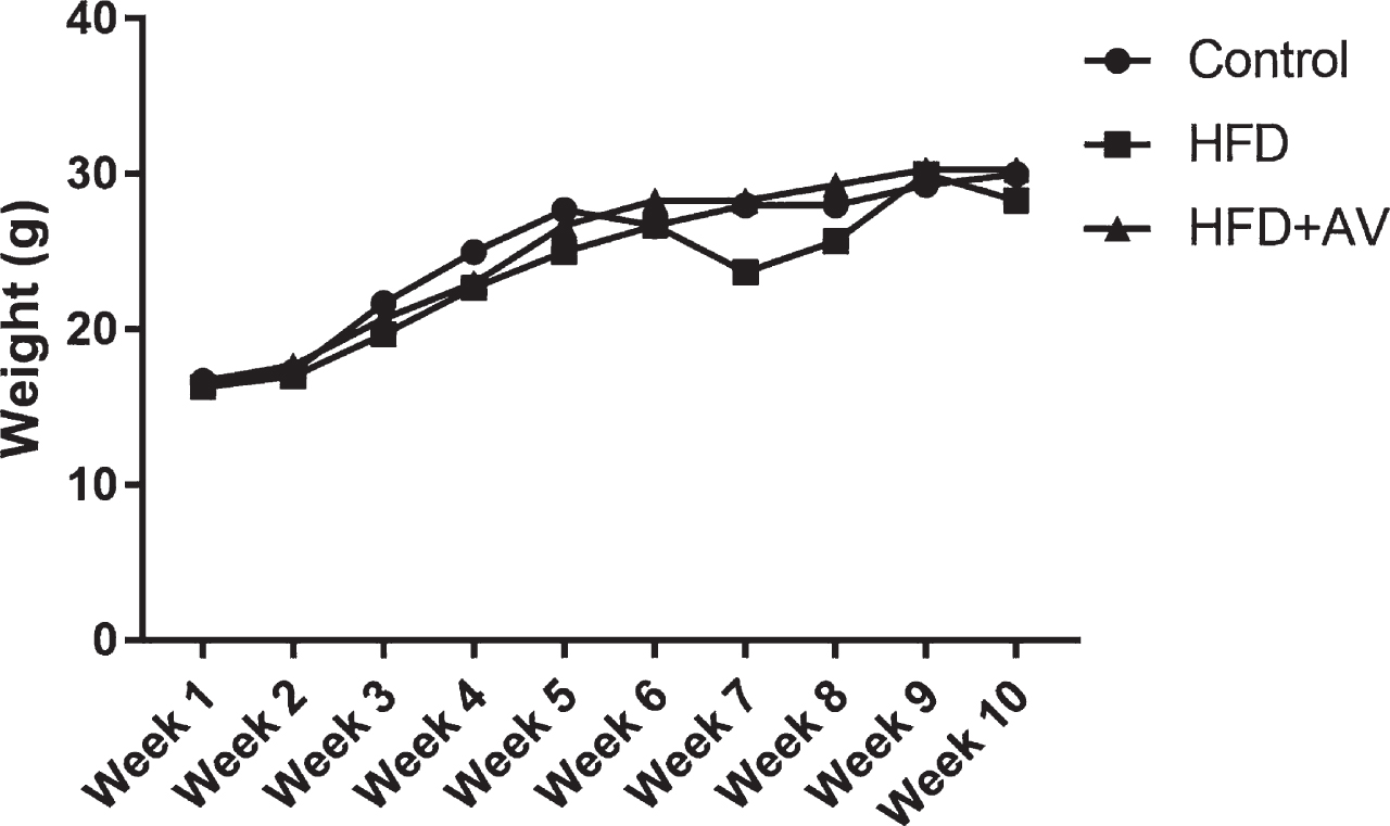
Weekly body weight changes of mice treated with high fat diet and Aloe vera. HFD = high fat diet, HFD+AV = high fat diet plus Aloe vera.
The serum albumin level of HFD+AV treated mice (0.33±0.07) was significantly higher (P > 0.05) relative to the control (0.17±0.03) and HFD (0.17±0.07) treated mice (Fig. 3a). The total protein concentration of HFD+AV treated mice (2.93±0.57) was significantly higher when compared to the control (0.93±0.03) and HFD (1.77±0.13) fed mice at P < 0.05 (Fig. 3b). Aspartate aminotransferase (AST) levels of HFD treated mice (89.67±12.67) was significantly higher relative to the control (15.17±0.17) and HFD+AV treated mice (18.33±1.70) at P < 0.05 (Fig. 4a). No significant change (P > 0.05) was observed in alanine aminotransferase (ALT) level of HFD (35.70±4.45) and HFD+AV (28.33±0.16) treated mice relative to the control (35.1±1.00) (Fig. 4b). The level of alkaline phosphatase (ALP) was significantly increased (P < 0.05) in HFD (44.20±0.20) and HFD+AV (40.23±1.37) treated mice compared to the control (20.80±0.10). The ALP levels of HFD treated mice (44.20±0.20) was significantly higher relative to HFD+AV treated mice (40.23±1.37) at P < 0.05 (Fig. 4c).

Albumin and total protein concentration of HFD and Aloe vera treated mice. HFD = high fat diet, HFD+AV = high fat diet plus Aloe vera. Values are expressed as Mean±SEM. @ and $ indicates significant difference with control and HFD respectively. SEM = standard error of mean
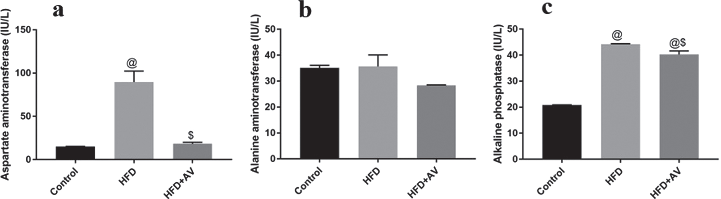
Liver function of mice treated with high fat diet and Aloe vera. HFD = high fat diet, HFD+AV = high fat diet plus Aloe vera. Values are expressed as Mean±SEM. @ and $ indicates significant difference with control and HFD respectively. SEM = standard error of mean
The levels of cholesterol, triglycerides and low density lipoprotein (LDL) were significantly increased (P < 0.05) in the HFD treated mice (111.50±1.40, 70.40±0.20 and 85.47±2.37) compared to the control (48.73±6.13, 49.50±0.50 and 35.60±0.30) (Fig. 5a–c). No significant change was observed in the levels of cholesterol, triglycerides and LDL in HFD+AV treated mice (56.73±8.37, 42.53±3.51 and 37.47±4.57) compared to the control mice (48.73±6.13, 49.50±0.50 and 35.60±0.30) at P > 0.05 (Fig. 5a–c). Serum level of high density lipoprotein (HDL) significantly increased (P < 0.05) in HFD+AV treated mice (23.33±4.41) and the control mice (21.67±4.41) compared to HFD treated mice (16.67±3.33) (Fig. 5d).
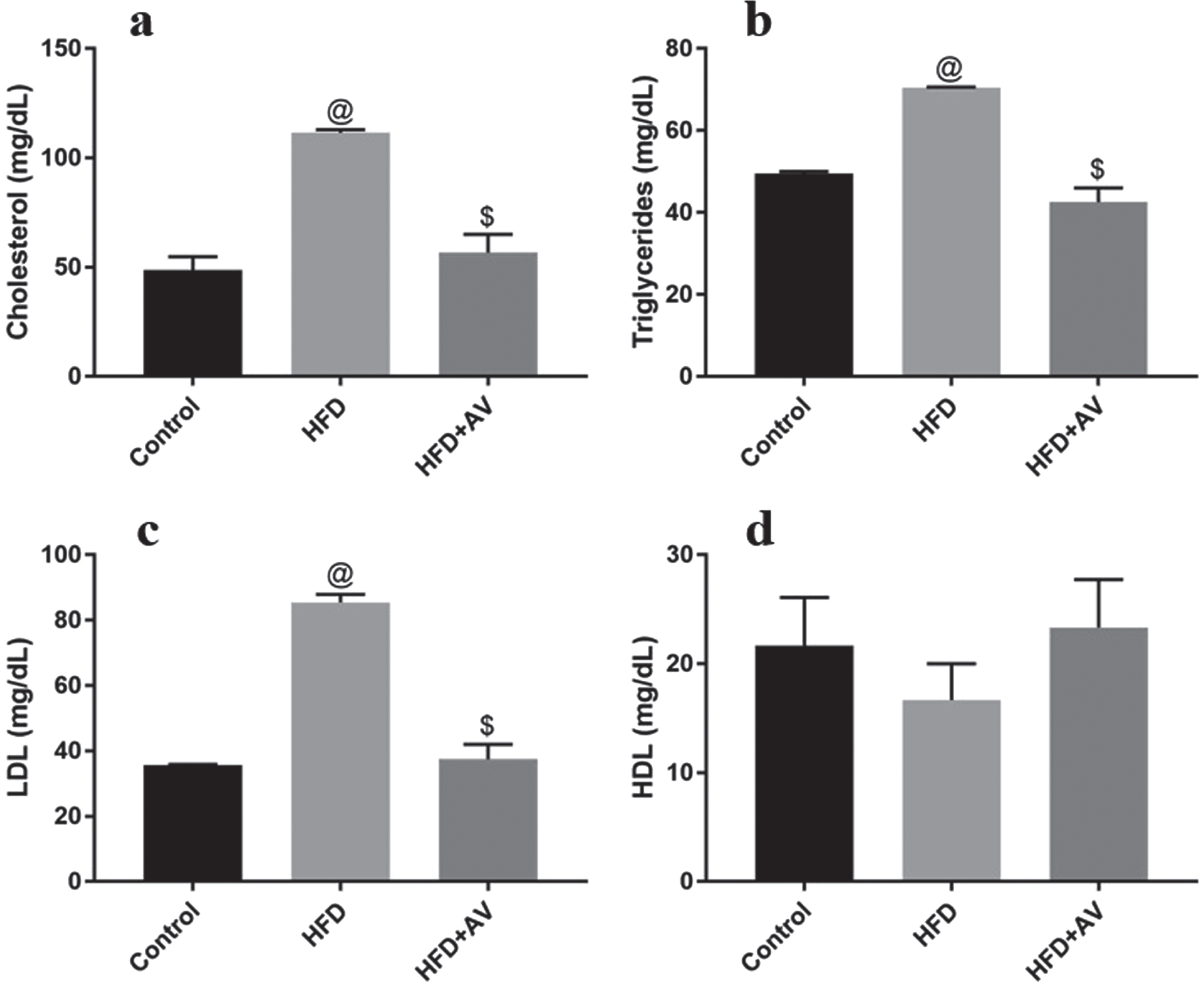
Lipid profile of HFD and Aloe vera treated mice. HFD = high fat diet, HFD+AV = high fat diet plus Aloe vera. Values are expressed as Mean±SEM. @ and $ indicates significant difference with control and HFD respectively. SEM = standard error of mean
Liver catalase and superoxide dismutase activities were significantly increased (P < 0.05) in HFD+AV treated mice (4.27±0.13 and 6.03±0.03) compared to the control (2.23±0.13 and 3.03±0.03) and HFD treated mice (0.96±0.07 and 2.63±0.07). However, a significant reduction (P < 0.05) in catalase and superoxide dismutase activities were observed in HFD treated mice (0.96±0.07 and 2.63±0.07) relative to the control (2.23±0.13 and 3.03±0.03) (Fig. 6). Furthermore, no statistically significant changes were observed in the activities of liver reduced glutathione among all the groups of mice (Fig. 6). On the other hand, a statistically significant decrease (P < 0.05) in brain catalase activity was observed in HFD+AV treated mice (1.43±0.23), when compared to the control group (2.77±0.13). Notwithstanding, no significant change was observed in brain superoxide dismutase and reduced glutathione activities of control mice (2.43±0.33 and 0.80±0.10) relative to the HFD (2.13±0.03 and 1.03±0.03) and HFD+AV treated mice (1.97±0.53 and 0.97±0.07) at P > 0.05 (Fig. 6).

Liver and brain antioxidant activities of mice treated with high fat diet and Aloe vera. HFD = high fat diet, HFD+AV = high fat diet plus Aloe vera. Values are expressed as Mean±SEM. @ and $ indicates significant difference with control and HFD respectively. SEM = standard error of mean
The liver micrograph of control mice and HFD+AV treated mice showed normal architecture while the liver of HFD treated mice showed numerous hepatic vacuoles indicative of fat droplets (Fig. 7). The micrograph of the heart of control mice as well as HFD and HFD +AV treated mice showed normal branching fibres of cardiac muscle (Fig. 7). Photomicrographs of the dentate gyrus region of the hippocampus in control mice and the mice treated with HFD and HFD +AV showed normal general architecture at x40 magnification with distinct pyramidal cells at x200 magnification (Fig. 8).

Liver and heart histology in HFD and Aloe vera treated mice. HFD = high fat diet, HFD+AV = high fat diet plus Aloe vera.

The dentate gyrus of mice treated with high fat diet and Aloe vera. HFD = high fat diet, HFD+AV = high fat diet plus Aloe vera.
Weight gain following high fat diet varies in different strains of mice. Some strains are highly susceptible to weight gain while others are resistant or might take a longer time [17]. Factors that influence the body weight changes include age, sex and composition of feed [18]. Previous study have reported a significant gain in body weight of C57BL/6J mice after 14 weeks exposure to HFD, when compared to mice fed on normal diet [19, 20]. Another study reported significant body weight gain in C57BL/6J mice after 12 weeks on HFD consumption compared to the same strain fed with normal fat diet [21]. In C57B16 mice, HFD led to significant increase in body weight at a faster rate (7 weeks) relative to low fat rodent chow [22]. The non-significant body weight changes in HFD fed mice relative to the control that was observed in the present study is an indication that BALB/c mice might need more time (>10 weeks) or a diet containing more than 40% fat for a period of about 10 weeks to manifest a significant weight gain compared to mice on normal diet.
Albumin constitutes 25% of the proteins synthesized by the liver and only about 25% of the hepatocytes are involved in albumin synthesis. Serum albumin can be lost from the circulation through the glomeruli, gut wall and altered blood vessels [23]. In the present study, Aloe vera was shown to increase albumin level in HFD fed mice. Previous reports have shown that Aloe vera could promote antioxidant activities and prevent hyperlipidemia in rats [24]. Albumin is an extracellular antioxidant and consists of a reduced and oxidized form, the reduced form contributes to the antioxidant ability of plasma and serve as major carriers of copper and free fatty acids [25]. The mechanism by which Aloe vera increases serum albumin level might be through promoting antioxidant activities and preventing hepatocyte damage.
ALT and AST are produced by hepatocytes and their serum levels are reported to increase in obesity and Non-alcoholic steatohepatitis [26, 27]. The result of the present study showed an elevated serum level of AST in HFD treated mice. It was correlated with the presence of hepatic vacuole in the liver of HFD treated mice which is an indicator of steatosis. Aloe vera was shown to prevent steatosis and the resultant effect was also observed in the normalized serum AST level in HFD + AV treated mice. A previous study reported that high fat diet induced liver damage including steatosis and fibrosis and also led to the elevation of both ALT and AST levels [28]. The normalized AST level that was observed in Aloe vera treated mice might be due to the protective role of Aloe vera on hepatocytes. The mechanism through which Aloe vera protects hepatocytes might be through enhancing lipid emulsification and digestion thereby preventing it accumulation in the liver tissue.
Obesity is one of the most common metabolic disorder in the world, it occurs as a result of sedentary lifestyle as well intake of high fat and glucose diet [29]. Obesity is associated with many chronic diseases such as hyperlipidemia, diabetes and cardiovascular disease [30]. High dietary fat consumption was reported to play an important role in the development of cardiovascular disease [31]. In the present study, cholesterol, triglycerides and low density lipoprotein were significantly increased in high fat diet treated mice. High fat diet was reported to increase lipid accumulation in mice [32]. Hyperlipidemia is an indicator of obesity and cardiovascular disease [24, 33]. Aloe vera supplementation normalized cholesterol, triglycerides and low density lipoprotein in the present study. A previous study reported the anti-obesity role of Aloe vera in mice through normalizing lipids level and up-regulating adiponectin gene [13]. Another study reported the ability of Aloe vera to reduce body fat and activate adipose lipolysis [24]. The mechanism through which Aloe vera normalized lipid profile in the present study might be by promoting lipolysis thereby enhancing lipid digestion and elimination rather than accumulation. High fat diet did not affect cardiac muscles in the present study but the hyperlipidemia might gradually lead to myocardial infarction if not controlled.
High fat diet elevated chylomicron levels in the intestine. Chylomicrons enter circulation and initiates the production of free fatty acids, which moves to the liver. These free fatty acids are either esterified into triglycerides and goes to the mitochondria for oxidation. The triglycerides may accumulate in hepatocytes as small droplets (steatosis) or produce very low density lipoprotein (VLDL), which is then converted into LDL [35]. Aloe vera prevented hyperlipidemia and steatosis by either preventing chylomicron production in the intestine or promoting the oxidation of free fatty acids in the mitochondria.
Oxidative stress plays a critical role in disease progression and is caused by reactive oxygen species (ROS) production. Increase in ROS is responsible for the initiation and progression of diseases such as obesity, liver and cardiovascular diseases [36]. In the present study, HFD was shown to decrease antioxidant levels (Catalase, superoxide dismutase and GSH) in the liver and brain. Others studies also reported that HFD decreased blood flow to the brain and antioxidant activities, increased blood brain barrier permeability thereby altering behavior and memory [37, 38]. Although the present study reported decreased antioxidant activities in HFD fed mice, it did not significantly affect dentate gyrus histology. This is an indication that the oxidative stress is temporal and may be prevented by dieting. Aloe vera was found to increase catalase and superoxide dismutase activities in the liver of mice. However, it did not improve the production of these antioxidants in the brain. This might be because catalase and superoxide dismutase are produced in cells other than the neuroglia and transported to the brain via blood vessels; crossing the blood brain barrier might take a longer time. The body is protected from ROS-induced cell injury and subsequent disease onset and progression by catalase, superoxide dismutase and GSH ([39]. Studies have shown that obesity induces the generation of white adipose tissue that secretes pro-inflammatory factors that activates immune cells. Activated immune cells enhance the production of ROS and subsequent development of oxidative stress [40]. The possible mechanism through which Aloe vera promotes antioxidant activity is by promoting catalase and superoxide dismutase production by body cells and/or preventing the generation of white adipose tissue.
Conclusions
Aloe vera supplementation for 10 weeks in HFD treated mice regulated liver function and prevented hyperlipidemia. The resultant effect increased antioxidant activities thereby preventing liver injury, steatosis and brain damage. Hence, Aloe vera supplementation in diet could prevent obesity and its associated diseases by preventing fat accumulation and promoting endogenous antioxidant activities.
Footnotes
Acknowledgment
None.
Funding
None.
Conflict of interest
The authors have no conflict of interest to report.
Author contributions
All authors conceived and design the study. FB, SMC & ZMG gave administrative support. NID, HEK, HS, UKJ, TJK, JA, AM, HSG, ZGF & AMM provided the study materials. NID, HEK, HS, UKJ, TJK, JA, AM, HSG, ZGF & AMM collected and assembled the data. NID, FB, SMC & ZMG analyzed and interpreted the data. NID was a major contributor in writing the manuscript. All authors read and approved the final manuscript.