
Abstract
Persistent pulmonary hypertension of newborn (PPHN) is a very rare manifestation of congenital cytomegalovirus (CMV) infection. PPHN associated with CMV can be severe but is usually transient and responds well to antiviral therapy. We report a rare case of PPHN that occurred in the setting of fulminant congenital CMV infection and successful treatment with antiviral therapy along with review of the very few cases reported in literature. A male appropriate for gestational Age (AGA) newborn developed rapidly progressive respiratory distress starting at 11 hours of life requiring ventilatory support and 100% oxygen. He developed hypotension and wide difference between pre and postductal saturations. Echocardiography revealed findings consistent with severe PPHN. Examination also revealed multiple purpuric skin lesions and soft hepatosplenomegaly. MRI Brain showed intraventricular hemorrhage, bilateral periventricular calcification, bilateral cerebral and cerebellar intraparenchymal hemorrhage. Complete Cell Count (CBC) revealed severe thrombocytopenia and blood serum showed positive Immunoglobulin M (IgM) for CMV and Urinary CMV was positive by nucleic acid test. He was treated with ganciclovir, inhaled nitric oxide and inotropes. He recovered and was discharged on day 24 of life. Severe PPHN is a rare manifestation of congenital CMV infection and carries a high risk of morbidity and mortality. Congenital CMV should be considered in neonates with PPHN of unknown etiology. Early institution of antiviral therapy in these babies is associated with favorable outcome.
Abbreviations
Cytomegalovirus
Persistent pulmonary hypertension
Inhaled nitric oxide
High frequency oscillatory ventilation
Introduction
Cytomegalovirus (CMV) is the commonest cause of congenital infection worldwide affecting approximately 1% of all live births [1]. CMV is also the most common congenital viral infection in the developed world, occurring in 0.4–2.3% of all live births [2].
CMV is an enveloped, double stranded DNA virus belonging to Herpesviridae family. CMV infections are ubiquitous in humans and usually cause symptomatic infection only in immunocompromised hosts. The pathogenesis of fetal injury in congenital CMV infection is poorly understood. Pathology has been attributed to lytic virus replication leading on to end-organ damage occurring either secondary to virus-mediated cell death or from host inflammatory responses targeting virus-infected cells [3].
Early presentation of congenital CMV usually manifests with fulminant multi-organ involvement. Characteristic features include growth restriction, pneumonitis, hepatitis, bleeding diathesis, chorioretinitis, neurologic abnormalities including microcephaly, seizures, ventriculomegaly, hypotonia, jitteriness, exaggerated reflexes and hematological manifestations like anemia and thrombocytopenia [1, 4]. Persistent pulmonary hypertension of newborn (PPHN) is an extremely rare manifestation of fulminant CMV with only few reported cases in literature till date. We report a late preterm, AGA baby with congenital CMV presenting as severe PPHN and successful treatment with anti-viral therapy in addition to pulmonary vasodilators.
Case report
A 28 year old primigravida delivered a baby boy at 36+6 weeks of gestation by vaginal delivery following an uneventful pregnancy. Maternal serologies like HIV, hepatitis B, syphilis, rubella, and varicella were protective and Group B streptococcus screen was negative. Antenatal scans were within normal limits except for the one done a day prior to delivery which showed Small for Gestational Age (SGA) baby with flow reversal in umbilical artery and borderline fetal ventriculomegaly. Baby was born with perinatal depression at birth and required positive pressure ventilation for 30 seconds and Continuous Positive Airway Pressure (CPAP) for 4 minutes immediately after birth. The Apgars at 1 and 5 minutes were 5 and 7 respectively and the arterial cord blood gas revealed a pH of 7.16 with base excess of - 4. Birthweight was 2.45 Kg (AGA) and postnatal examination revealed multiple purpuric skin lesions (Fig. 1), soft hepatosplenomegaly, active precordium with grade 3/6 ejection systolic murmur in left parasternal area associated with mild respiratory distress.
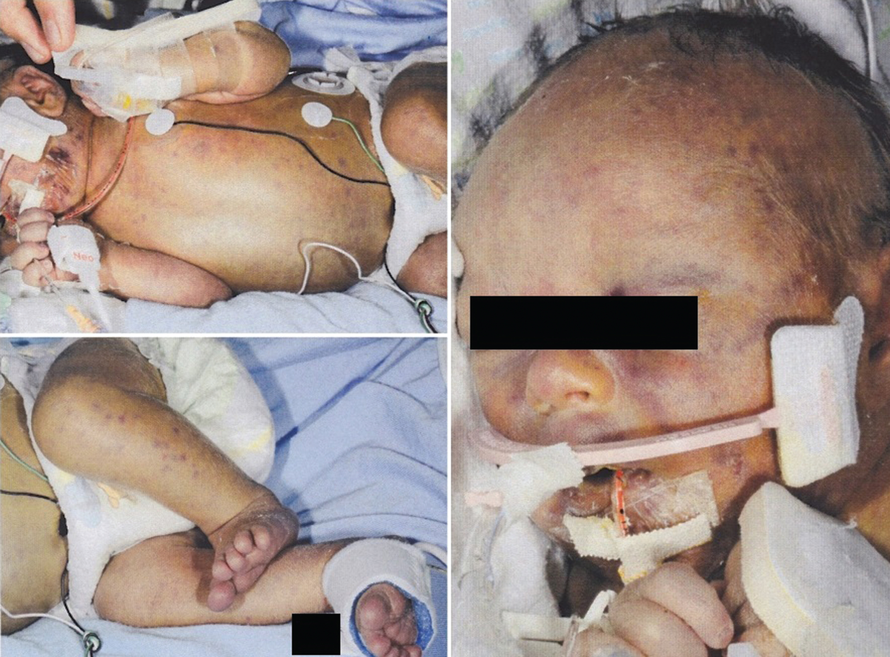
Distribution of multiple purpuric skin lesions classically described as ‘Blue berry muffin syndrome’ which are the areas of extra-medullary dermal hematopoiesis.
At 11 hours of age, he had increased work of breathing and desaturations requiring 30% oxygen by nasal cannula. He was intubated and started on conventional ventilation due to increasing oxygen requirements and respiratory failure. The oxygen requirement gradually increased to 100% within 3 hours in order to maintain saturation of more than 92% . Arterial blood gas showed severe hypoxemia in spite of 100% oxygen and hypercarbia which required high frequency oscillatory ventilation (HFOV).
On HFOV, he developed hypotension and wide difference (>20%) between pre and postductal saturations. Echocardiography revealed dilated and hypertrophied right ventricle, flattening interventricular septum and suprasystemic right sided pressures, severe tricuspid regurgitation, Right Ventricular Systemic Pressure (RVSP) at 77 with right to left shunt at atrial and ductal level suggestive of severe PPHN. He was subsequently started on inhaled nitric oxide (iNO) (Max dose - 20 ppm), Milrinone infusion (Max dose - 0.45 mcg/kg/min) and Dopamine infusion (Max dose - 20 mcg/kg/min). His mean arterial blood pressure was low (below the corrected gestational age), arterial blood revealed lactic acidosis in spite of high doses of inotropes and optimal settings of HFOV for 48 hours. ACTH stimulation test revealed low cortisol level and stress dose of Hydrocortisone (1 mg/kg/dose every 6 hours) was added.
He also developed hypoglycemia (point of care glucose level <2.6) and severe thrombocytopenia on Day 1 of life (platelet count 13,000), elevated International Normalized Ratio (INR) and activated partial thromboplastin time (aPTT). Liver enzymes were markedly elevated along with severe direct hyperbilirubinemia. He received total of 8 units of platelet transfusion over 4 days. Blood tests on day one of life revealed anemia (hemoglobin of 141 g/L), Neutropenia (3.9 10E9/L) and severe thrombocytopenia.(PLT count of 13000) Peripheral smear revealed >10% atypical lymphocytes, microcytic anemia, spherocytes, echinocytes with brisk erythroblastic response.
Head ultrasound done on Day 2 showed multiple linear calcifications in basal ganglia and bilateral periventricular region and bilateral grade 2 intraventricular hemorrhages.
In view of multiorgan involvement, blueberry muffin skin lesions, thrombocytopenia, hepatosplenomegaly, a diagnosis of congenital CMV was considered. IgM CMV serology in serum blood and Urine Nucleic acid test were positive for Cytomegalovirus on day one of life. He was started on IV ganciclovir (6 mg/kg/dose q12 h IV) along with a dose of Intravenous Immunoglobulin (IVIG). He was later switched to oral valganciclovir (16 mg/kg/dose q 12 hourly) after initiating feeds. Serial echocardiographic evaluations revealed improving PPHN and ventricular functions from day 5 of life. The iNO and vasopressors were weaned gradually and stopped on Day 7 of life. Oxygen requirements gradually decreased and he was switched back to conventional ventilation on day 4 of life and extubated on 8 of life. MRI brain done on day 7 of life revealed intraventricular hemorrhage, bilateral periventricular calcification, bilateral cerebral intraparenchymal hemorrhage, bilateral cerebellar hemorrhage and multiple infarcts (Fig. 2A, B).
Echo done pre-discharge revealed good biventricular functions and mild PPHN, but did not require any supplemental oxygen on day 24 of life. Ophthalmological examination showed multiple retinal hemorrhages, but not obstructing the visual axis. No retinitis or chorioretinitis of CMV. Auditory brainstem response done on follow up was normal. Baby is currently on follow up and will receive oral valganciclovir for 6 months.

A. MRI Brain, Coronal view (T2) shows bilateral parenchymal hemorrhage, bilateral ventricular dilatation and periventricular calcifications. B. MRI Brain, Axial view (GRE) showing same findings.
The exact mechanism of CMV induced PPHN is poorly understood. However CMV pneumonitis and vasculitis are thought to play a major role. CMV interstitial pneumonitis can lead to pulmonary hypertension secondary to severe parenchymal damage [5]. CMV infection alters the normal migration of pulmonary vascular smooth muscle cells from vessel media to the intima which is in turn followed by cellular proliferation and inflammation. Vasculitis can also result from direct endothelial cell injury affecting pulmonary microvasculature causing significant narrowing and increase in pulmonary arterial resistance [2]. Other postulated mechanisms include suppression of nitric oxide (NO) production, endotoxin mediated myocardial depression and pulmonary vasoconstriction associated with release of thromboxanes and leukotrienes [6]. Autopsy of newborn with CMV induced PPHN revealed excessive muscularization of pulmonary arterioles [5].
Echocardiography is the gold standard investigation to diagnose PPHN. Diagnosis of pulmonary hypertension is not as straightforward as in older children and adults because even in physiological conditions, the pulmonary pressures are high at birth and slowly decrease thereafter. So the currently accepted echocardiographic diagnostic criteria for PPHN in neonates are aimed at demonstrating suprasystemic pulmonary pressures [7]. The commonly used parameters to diagnose PPHN include peak systolic right ventricular pressure computed from the velocity of tricuspid regurgitant jet, presence of pure right-to-left shunt at the atrial or ductal level and paradoxical interventricular septal motion at end of systole [7]. Tricuspid Regurgitation (TR) can be absent in 30% of cases owing to poor RV contractility [8]. Comparative analysis of clinical characteristics of other similar reported cases in literature are summarized in Table 1 [5, 10]. In addition to PPHN, other classic markers of CMV like hepatosplenomegaly, hepatitis, hematological abnormalities, coagulopathy etc. were also present in these babies. Most babies required iNO and HFOV as first line management but there was considerable variation in the choice of pulmonary vasodilators and inotropes.
Comparative analysis of clinical characteristics of other published cases of PPHN associated with congenital CMV reported in literature
Comparative analysis of clinical characteristics of other published cases of PPHN associated with congenital CMV reported in literature
HSM – hepatosplenomegaly; CH- conjuagaed hyperbilirubinemia; ELE - elevelated liver enzymes; BM - blueberry muffin; CFT – complement fixation tests.
Ganciclovir has been used in children for more than 20 years, however, the optimal dose, duration, and route of administration remains controversial [9]. Therapy with intravenous ganciclovir or oral valganciclovir, which is a prodrug of ganciclovir for 6 weeks was an accepted treatment for symptomatic congenital CMV disease involving the CNS till recently. However a recently published trial showed oral valganciclovir for 6 months is superior to 6 weeks therapy in terms of improving long term hearing and developmental outcomes [11].
PPHN in CMV is severe but usually transient and responds well to anti-viral therapy, as seen with other reported cases in literature including ours [9, 10]. So it is crucial to initiate antiviral therapy as early as possible once the diagnosis is established. In one published case, antiviral therapy was not offered since the diagnosis was confirmed post-mortem [5].
Moreover, 2 out of 6 cases of CMV induced PPHN reported till date, died during the course, making this one of the most life threatening complications of CMV. So it is extremely important to suspect the possibility of CMV in babies presenting with idiopathic PPHN. Few cases of congenital CMV have been treated with immunoglobulin therapy though there is no current evidence to support its use [12].
Despite the CMV positive diagnosis, response to ganciclovir treatment, and absence of meconium aspiration signs on chest XRY, we did not have definite diagnosis of CMV pneumonitis by lung biopsy or CT scan. The PPHN may be explained in part by perinatal depression.
Conclusion
In conclusion, severe PPHN is a rare manifestation of congenital CMV infection and carries a high chance of mortality. In cases of PPHN with unknown etiology, congenital CMV should be considered. Early institution of antiviral therapy in addition to pulmonary vasodilators results in prompt resolution of pulmonary hypertension.
Funding source
No funding was secured for this study.
Financial disclosure
All authors have no financial relationships relevant to this article to disclose.
Conflict of interest
All authors have no conflicts of interest to disclose.