
Abstract
BACKGROUND:
End-effector robots allow intensive gait training in stroke subjects and promote a successful rehabilitation. A comparison between conventional and end-effector Robot-Assisted Gait Training (RAGT) in subacute stroke patients is needed.
OBJECTIVE:
To investigate the efficacy of end-effector RAGT in subacute stroke patients.
METHODS:
Twenty-six subacute stroke patients were divided into two group: 14 patients performed RAGT (RG); 12 patients performed conventional gait training (CG). Clinical assessment and gait analysis were performed at the beginning (T0) and at the end (T1) of the rehabilitation.
RESULTS:
The RG revealed a significant improvement in body function, activities, participation scales, and in the distance measured with the 6 MWT. The affected lower limb’s spasticity significantly decreased at T1. In gait analysis, RG showed significantly increases in many parameters. The CG significantly improved clinical assessments but showed no significant changes in gait parameters. Statistically significant differences between RG and CG were found in MRC-HE, TCT, 10 MWT, 6 MWT, and TUG. No significant difference between groups was registered in gait kinematics.
CONCLUSIONS:
Both rehabilitation treatments produce promising effects in subacute stroke patients. RAGT device offers a more intensive, controlled, and physiological gait training and significantly improved deambulation.
Keywords
Introduction
Stroke is not only the third cause of death after cardiovascular disease and cancer, but also the first cause of disability in the world with a significant impact on individuals, their families and finances (Palmieri et al., 2007). Post-stroke disability involves mobility and balance, muscle strength, control of movement, and gait pattern functions (Swinnen et al., 2007). Although the majority of stroke patients learns to walk independently by 6 months after stroke, gait and balance problems persist through the chronic stage and may have a significant impact on patients’ quality of life (Eng & Tang, 2014). Accordingly, the restoration and improvement of walking functions is a primary concern to obtain independence in daily life. For this reason, gait recovery is a realist goal in the rehabilitation of almost all patients with stroke (Langhorne et al., 2009; Pournajaf et al., 2018). The recovery of a more fluid, safe and correct execution of motor tasks such as gait and stair climbing are a prerequisite for the patients to become autonomous in the activities of daily living.
There is a relevant evidence on the efficacy of high dose therapies, intensive and repetitive task practice, and patient’s active participation for a successful gait rehabilitation. In this context, the introduction of robotic technologies in gait rehabilitation of stroke patients has had a great interest (Morone et al., 2011). Robotic devices have several advantages: they require a smaller workforce, they allow more enduring and intensive treatment with multi-sensory stimuli, and they allow to assess objectively and quantitatively the patient’s disability and its development (Mehrholz et al., 2017).
Robot-Assisted Gait Training (RAGT) can be categorised, with respect to the technology and the physical interface between the subject and the robot (Pons et al., 2008), into end-effector and exoskeleton devices. End-effectors are robots in which patient’s feet are placed on foot-plates, whose trajectories simulate the stance and swing phases during the gait training giving inputs of a correct walk pattern. On the other hand, the exoskeletons are outfitted with programmable drives or passive elements, which move the knees and hips during the various phases of gait (Hesse et al., 2010). In 2012, Mehrholz and Pohl published a systematic review and compared the effects of end-effector and exoskeleton devices for RAGT after stroke and they found significantly higher rates of independent walking in end-effector compared with exoskeleton-based training (Mehrholz & Pohl, 2012). Such findings were recently confirmed by Bruni et al (Bruni et al., 2018).
Both exoskeleton and end-effector robots have been used for gait training in neurological disorders, including stroke, spinal cord injury and multiple sclerosis, yielding good results in gait recovery (Kelley et al., 2013; Bonnyaud et al., 2014; Gandolfi et al., 2014; Cho et al., 2015; Li et al., 2015; Lonini et al., 2016; Sale et al., 2016; Goffredo et al., 2019).
In chronic stroke patients, the effects of RAGT compared to gait conventional rehabilitation were studied with encouraging preliminary results (Hornby et al., 2008; Dundar et al., 2014; Aprile et al., 2017).
In subacute stroke patients, few results obtained using robotic exoskeletons (Swinnen et al., 2014) or treadmill-base devices (Werner et al., 2002; Peurala et al., 2009; Tong et al., 2006; Taveggia et al., 2016;) are available. Furthermore, a small amount of studies employed gait analysis to quantitatively assess improvements in gait parameters after rehabilitation (robotic and conventional treatment) in subacute stroke patients (Mao et al., 2015).
To our knowledge, no studies compared conventional gait rehabilitation program with end-effector RAGT in subacute stroke patients by analysing the variations of gait kinematics beyond clinical multi prospective outcomes.
The aim of this pilot study was to evaluate the efficacy of end-effector RAGT in subacute stroke patients in terms of clinical outcomes and gait kinematics, comparing them with conventional gait rehabilitation program.
Materials and methods
This was a case-control pre-post pilot study on subacute stroke subjects, where RAGT with an end-effector device was compared to conventional gait rehabilitation program. The results presented in this study are a sub-set of data included in a study registered on Clinical Trials with the code NCT03805009.
Participants’ recruitment
Inclusion criteria: first cerebral stroke; 2 weeks up to 6 months post the acute event (subacute patients); age between 18–80 years; ability to fit into the end-effector footplates; no significant limitation of joint range of motion; ability to tolerate upright standing for 60 seconds; ability to walk unassisted or with little assistance; ability to give written consent and comply with the study procedures.
Exclusion criteria: contractures of the hip, knee, or ankle joints that might limit the range of motion during gait; medical issue that precludes full weight bearing and ambulation (e.g. orthopaedic injuries, pain, severe osteoporosis, or severe spasticity); cognitive and/or communicative disability (e.g. due to brain injury): inability to understand the instructions required for the study; cardiac pathologies, anxiety or psychosis that might interfere with the use of the equipment or testing.
Written informed consent was obtained from each subject. Ethical approval of the treatment and of the evaluation protocol was granted by the Ethics Committee of the coordinator centre (date: 19/03/2013; code number: 15/13).
A total of 26 subacute stroke patients were recruited in two Italian rehabilitation centres from 01/2013 until now. The main characteristics of 26 enrolled subjects were: mean age 58.81±11.38 years; 19 male, 7 female; 19 ischemic and 7 haemorrhagic stroke; and 14 with left hemiparesis and 12 with right hemiparesis. Time post the acute event ranged from 17 to 176 days (mean days 64.15±42.55).
The patients were divided into two groups and conducted two different type of gait training: one group (N = 14) was recruited by the coordinator centre and performed, in addition to conventional therapy, gait training using an end-effector robotic device for RAGT (Robotic Group, RG); and another group (N = 12) was recruited by the second rehabilitation centre, and performed conventional gait rehabilitation program (Conventional Group, CG).The demographic and clinical characteristics at baseline of the CG and the RG are shown in Table 1.
Description of the CG and RG at T0. Characteristics of the sample and clinical outcomes at T0 (N = 26)
Description of the CG and RG at T0. Characteristics of the sample and clinical outcomes at T0 (N = 26)
Abbreviations: CG – Conventional Group; RG – Robotic Group; T0 – before the treatment; FMA – Fugl-Meyer Assessment; MI-LL – Motricity Index affected Lower Limb; MRC-LL – Total Medical Research Council affected Lower Limb; MAS-LL – Total Modified Ashworth Scale affected Lower Limb; FAC – Functional Ambulatory Classification; TIN-B – Tinetti Scale Balance; TIN-W – Tinetti Scale Walking; TCT – Trunk Control Test; WHS – Walking Handicap Scale; 10 MWT – Ten-Meter Walking Test; 6 MWT – Six-Minute Walking Test; TUG – Timed Up and Go Test.
The RG included patients who received RAGT by using an end-effector device (G-EO system; Reha Technology AG; Olten, Switzerland), 3 times a week, in 20 sessions. The end-effector robot is characterized by a Body Weight Support (BWS) and 2 footplates placed on a double crank and a rocker gear system, with 3 Degrees of Freedom (DoF) each, which allow the step length and height to be controlled. The trajectories of the footplates and the vertical and horizontal movements of the centre of mass were fully programmable, thus allowing the simulated floor walking to be simulated repetitively. During the training, the patients were asked to walk, at a varying speed, for 45 minutes, with a partial BWS. All the participants started with 30–40% of BWS and an initial speed of 1.5 km/h; thereafter, speed was increased to a maximum of between 2.2 and 2.5 km/h and the initial BWS was reduced to 15%. The therapist stood in front of the patient during the treatment session to provide any help if required. Over 45 minutes, the patient simulated a minimum of 300 steps (Hesse et al., 2012); patients could rest during the session, though they were required to walk continuously for a minimum of 5 minutes during each session. A representative patient setup and RAGT is shown in Fig. 1.
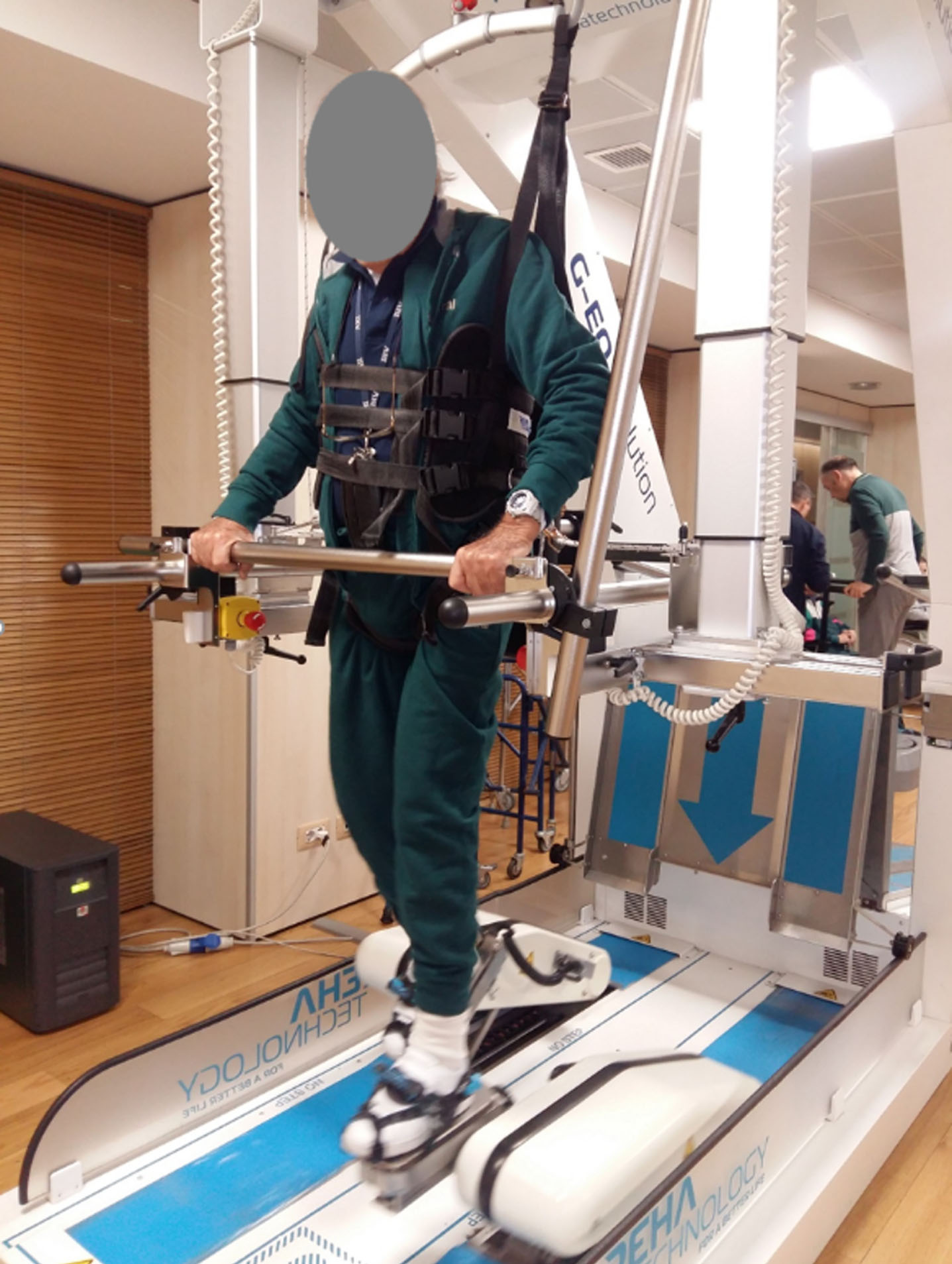
A representative patient setup and RAGT.
The CG included patients treated by means of a conventional gait rehabilitation program, 3 times a week, in 20 sessions. The treatment included: muscle strengthening exercises and stretching of the lower limb, and static and dynamic exercises for the recovery of balance in the supine and standing positions using assistive devices; training gait exercises with parallel bars or in open spaces performed both with and without assistive devices; training to climb up and down stairs; exercises to improve proprioception in the supine, sitting and standing positions, using a proprioceptive footboard; exercises to improve trunk control.
In both groups, the gait training was combined with daily conventional therapy including: functional task practice, muscle strengthening, speech therapy, and occupational therapy.
A clinical assessment based on the International Classification of Functioning, disability and health (ICF) was carried out at the beginning (T0) and at the end (T1) of the training period.
For the body function and structure ICF domain, the following clinical scales were used: Fugl-Meyer Assessment (FMA) scale; Motricity Index affected Ankle Dorsiflexion (MI-AD); Motricity Index affected Knee Extension (MI-KE); Motricity Index affected Hip Flexion (MI-HF); Motricity Index affected Lower Limb (MI-LL); Medical Research Council affected Hip flexion (MRC-HF); Medical Research Council affected Hip Extension (MRC-HE); Medical Research Council affected Knee Flexion (MRC-KF); Medical Research Council affected Knee Extension (MRC-KE); Medical Research Council affected Ankle Flexion (MRC-AF); Medical Research Council affected Ankle Extension (MRC-AE);Medical Research Council affected lower limb (MRC-LL); Modified Ashworth Scale affected Hip (MAS-H); Modified Ashworth Scale affected Knee (MAS-K); Modified Ashworth Scale affected Ankle (MAS-A); Modified Ashworth Scale affected Lower Limb (MAS-LL).
The following scales were used to measure activity ICF domain: Functional Ambulatory Classification (FAC); Tinetti Scale Balance (TIN-B); Tinetti Scale Walking (TIN-W); Trunk Control Test (TCT); Ten-Meter Walking Test (10 MWT); Six-Minute Walking Test (6 MWT); Timed Up and Go Test (TUG).
For the participation ICF domain, the Walking Handicap Scale (WHS) was used.
The primary outcome was the distance covered over a time of 6 minutes (6 MWT).
Gait analysis
Biomechanical data were collected by using the 8-camera SMART-DX motion capture system (BTS Bioengineering, Milano, Italy) sampling at 200 Hz. The Davis marker set (Davis et al., 1991), which includes 22 retro-reflective markers was adopted, and anthropometric data were collected for each subject (Winter, 2009). Each patient was asked to perform ten linear walking trials, barefoot and at a self-selected speed, straight ahead along a level surface that was approximately 6 meters long. Before formal measurements were started, practice sessions were performed to familiarize the participants with the procedure. We computed the average value of the parameters selected and the average pattern of the biomechanical gait variables across five trials for each patient. Owing to the asymmetric nature of the pathology, we analysed the affected and the unaffected sides separately. Three-dimensional marker trajectories were tracked using a frame-by-frame tracking system (Smart Tracker, BTS Bioengineering, Milan, Italy). Data were processed using 3D reconstruction software (SMART Analyzer, BTS, Milan, Italy).
In order to describe the characteristics of the gait, the following spatiotemporal parameters were analysed: step width (mm) - mediolateral distance between the two feet during double support; step length (mm) - longitudinal distance from one foot strike to the next one; stride length (mm); cadence (steps/min) - number of steps in a unit of time; mean velocity (m/s) - the mean velocity of progression for each limb; swing velocity (m/s) - the mean velocity of the swing phase for each limb; gait cycle (ms) - mean temporal duration of the gait cycle that begins with initial heel contact and ends with the subsequent heel contact of the same limb; stance time (as a % of the gait cycle) - % of the gait cycle that begins with initial contact and ends at toe off of the same limb; swing time (as a % of the gait cycle) - % of the gait cycle that begins with the toe off and ends at heel strike of the same limb; double support (as a % of the gait cycle) - % of the gait cycle feet are on the ground.
Moreover, to assess the lower limb joint kinematics were also calculated hip, knee, and ankle flexion/extension and the Range Of Motion (ROM) was defined for each joint on these graphs in the sagittal plane.
Statistical analysis
The within-group analysis was based on the application of the Wilcoxon Signed Rank test for each clinical and gait outcome registered and T0 and T1. The between-group differences were analysed by comparing the percentage increase of each outcome, defined as:
No drop outs were recorded during the treatment in both groups, and all the subjects correctly completed the protocol (compliant subjects: N = 26). Subjects of the RG tolerated the RAGT well and no adverse events were reported. The distribution of the subjects by demographic characteristics and main clinical scales at baseline did not show significant differences between the RG and the CG (Table 1).
The clinical outcomes are depicted in Table 2, which includes the results of the within-group and between-group analyses.
Clinical outcomes (median, 5th and 95th percentiles) obtained at T0 and T1, for both the CG and RG, together with the results of the statistical analysis. p-values are reported for the within-group analysis: T0score vs T1score, for the two groups separately. *(p < 0.05) and †(p < 0.0005) indicate a significant between-group difference calculated by comparing the percentage increase of each clinical outcome
Clinical outcomes (median, 5th and 95th percentiles) obtained at T0 and T1, for both the CG and RG, together with the results of the statistical analysis. p-values are reported for the within-group analysis: T0score vs T1score, for the two groups separately. *(p < 0.05) and †(p < 0.0005) indicate a significant between-group difference calculated by comparing the percentage increase of each clinical outcome
Abbreviations: CG – Conventional Group; RG – Robotic Group; T0 – before the treatment; T1 – at the end of the treatment; FMA – Fugl-Meyer Assessment; MI-AD – Motricity Index affected Ankle Dorsiflexion; MI-KE – Motricity Index affected Knee Extension; MI-HF – Motricity Index affected Hip Flexion; MI-LL – Motricity Index affected Lower Limb; MRC-HF - Medical Research Council affected Hip Flexion; MRC-HE - Medical Research Council affected Hip Extension; MRC-KF - Medical Research Council affected Knee Flexion; MRC-KE - Medical Research Council affected Knee Extension; MRC-AF - Medical Research Council affected Ankle Flexion; MRC-AE - Medical Research Council affected Ankle Extension; MRC-LL – Medical Research Council affected Lower Limb; MAS-H - Modified Ashworth Scale affected Hip; MAS-K - Modified Ashworth Scale affected Knee; MAS-A - Modified Ashworth Scale affected Ankle; MAS-LL – Modified Ashworth Scale affected Lower Limb; FAC – Functional Ambulatory Classification; TIN-B – Tinetti Scale Balance; TIN-W – Tinetti Scale Walking; TCT – Trunk Control Test; WHS – Walking Handicap Scale; 10 MWT – Ten-Meter Walking Test; 6 MWT – Six-Minute Walking Test; TUG – Timed Up and Go Test.
In the within-group analysis, the RG revealed statistically significant changes in all clinical scales except for the FMA and the MAS-A. The variations between T0 and T1 revealed an improvement in body function (MI, MRC), activities (FAC, TIN-B, TIN-W, TCT), and participation (WHS). The spasticity of the affected lower limb (MAS-LL) decreased at the end of the RAGT with a p-value <0.005, although the ankle did not change it significantly. The clinical outcomes that assessed the execution of motor tasks, revealed a significantly increase in the distance covered by RG during the 6 MWT, whose median value at T0 was 155 m and at T1 was 289.50 m. The patients in RG obtained a significant increase in the velocity and in the time measured during the 10 MWT (T0 = 0.60 m/s; T1 = 0.91 m/s; p-value = 0.021) and TUG (T0 = 17.20 s; T1 = 13.00 s; p-value = 0.003) respectively.
In the within-group analysis, the CG significantly improved all clinical scales except for the MI-HF, MRC-HE, MRC-AF, MRC-AE, and MAS. Despite the MRC-HE, MRC-AF, MRC-AE did not change significantly, the total muscular strength of the affected lower limb (MRC-LL) revealed a significant increase at T1. The spasticity showed no statistically significant changes in any of the considered joints (MAS-H, MAS-K, MAS-A). The variations between T0 and T1 revealed significant improvements in the Motricity index of the affected ankle dorsiflexion (MI-AD), knee extension (MI-KE), and in the total score of the affected lower limb (MI-LL). Positive significant variations between T0 and T1 were found in the activity (FAC, TIN-B, TIN-W, TCT) and participation (WHS) domains. The performances during the 10 MWT and TUG revealed a significant improvement at the end of the treatment. However, the distance covered during the 6 MWT did not increase significantly.
In the between-group analysis of the clinical outcomes, statistically significant differences between RG and CG were found in MRC-HE, TCT, 10 MWT, 6 MWT, and TUG. In particular, the RG revealed an increase of muscular strength in hip extension, while such outcome did not change in the CG. Moreover, RG’ gain in performances during 10 MWT, 6 MWT and TUG tasks was higher than CG.
The gait outcomes are illustrated in Table 3, which includes the results of the within-group analysis. The CG showed no statistical significant changes in any of the gait parameters. The RG significantly increased the step length of the affected side, the length and time of the gait cycles, the cadence, the mean velocities, the swing velocity of the affected side, the stance time of the unaffected side, and the ROM of the affected knee. The between-group analysis did not reveal a statistical significant difference between CG and RG in gait outcomes.
Gait outcomes (median, 5th and 95th percentiles) obtained from the instrumental gait analysis at T0 and T1, from both the CG and RG, together with the results of the statistical analysis. p-values are reported for the within-group analysis: T0score vs T1score, for the two groups separately. No significant between-group differences were obtained by comparing the percentage increase of each clinical outcome
Abbreviations: CG - Conventional Group; RG - Robotic Group; T0 - before the treatment; T1 - at the end of the treatment; AS – Affected Side; US – Unaffected Side.
Literature on stroke rehabilitation suggests high-dose therapy, intensive and repetitive task oriented practice as strategies for successful active motor relearning of ambulation (Nichols-Larsen et al., 2005). Such features are typical of RAGT. Published studies assessing the efficacy of RAGT in stroke rehabilitation found that RAGT, when combined with conventional therapy, improves functional ambulation outcomes (Freivogel et al., 2008; Hesse et al., 2012; Aprile et al., 2017). Moreover, subjects who received RAGT were more likely to achieve independent walking than their peers who received conventional therapy only (Mehrolz et al., 2012; 2013; 2017). Systematic reviews have not found any difference in gait speed and endurance when RAGT was administered with the same intensity and duration than conventional one (Mehrolz et al., 2017; Bruni et al., 2018). Comparisons between end-effector and exoskeleton RAGT have reported significantly higher rates of independent walking in patients who conducted RAGT with end-effector devices (Mehrholz & Pohl, 2012). However, literature on the effects of RAGT in terms of variation of gait parameters is rather limited (Mao et al., 2015; Aprile et al., 2017; Esquenazi et al., 2017). In our previous study on chronic stroke patients, the RAGT increased the gait endurance and decreased spasticity in the lower limb, compared with traditional therapy (Aprile et al., 2017).
The aim of this pilot study was to evaluate the efficacy of end-effector RAGT in subacute stroke patients in terms of clinical and gait outcomes, comparing them with their peers who conducted a conventional gait rehabilitation program.
We recruited 26 subacute stroke patients: all subjects tolerated the gait training well, and nobody dropped out. The RG perceived the RAGT positively and considered the treatment comfortable and useful.
The primary outcome of the study was the walking endurance measured with the 6MWT. In RG, the distance travelled in 6-minute time increased of a mean value of 106 m at the end of RAGT: this pre-post difference is more than double of the Minimal Clinically Important Difference (MCID) for subacute stroke patients of 50 m (Holden et al., 1984). Conversely, in CG, the distance travelled in 6-minute time increased of a mean value of 20 m at the end of the therapy and such outcome is lower than the MCID. Moreover, the pre-post values of 6 MWT were significantly different in EG and not significant in CG. Such findings are consistent with studies on subacute stroke subjects (Tong et al., 2006; Peurala et al., 2009; Taveggia et al., 2016) and on the chronic ones (Aprile et al., 2017) who conducted RAGT with similar devices.
For the body function and structure ICF domain, the FMA showed a positive increase in both groups, although it was significant in the CG that had a lower value at T0 than RG. The total and partial scores of MI significantly improved at T1, except for the MI ad hip level in the CG. Literature on RAGT confirms these results: at the end of RAGT patients restored a complete active range of motion against gravity at ankle, knee and hip level (Chen et al., 2013). Such findings are different form the ones obtained on chronic stroke subjects, who did not achieve significant changes in the MI (Aprile et al., 2017). The total score representing the muscular strength of the affected lower limb (MRC-LL) significantly improved in both groups at the end of the gait training: in RG there was an improvement in the muscular strength of all joints (MRC-HF, MRC-HE, MRC-KF, MRC-KE, MRC-AF, MRC-AE); in CG there was a statistically significant improvement only in MRC-HF, MRC-KF and MRC-KE. In particular, the between-group analysis revealed a significant difference between the CG and the RG in the muscular strength during hip extension (MRC-HE). The spasticity did not change in the CG, while the subjects belonging to the RG experienced a significant decrease of their spasticity at hip and knee level. Such findings are partially in accordance with literature on the effects of RAGT with treadmill-based devices (Mehrholz & Pohl, 2012; Mehrholz et al. 2013; 2017; Bruni et al., 2018).
For the activity ICF domain, all subjects significantly increased the degree of independence and the motor skills necessary for functional ambulation (FAC scores significantly increased in both groups). Such finding is in line with the systematic reviews on the topic (Mehrholz & Pohl, 2012; Bruni et al., 2018) and it is particularly important considering that the FAC outcome predicts independent community ambulation 6 months after stroke (Mehrholz et al., 2007). The trunk control (TCT scores) positively increased at the end of the treatment in all patients. However, the RT showed a higher percentage increase of TCT scores (21.7%) than the CG (9.6%) and such between-group difference was significant (p < 0.05). Similarly, the performance related to the 10 MWT the TUG positively changed in both group, with a higher significant increment in the RG. Such outcomes are in accordance with the ones by Taveggia et al. (Taveggia et al., 2016).
For the participation ICF domain, a significant improvement in the WHS was observed in both groups, thus indicating an increased walking ability at home and in the community. At T0, the WHS was evaluated by considering the patients’ participation in the common areas of the hospitals. These results are similar to the ones obtained in chronic stroke patients who participated to a multicentric clinical study (Mazzoleni et al., 2017).
Therefore, the obtained clinical outcomes suggest that RAGT produce encouraging changes in body function, activity and participation in subacute stroke patients. Moreover, our findings are in agreement with a previous review by Bruni et al (Bruni et al., 2018), who suggested that earlier RAGT produces higher recovery rates.
The gait performance data obtained from the biomechanical analysis of ambulation are consistent with findings reported in persons with hemiplegic pattern (Boudarham et al., 2016). The gait outcomes showed significant pre-post changes (improved values) in the RG (step length of the affected side, the length and time of the gait cycles, the cadence, the mean velocities, the swing velocity of the affected side, the stance time of the unaffected side, and the ROM of the affected knee) after RAGT. On the contrary, the CG did not reveal any significant variation in gait parameters at the end of the therapy. These findings are in accordance with the ones obtained by Mao et al. (Mao et al., 2015) on the biomechanical effects of body weight support treadmill training on gait in subacute stroke subjects.
The gait cycle length of both sides significantly increased in RG (AS: T0 = 688.0 mm T1 = 793.5 mm; US: T0 = 734.0 mm T1 = 790.0 mm): this is particularly relevant if we consider that the main gait cycle length for healthy adults is of about 1000 mm.
The spatiotemporal parameters that describe the speed in performing the deambulator motor task evidenced increases at the end of the gait training in both groups, although only in the RG such variation was significant. At T0, the CG registered mean velocities of both sides over 0.4 m/s respectively, while the RG showed values under 0.4 m/s. Since Perry et al. (Perry et al., 1995) classified subjects with self-selected gait velocities <0.4 m/s as “household ambulators”, and with values between 0.4 and 0.8 m/s as “limited community ambulators”, our subjects could be classified accordingly. Interestingly, although the participants of the RG had a lowers walking speed at T0, they significantly increased it, and moved forward to the next ambulatory level, i.e. “limited community ambulators” at the end of RAGT.
As regards the gait kinematics, significant changes were produced by robot training at knee level of the affected side, with a mean percentage increase of 3.8%. The other ROM calculated for each joint did not reveal any significant pre-post difference in both groups.
The gait outcomes did not demonstrate any significant differences between groups. A similar finding was obtained by Esquenazi (Esquenazi et al., 2017), where a comparison of end-effector, exoskeleton and treadmill based gait training was presented.
Training based on the robotic device thus offers the patient a more intensive, repetitive and automatic form of exercise that more closely reflects the characteristics of the physiological deambulation and their effects are more incisive in subacute than in chronic stroke patients.
These results suggest that with an intensive and appropriate RAGT, subacute stroke patients can increase the walking performance and the quality of their deambulation.
To our knowledge, our study is the first attempt to compare robotic versus conventional gait training in subacute stroke patients that considers both clinical and gait outcomes (spatiotemporal parameters and gait kinematics). The obtained findings, even if are preliminary data, are interesting for clinical practice because they suggested that this type of RAGT could significantly improved gait pattern. With the conventional gait training, on the other hand, no gait outcomes changed at the end of the therapy.
The simplicity of the treatment, the lack of side effects, and the obtained results suggest that end-effector RAGT seems to be effective for gait rehabilitation in subacute stroke subjects.
A limitation of this study was the small number of enrolled subjects and the case-control nature of the study. However, this work represents one of the first attempts to describe the clinical and biomechanical effects provided by end-effector RAGT in subacute stroke subjects and demonstrates that future RCT studies with a larger population are recommended. Another limitation is the lack of a long-term follow-up, to detect the time after which the recovery of walking can be considered completed and gait strategies not changeable.
However, our preliminary data highlights that both types of gait rehabilitation yielded significantly positive results in subacute stroke patients: both conventional and robot-assisted gait training produced promising effects on clinical outcomes, but only RG showed significant improvement in the gait parameters. Comparing the two groups, clinical outcomes improved more in the RG than the CG at the end of the therapy. These results are obtained probably because the end-effector device offers a more intensive, controlled, and physiological gait training from the beginning of the rehabilitation program without to wait that the patient reaches a trunk control like happen during conventional treatment.
Conclusions
This study showed that end-effector robotic-assisted gait training may lead improvements in clinical and gait outcomes in subacute stroke patients. The comparison with conventional gait training depicts that the robotic group improved more their functional and motor status. The obtained results suggest future research for optimizing and personalising the robotic treatment.
Conflict of interest
The manuscript has not been submitted to other journal for simultaneous consideration.
The manuscript has not been published previously. No data have been fabricated or manipulated (including images). No data, text, or theories by others are presented as if they were the author’s own.
Ethics
Availability of data and material
The authors are available to send data to those who request it.