
Abstract
BACKGROUND
We have shown elevated levels of p53 and active caspase-3 in the heart with Parkinson disease (PD). The main aim of this study is to examine the effect of treadmill training on the cardiac expression of p53 and active caspase-3 in the mouse with induced Parkinsonism.
METHODS:
Thirty randomly selected normal albino mice were equally divided into the following 3 groups: sedentary control (SC), sedentary Parkinson diseased (SPD), and exercised Parkinson diseased (EPD). 1-methyl-4-phenyl-1,2,3,6-tetrahydropyridine and probenecid (MPTP/p) were used to induce chronic Parkinson disease in the SPD and EPD animals. The expression of p53 and active caspase-3 was investigated, using immunohistochemistry, in the heart in each animal group.
RESULTS:
Both p53 and active caspase-3 expression was significantly (p value < 0.05) reduced in the PD heart following endurance exercise training.
CONCLUSION:
Our present data suggest that chronic exercise training reduced PD-induced upregulation of p53 and active caspase-3 in the heart. Thus, our study suggests that inhibiting p53 and/or active caspase-3 may be considered as a therapeutic approach to ameliorate PD cardiomyopathy.
Introduction
Parkinson disease is a progressive neuromuscular disorder (Delenclos, Jones, McLean, & Uitti, 2016). It is caused by dopaminergic neurodegeneration that is suggested to be due to apoptosis of the dopaminergic neurons leading to subsequent considerable reduction in dopamine (Hu et al., 2019). Apoptosis is a programmed cell death that utilizes caspases, which can be either initiator or executioner caspases in the apoptotic pathways (Crowley & Waterhouse, 2016; Erekat, 2018a). Caspases normally exist in cells as inactive zymogens that become proteolytically active only upon apoptotic stimuli (Crowley & Waterhouse, 2016). Active caspase-3 is an executioner caspase that is regarded as key player in apoptosis, and that has been shown in PD brains and skeletal muscles (M. D. Al-Jarrah & Erekat, 2017; N. S. Erekat, 2015, 2017, 2019). Caspase activation has been shown to be caused by tumor suppressor protein p53 (Amaral, Xavier, Steer, & Rodrigues, 2010). P53 upregulation has been shown in PD brain and skeletal muscles (M. D. Al-Jarrah & Erekat, 2017; Perier, Bove, & Vila, 2012).
Beside the motor symptoms, non-motor symptoms, such as cardiac autonomic dysfunction, charac-terize PD (Scorza, Fiorini, Scorza, & Finsterer, 2018). Exercise training has been considered as complementary strategy in the management of PD, since it has been shown to exert neuroprotective effects, and to promote both motor and non-motor symptoms in PD (Schenkman et al., 2018; Xu, Fu, & Le, 2019).
Upregulation of p53 and active caspase-3 has been shown in PD heart (N. S. Erekat & M. D. Al-Jarrah, 2018a). However, to our knowledge, alterations in the cardiac expression of p53 and active caspase-3 in PD following endurance exercise training have never been examined before. Therefore, the goal of this study was to investigate the effect of chronic exercise training on p53 and active caspase-3 expression in the heart from mice with chronic MPTP-induced PD.
Materials and methods
Animals
Thirty randomly chosen normal Albino mice were separated into 3 equal groups, which were sedentary control (SC), sedentary Parkinson diseased (SPD), and exercised Parkinson diseased (EPD) groups. Identical conditions, including 22±1°C temperature, 12 hours dark/light cycle, and free access to standard chow and water, were used to house mice in individual cages. Animal-related protocols were carried out in accordance with the guidelines of the Institutional Animal Care and Use Committee at Jordan University of Science and Technology. A formerly described protocol was used to induce Parkinson disease in mice in the SPD and EPD groups, by injecting them with 10 doses of 1-methyl-4-1,2,3,6-tetrahydropyridine hydrochloride (25 mg/kg) subcutaneously and probencid (250 mg/kg, in Tris-HCl buffer) intraperitoneally, over 5 weeks, three days and half apart (Meredith, Totterdell, Potashkin, & Surmeier, 2008, Erekat & M. D. Al-Jarrah, 2018b). Simultaneously, intraperitoneal saline (25 mg/kg) injections were administered in control mice. Cervical dislocation was used to sacrifice all mice at the end of the exercise training, which was 4 weeks post MPTP/p treatment.
Exercise protocol
Mice in the EPD group were trained according to the previously described exercise training protocol that was reported to afford sufficient systemic and cellular adaptations (M. D. Al-Jarrah et al., 2007). Briefly, mice in the EPD group ran, at a speed of 18 m/min, 40 min/day for 5 days/week, on a custom treadmill with 10 separate lanes (M. D. Al-Jarrah & Erekat, 2018b; N. S. Erekat, Al-Khatib, & Al-Jarrah, 2014; N. S. Erekat, Al-Jarrah, & Al Khatib, 2014).
Immunohistochemistry of p53 and active caspase-3 in the heart
Immunohistochemistry for p53 and active caspase-3 was executed using the previously explained protocol (Erekat et al. 2018; N. S. Erekat, 2018b). Briefly, deparaffinization and rehydration were performed to the prepared 4μm thick paraffin-embedded heart sections. After that, 3%hydrogen peroxide in methanol was used to block endogenous peroxidase activity in the sections, which were subsequently washed in phosphate buffered saline (PBS). Next, some of the sections were incubated with anti-p53 antibody (Santa Cruz Biotechnology, Santa Cruz, CA, USA), and the others were incubated with anti-active caspase-3 antibody (abcam, Cambridge, MA, USA), according to the dilutions recommended by the manufacturers. Afterward, after being washed with PBS, the sections were incubated with biotinylated secondary antibody (LSAB kit, Dako, Carpinteria, CA, USA). Next, the sections were rinsed off with PBS, and they were subsequently incubated with streptavidin horse radish peroxidase (LSAB kit, Dako, Carpinteria, CA, USA). Successively, the sections were washed with PBS, and they were successively treated with 3, 3’-Diaminobenzidine (DAB) substrate until the wanted color intensity was viewed. Finally, the sections were washed with tap water in order to stop the reaction. Then, hematoxylin was used to counterstain sections. Primary antibody was omitted from the processing of the negative control slides. Sections of PD heart were used as the positive controls for p53 and active caspase-3. Sections were observed under the light microscopy.
Data collection and analysis
Ten slides from each animal in each of the three groups were inspected under the light microscopy. Ten arbitrarily selected areas from every section were photographed using digital camera, and they were consequently studied for p53 and active caspase-3 expression in the heart. Then, the overall pixels area occupied by positive staining in relation to the entire pixels area was calculated in every photographed area in each section using Adobe Photoshop software, as explained previously (Al-Jarrah, Erekat, & Al-Khatib, 2014, Erekat, Al-Khatib, & Al-Jarrah, 2013, Erekat, Rababa’h, & Al-Jarrah, 2019). Subsequently, the average of the proportions of the pixels area engaged by positive staining relative to the total pixels area was calculated for every animal in every group.
Statistical analysis
P53 and active caspase-3 expression was assessed in the hearts, and statistically compared among the three various groups (n = 10 animals per group) via one way ANOVA using SPSS software version 19.0 (SPSS Inc., USA). Differences in p53 expression and active caspase-3 expression at p value < 0.05 were considered statistically significant.
Results
Immunostaining of p53 (Figs. 1A) and of active caspase-3 (Figs. 1D) could be barely noticed in cardiac muscle from SC. Conversely, p53 and active caspase-3 immunoreactivities were noticeably improved in cardiac muscles from the SPD group (Fig. 1B and 1E, respectively), but they were substantially reduced in cardiac muscle sections from EPD mice (Fig. 1C and 1F, respectively).
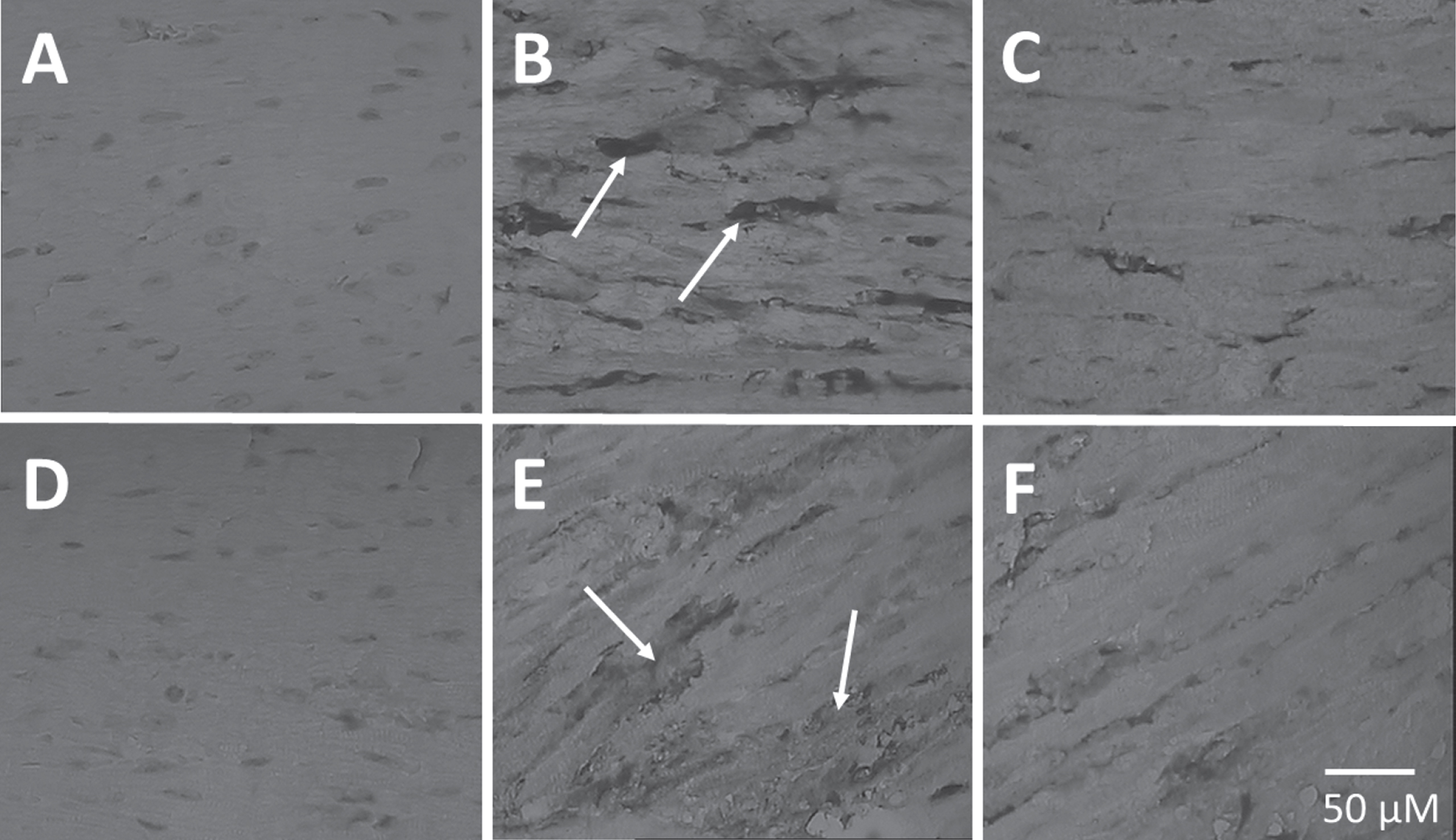
Immunohistochemical staining of p53 and active caspase-3 in 4μm thick paraffin-embedded heart sections. (A&D) From SC. (B&E) From SPD. (C&F) From EPD. SC: Sedentary Control. EC: Exercised Control. SPD: Sedentary Parkinson diseased. EPD: Exercised Parkinson diseased. Scale bar shown in (F) applies to all images in the figure.
P53 immunoreactivity is barely detected in the SC heart. However, p53 immunostaining is conspicuously noticeable in the heart from SPD group (at the tip of the arrows), but it is apparently reduced following endurance exercise training in the EPD mice.
Similarly, active caspase-3 immunoreactivity is scarcely noticed in the SC heart. Nevertheless, active caspase-3 immunostaining is very noticeable in the SPD group (at the tip of the arrows), but it is observably reduced subsequent to endurance exercise training in the EPD mice.
P53 (p < 0.01) (Fig. 2) and active caspase-3 (p < 0.05) (Fig. 3) expression is statistically significantly higher in SPD cardiac muscle than that in the control cardiac muscle. However, p53 (p < 0.01) (Fig. 2) and active caspase-3 (p < 0.05) (Fig. 3) expression in the cardiac muscle is statistically significantly lower in the EPD group than in the SPD group.
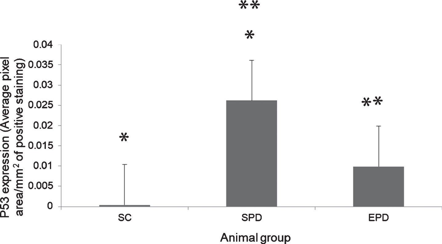
Expression of p53 in the heart. The level of P53 expression was significantly larger in the SPD group compared to the SC group (P < 0.01*). P53 expression level significantly declined (P< 0.01, **) in the EPD mice. SC: Sedentary Control. SPD: Sedentary Parkinson diseased. EPD: Exercised Parkinson diseased.
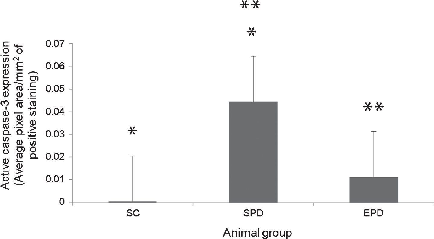
Expression of active caspase-3 in the heart. The level of active caspsase-3 expression was significantly larger in the SPD group compared to the SC group (P < 0.01*). Active caspase-3 expression level significantly declined (P < 0.01, **) in the EPD mice. SC: Sedentary Control. SPD: Sedentary Parkinson diseased. EPD: Exercised Parkinson diseased.
This study is the first to elucidate the effect of endurance exercise training on the cardiac expression of the apoptotic mediators, namely p53 and active caspase-3, in PD. The consequent analysis suggests that endurance exercise training could considerably suppress the cardiac overexpression of p53 and active caspase-3 in mice with MPTP-induced parkinsonism.
Cardiac upregulation of p53 and active caspase-3 has been previously shown in PD 4 weeks following MPTP/p treatment indicating apoptosis as a potential mechanism underlying cardiac abnormalities characterizing PD (M. D. Al-Jarrah & Erekat, 2017). Concomitantly, substantial reduction in striatal dopamine was illustrated 4 weeks following MPTP/p treatment (Gdynia et al., 2009; Meredith et al., 2008; Potashkin et al., 2007).
Exercise training has been assumed to improve motor and non-motor symptoms in PD patients (Cugusi et al., 2015; Dashtipour et al., 2015). And since the apoptotic mediators p53 and active caspase-3 have potential involvement in the progression of PD cardiac abnormalities (N. S. Erekat & M. D. Al-Jarrah, 2018a), we sought to examine the impact of chronic exercise training on p53 and active caspase-3 expression in PD heart in order to investigate the potential mechanism by which exercise training may beneficially affect PD patients.
Endurance exercise training has reduced p53 and active caspase-3 upregulation in PD skeletal muscle (M. D. Al-Jarrah & Erekat, 2017). Exercise training has also been reported to reduce apoptosis in the heart (Lin et al., 2018). Similarly, our results illustrate significant attenuation in the upregualtion of the apoptotic mediators p53 and active caspase-3 in cardiac muscles of mice with MPTP-induced chronic Parkinson’s disease following endurance exercise training (Figs. 2 3, respectively), indicating potentially attenuated apoptosis following endurance exercise training.
Exercise training promotes mitochondrial biogenesis in the heart (Vettor et al., 2014). Moreover, exercise training attenuates oxidative stress in the heart (Darband et al., 2020). It has been suggested that oxidative stress mediates apoptosis (Chen et al., 2019; Li et al., 2017). Additionally, exercise suppressed inflammatory responses, which have been shown to induce apoptosis (Darband et al., 2020; Handschin & Spiegelman, 2008; Ramesh, Benge, Pahar, & Philipp, 2012). Hence, it can be inferred that attenuated p53 and active caspase-3 upregulation in the PD heart, indicative of attenuated apoptosis, following endurance exercise training, revealed by our results (Figs. 2 3), may explain the beneficial effects of endurance exercise training on cardiac function in PD (Montero Ferro et al., 2019).
Conclusion
In conclusion, this is the first study to illustrate the impact of endurance exercise training on the apoptotic mediators p53 and active caspase-3 expression in PD heart. In summary, chronic exercise training suppressed the upregulation of p53 and active caspase-3 in chronic PD heart subsequent to MPTP/p treatment. Thus, this study confirms the previous suggestion that inhibiting p53 and/or active caspase-3 might be considered as a new therapeutic approach in improving PD symptoms.
Author contributions
All authors participated in the design, interpretation of the studies, analysis of the data and review of the manuscript.
Conflict of interest
The authors have no conflict of interest to declare.
Funding
This study was financially supported by The Deanship of Research at Jordan University of Science and Technology, Irbid, Jordan. Grant # 20130252.