
Abstract
The most common visual defect to follow a lesion of the retrochiasmal pathways is homonymous hemianopia (HH), whereby patients are blind to the contralesional visual field of each eye. Homonymous hemianopia has been studied in terms of its deleterious consequences on perceptual, cognitive and motor tasks as well as because it represents an interesting model of vision loss after a unilateral lesion of the occipital lobe. From a behavioral perspective, in addition to exhibiting a severe deficit in their contralesional visual field, HH patients can also exhibit dissociations between perception and awareness. Firstly, HH patients suffering from anosognosia may be unaware of their visual field defect. Secondly, HH patients can present with unconscious visual abilities in the blind hemifield, a phenomenon referred to as blindsight. Thirdly, recent reports demonstrate that HH patients can suffer from a subtle deficit in their ipsilesional visual field that they are unaware of, a condition called sightblindness (i.e. the reverse case of ‘blindsight’). Finally, HH patients may also exhibit visual hallucinations in their blind field; however, such patients are not systematically aware that their perceptions are unreal. In this review, we provide an overview of the visual-field losses in HH patients after a left or right unilateral occipital lesion. Furthermore, we explore the implications of these four phenomena for models of visual processing and rehabilitation of visual field defects in HH patients. Finally, in contrast to the traditional view that HH is solely a visual-field defect, we discuss why this deficit is an interesting model for studying the dissociation between perception and awareness.
Homonymous hemianopia: definition, etiology and lesion location
Clinically, the most common visual field defect to follow a retrochiasmatic lesion is homonymous hemianopia (HH) (Zihl, 2011). In most cases, the hemianopic defect is congruent: the losses to the contralesional field of each eye are symmetric to the point that they can be superimposed (Zihl, 2011) (see Fig. 1). Interestingly, 70% of strokes that involve the posterior cerebral arteries lead to HH (Pambakian & Kennard, 1997) and HH occurs in 30% of patients that have suffered a stroke (Zhang et al., 2006). Regarding lesion side, our group recently demonstrated that the defects in HH patients, as well as the cortical reorganization that follows a V1 lesion, can depend on the hemisphere in which the lesion occurs (Cavezian et al., 2010; and Perez et al., 2013). These results suggest the existence of hemispheric specialization at the occipital level, which could influence the adaptive and reorganizational phenomena that follow visual-cortex lesions and visual-field defects (for a discussion, see: Perez et al., 2013 and Cavezian et al., 2015). In this review, we provide an overview of the visual-field losses in HH patients after a left or right unilateral occipital lesion. Furthermore, in contrast to the traditional view that HH is solely a visual-field defect, we discuss why this deficit is an interesting model for studying the dissociation between perception and awareness.

Illustration of the visual-field deficits in left HH (
Dissociations between perception and awareness in homonymous hemianopic (HH) patients after a unilateral lesion of V1.
Lesions in the primary visual cortex (V1) cause a loss of conscious access to most visual information in the contralesional visual field (Holmes, 1918; and Weiskrantz et al., 1974). However, some HH patients are unaware of their visual field defect. In 1885, Von Monakow published the first description of unawareness of disorders due to brain lesions. Subsequently, Babinski (1918) coined the term anosognosia to describe a phenomenon that ranges from non-critical underestimation to explicit, intractable denial of clinical signs that include cortical blindness, spatial neglect, cortical deafness, hemiplegia, word deafness, dyslexia and dysphasia (for a review and discussion, see: Gainotti, 2019). Anosognosia is generally associated with a poorer prognosis because its presence prevents the patient from getting involved in rehabilitation and most likely lengthens it. This is the reason why in most rehabilitation programs the first step aims at reducing anosognosia. In a seminal paper on this topic, Bisiach et al. (1986) investigated awareness of motor and visual-field defects in 97 right brain-damaged subjects. The researchers found that anosognosia to motor disorders and anosognosia to visual disorders were double-dissociated and that anosognosia for HH failed to show any association to unilateral neglect. Additionally, four (40%) of the ten patients with severe anosognosia for HH had minimal (if any) anosognosia for hemiplegia. These findings confirmed an earlier. crucial observation by Anton (1899) that his patient Mercz, although quite unaware of her blindness, was painfully conscious of her mild verbal disorders. Moreover, Bisiach et al. (1986), in their study, reported that severe unawareness of hemianopia was much more frequent than was unawareness of motor impairment. These authors proposed that lesions involving the visual pathways could be very close to the damaged brain area responsible for disordered monitoring of the resulting dysfunction. Indeed, as Breitmeyer (2014) proposed patients with visual-field defects, may thus fail to acknowledge their visual deficits and appear as being cognitively blind to their neurological blindness. Critchley (1949) identified six degrees of awareness of visual-field defects: the first, a total lack of awareness of the deficit; the second, unawareness of the defect, albeit with recognition of the consequences of the defect (e.g. frequently bumping into objects or persons); the third, attribution of the deficit to external causes (e.g. a lack of illumination); the fourth, an awareness that something is “wrong” with vision, but an incapacity to precisely define or describe the defect; the fifth, an awareness of a lateralized visual defect, yet with the belief that this is due to an eye problem rather than a visual-field defect; and finally, full awareness of the HH defect.
Most HH patients do not experience a “black spot” in place of their blind visual field. Indeed, HH can be considered as a reduction of the visual field (i.e. a field with new peripheral borders) that leads to conscious perception in only half of the original visual field. Accordingly, awareness of an absence of visual stimulation in the contralesional visual field might be particularly difficult for patients, thus leading to anosognosia of the visual-field loss. Along these lines, Levine et al. (1990) proposed that a post-chiasmatic lesion does not necessarily provide any specific information about the direct experience that uniquely specifies the defect. As such, Zihl and colleagues (2000) suggested that unawareness of visual-field defects is common among patients with normal intellectual function. Indeed, the authors insisted that, due to the absence of immediate sensations, HH patients must infer the presence of the visual-field defect based on failures that derive from it (e.g. car accidents, falls, difficulties in reading, etc.). Moreover, according to Levine (1990), discovery of visual loss requires “the ability to change the mental set or outlook based on evidence from the external world” and, according to Zihl (2000), it depends mainly on mental flexibility.
Regarding the neuro-anatomic correlates of anosognosia in HH, there are clinical and experimental reports that various types of lesions involving different anatomic regions can lead to anosognosia in HH and that, conversely, similar lesions are not always associated with anosognosia (see, for example: Bisiach et al., 1986). In 1997, Celesia, Brigell & Vaphiades reported that 20 of 32 (62%) HH patients expressed anosognosia of their visual deficit. Interestingly, they observed anosognosia in 16 of the 26 (62%) patients with right-brain lesions and in four of the six (67%) patients with left-brain lesions. Consistent with the findings of Bisiach et al. (1986), the authors found that anosognosia for hemianopia was clearly dissociated from anosognosia for neglect and appeared in the absence of cognitive impairment. Interestingly, Celesia, Brigell & Vaphiades also found that most patients with positive spontaneous visual phenomena (PSVP), including phosphenes, photopsia, visual hallucinations or palinopsia, were aware of their deficit: only three (25%) of twelve PSVP patients exhibited visual anosognosia. We further discuss this issue below. Regarding the neuroanatomic correlates of anosognosia for hemianopia, Celesia et al. (1997) were unable to identify any cortical areas specific for awareness of the visual deficit. Lesions limited to V1, V2 and portions of V3, or their connections from the lateral geniculate nucleus, were sufficient to produce anosognosia for the visual-field defect. These findings are consistent with those of an earlier study by Koehler et al. (1986) who, based on CT correlations, proposed that patients aware of their HH had purely occipital lesions, whereas patients unaware of their hemianopia had more anterior lesions such as parietal lesions or lesions interrupting the associative pathways from the primary visual cortex. Interestingly, endeavoring to identify hemispheric specialization for unawareness of the visual deficit, Celesia et al. (1997) observed anosognosia for hemianopia in sixteen of 20 patients (80%) with lesions in the right hemisphere, but in four of seven patients (57%) with lesions in the left hemisphere. Accordingly, anosognosia for HH might be preferentially linked to right hemisphere lesions, analogously to what had previously been proposed for left unilateral spatial neglect (Heilman and Abell, 1980). As proposed by Celesia et al. (1997), and as we discuss at the end of this review, anosognosia for HH could also be linked to other behavioral phenomena that we describe below: blindsight, sightblindness and visual hallucinations (Table 1).
Dissociations between perception and awareness in homonymous hemianopia
Dissociations between perception and awareness in homonymous hemianopia
Definition
In a seminal study, Pöppel, Held and Frost (1973) found that some HH patients could accurately steer their gaze toward a flashing light presented in their blind field, yet did not report having perceived the stimulation. Shortly afterwards, the pioneering work of Weizkrantz et al. (1974) revealed that, from a behavioral perspective, HH patients presenting with a severe visual-field defect could indeed exhibit implicit residual capacities in the contralesional (so-called “blind”) visual field. Subsequently, the same group explored several residual visual functions in the patient D.B. through forced-choice methods (Sanders et al., 1974). Specifically, they asked D.B. to distinguish the orientation of stripes (horizontal vs. vertical), to identify letters (X vs. O), and to reach, grasp, or steer his gaze towards a stimulus in his contralesional visual field. Intriguingly, in addition to performing significantly above chance level in all tasks, the patient never reported any conscious perception of the stimuli in his blind field. Based on their findings, the authors collectively referred to these capacities as blindsight. Nearly 2 decades later, the same group described another blindsight patient, G.Y., who reported awareness of certain stimuli despite not being able to attest to any concrete visual perception of them (Weiskrantz, Barbur & Sahraie, 1995). Consequently, the authors classified blindsight into two types: type I, the ability to discriminate specific attributes of a stimulus in the contralesional visual field without any awareness of it; and type II, exhibiting objective performance and subjective performance in the blind visual field above chance level, without any conscious detection of the stimuli (Weiskrantz, 1998; and Brogaard, 2015). Typically, type II blindsight corresponds to patients that exhibit significant objective performance scores in their blind visual field and report having felt something in the absence of conscious perception (for a review, see: Brogaard, 2015).
Experimental design is crucial for enabling researchers to ascertain and classify blindsight among patients. Diagnosing blindsight implies not only an accurate measure of objective performance, but also a well-controlled and interpretable measure of subjective experience. On one hand, the stimuli-presentation parameters (e.g. duration, contrast, size or location) and the response mode (e.g. verbal or motor; for a review, see: Fayel et al., 2014) of a given experiment activate dissimilar objective abilities that may differ among subjects. On the other hand, although blindsight is defined by an absence of subjective experience, the literature reflects debates on the methodology used to assess patients’ awareness (see, for example: Overgaard et al., 2008). For example, use of a binary scale (i.e. Aware vs. Unaware) may compel a subject to report being “unaware” of a stimulus despite having experienced a minor degree of awareness (Overgaard et al., 2008; and Overgaard, 2011). In their study of the patient G.R., Ramsøy & Overgaard (2004) identified distinct responses for level of awareness in the blind visual field, depending on whether the patient was tested using a binary scale (Seen vs. Not Seen) or the Perceptual Awareness Scale (PAS), a four-level scale that they developed, which comprises Clear Experience, Almost Clear Experience, Weak Glimpse and Not Seen. Indeed, when they asked G.R. to process a stimulus in his contralesional visual field and subsequently report a dichotomous answer (Seen vs. Not Seen), G.R. exhibited a typical type-I profile (i.e. implicit processing without awareness of the stimuli). However, when they assessed G.R. according to the PAS, G.R. significantly reported some degree of awareness in the presence of stimuli in his contralesional visual field, thus exhibiting a type-II profile. Despite this proven drawback, binary questionnaires are still widely used to study blindsight; indeed, very few researchers use graduated measurements of perception (see for example, Overgaard et al., 2008; and Mazzi, Bagattini, & Savazzi, 2016). Thus, understanding the nature of patients’ perceptual experience in their blind field remains a critical issue, as we discuss below.
Perceptual experience in the blind visual field
Three distinct hypotheses have been proposed to explain blindsight. Firstly, some researchers have suggested that blindsight is based on unconscious perceptual abilities (see, for example: Sanders et al., 1974; and Leh, 2006). Secondly, others have proposed that blindsight relies on residual remaining “normal” vision (see, for example: Cowey, 2009; and Hadid & Lepore, 2017). In this view, blindsight is simply considered as a reduced form of normal vision. Finally, other researchers have theorized that blindsight is a type of degraded, abnormal vision that differs from “normal” visual perception, akin to patients looking through glasses with deformed lenses. This third hypothesis arose from studies on type II blindsight and deals with a phenomenon that has been described as a “conscious experience, though of a very different nature to that of normal vision” (Mazzi et al., 2016; Weiskrantz, 2009; and Kentridge, 2015).
The three aforementioned hypotheses remain the subject of debate and disentangling them is not trivial, as most of the research in this area comprises case studies that reflect either type I or type II blindsight. Indeed, group studies using objective visual detection combined with discrimination tasks and subjective perceptual scales might enable better assessment of the frequency of specific perceptual experiences among HH patients. Moreover, measuring the occurrence of blindsight requires group studies. Given the fact that blindsight stimulation has been proven to induce visual restoration (for a review, see: Chokron et al., 2008; and Perez & Chokron, 2014), clinicians should be aware of the type and the frequency of perceptual experiences among HH patients, in order to choose the most appropriate course of therapy for each patient. However, only a few studies have been conducted on patient cohorts, and most of these have provided only indirect information on the occurrence of blindsight. For example, Perenin & Jeannerod (1975) studied eight HH patients and found blindsight in six (75%) of them, but they did not use any subjective perceptual scale. Marzi et al. (1986), studied 20 HH patients using the Redundant Signal Effect (RSE) task, which involves indirect flash presentation to assess visual information processing in the blind and in the healthy visual field. They found that only one (5%) of the 20 patients exhibited faster responses after simultaneous presentation of visual stimuli in the two visual fields, compared to single visual stimulation in the preserved field, as expected for blindsight. Thus, in this study, the incidence of blindsight was only 5%. In yet another study, (Sahraie et al., 2013), blindsight incidence was evaluated among nineteen patients by measuring their pupil responses to visual stimuli presented in the hemianopic visual field, which reflects residual visual reflexes. These authors indirectly calculated a blindsight incidence of 70% (Sahraie et al., 2013); however, this was not a direct measurement of unconscious visual capacities in the blind visual field. Ajina et al. (2015) studied 17 HH patients and distinguished between a set of twelve patients, whom they classified as “blindsight positive”, and another set of five, whom they classified as “blindsight negative”, based on two, alternative forced-choice detection tasks related to the contralesional visual field. Thus, these examples of cohort studies, in which blindsight incidence ranged from as low as 5% (Marzi et al., 1986) to as high as 75% (Perenin & Jeannerod, 1975), reflect the complexity of measuring blindsight incidence in cohorts of HH patients and the importance of experimental design.
Recently, our group sought to investigate objective and subjective perceptual experience in a cohort of eight post-stroke complete-HH patients (Garric et al., 2019). To evaluate objective perceptual abilities in the two visual fields, we first presented the patients with a letter on a screen, and then asked them to perform letter-detection (“Do you see anything?”) and identification (“Is it an X or a O”?) tasks. Here, the objective detection task was performed on a classical binary scale (“Something was presented” vs. “Nothing was presented”). Secondly, to investigate the nature of the perceptual experience in their blind visual field, a topic which had previously been examined only in single-case studies, we assessed the patients by a five-level scale, the Sensation Awareness Scale (SAS). In contrast to the aforementioned four-level PAS scale of Ramsøy & Overgaard (2004), we added a fifth level of “I felt something” to allow patients to express that they had experienced a sensation that might differ from visual sensation. Indeed, in clinical practice, patients often communicate that they “feel” the presence of something but cannot report a visual sensation per se. Thus, after each trial of the detection and identification tasks, we asked the patients to choose the most appropriate assertion: [1] I did not see anything; [2] I don’t think that I saw anything, but I am not sure; [3] I felt something; [4] I saw something; and [5] I clearly saw something and can identify it. Having measured objective performance by the forced-choice detection and identification tasks, and subjective performance by the SAS, we then compared the two scores for each patient. Consequently, we were able to reclassify our HH patients according to the dissociation between objective and subjective performance and by different concepts of blindsight described in the literature. Based on our results, we identified the following four distinct profiles:
Absence of blindsight: objective performance and subjective performance both at chance level;
Type I blindsight: objective performance above chance level, combined with a lack of statistically significant subjective sensitivity;
Type II blindsight: objective performance and subjective performance both above chance level; and
Blindsense: objective performance at chance level combined with a statistically significant subjective performance (a phenomenon that, to the best of our knowledge, had never previously been reported).
Interestingly, our findings showed that only one (12.5%) of the eight complete-HH patients met the criteria for blindsight. More importantly, our SAS enabled us to identify a previously unreported dissociation, which we characterized as blindsense, in four (50%) of the eight patients. Specifically, these four patients exhibited better-than-chance sensitivity to the presence of a stimulus on the subjective scale, despite being unable to identify the stimulus during the forced-choice task. Moreover, three (75%) of these four blindsense patients presented a significant subjective sensitivity using the gradual SAS scale, while performing at chance level on the binary detection task. Our findings from that study underline the need to investigate not only objective perceptual abilities in the blind visual field of HH patients, but also their degree of awareness. Furthermore, they suggest that blindsight in complete post-stroke HH patients is not as frequent as previously thought and in any case, is far from being the most common dissociation between perception and awareness.
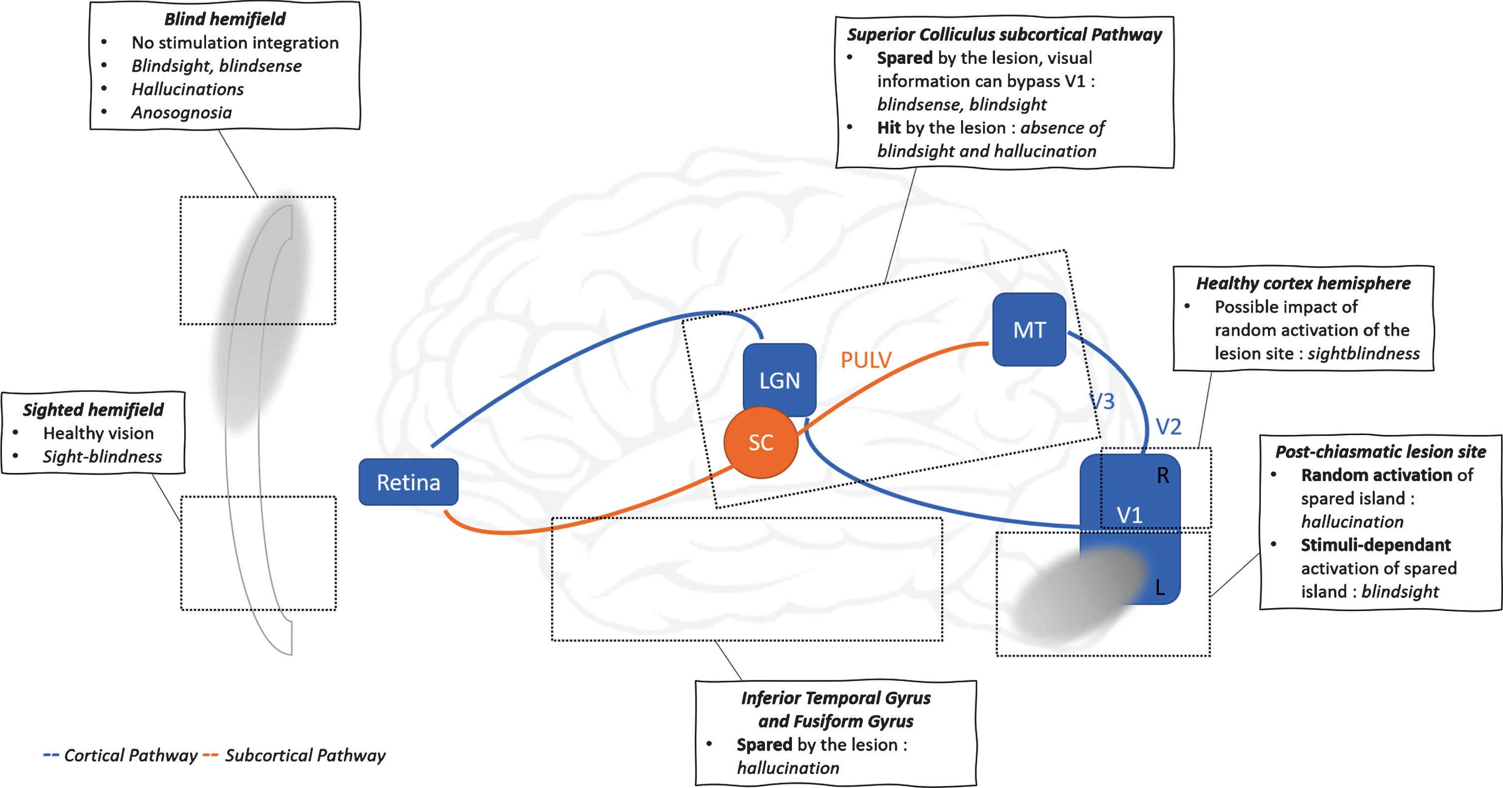
According to several studies, blindsight could depend on sparse surviving islands in the primary visual cortex (V1) (Campion, Latto & Smith, 1983; and Fendrich, Wessinger & Gazzaniga, 2001). However, blindsight has also been observed in patients lacking a functional primary visual cortex (Ajina et al., 2015; and Mazzi et al., 2016) as well as in monkeys with complete ablation of V1 (Stoerig & Cowey, 1997). Indeed, Stoerig and colleagues (2006) concluded that blindsight can occur without any functional portion of the primary visual cortex. Confirming this hypothesis, researchers have shown that blindsight could be mediated by subcortical pathways that bypass V1, such as the superior colliculus pathway and the dorsal lateral geniculate nucleus (dLGN) pathways. Moreover, several recent studies have underscored the roles of the ipsilateral hemisphere and the corpus callosum in processing of stimuli in the contralesional visual field of HH patients (Bridge et al., 2008; Celeghin et al., 2017; and Leh, 2006).
Sightblindness
Contrary to the case of blindsight, which has been extensively studied in HH patients, vision quality in the central visual field and in the ipsilesional visual field of these patients has scarcely been assessed, and moreover, has traditionally been assumed to be fully preserved. However, as we discuss ins this section, HH patients may present a subtle deficit in their ipsilesional (presumed to healthy) visual field that they are unaware of.
Hess and Pointer (1989) were among the first authors to demonstrate that spatial and temporal sensitivities were lower in the ipsilesional visual field of HH patients than in control subjects. Similarly, Rizzo & Robin (1996), followed by Poggel, Treutwein & Strasburger (2011), confirmed that HH patients can exhibit lower sensitivity to signals, compromised processing of temporal information and longer reaction times in their contralesional and ipsilesional visual fields, as compared to healthy controls. Our group investigated the right ipsilesional visual field performance of a patient 1 week before and 6 months after a neurosurgical intervention (embolization of an arteriovenous malformation in the right occipital lobe) (Peyrin et al., 2006). We addressed the role of the right visual cortex on local analysis (based on the high spatial frequency content of scene stimuli) and global analysis (based on the low spatial frequency content) of visual information in scenes. We found that damage to her right primary visual cortex (V1) had degraded her performance in her right ipsilesional visual field. Interestingly, even before surgery, she responded with less accuracy and longer reaction times in her right ipsilesional visual field for all types of scenes compared to healthy controls. Moreover, after surgery, she exhibited an additional deficit for global analysis (based on low spatial frequencies) in her right ipsilesional visual field. Based on these findings, we hypothesized that the right occipital lobe could be involved in the processing of the global aspects of a visual scene (low spatial frequencies) in both visual fields, thereby leading to a massive visual-field defect in the contralesional visual field and to a weaker visual deficit in the ipsilesional visual field.
Schadow et al. (2009) found deficits in the early and late visual processing of Gestalt patterns in the ipsilesional visual field of HH patients, whereas Paramei & Sabel (2008) reported that HH patients exhibited a diminished ability to detect fragmented targets among a noisy background in the ipsilesional visual field. Additionally, Bola, Gall & Sabel (2013a) confirmed these findings and reported processing-speed deficits in a simple detection task in the ipsilesional visual field. The authors termed this phenomenon sightblindness, as the reverse situation of blindsight (Bola, Gall, & Sabel, 2013b): thus, sightblindness refers to visuo-attentional deficits in the ipsilesional visual field, rather than to residual (albeit implicit, as discussed above) visual abilities in the contralateral visual field that are highlighted in forced-choice tasks (see, for example: Weiskrantz et al., 1974; or Leopold, 2012). Interestingly, HH patients are supposed to be unaware of residual capacities in their contralesional visual field (blindsight) and/or of their subtle visual deficit in the ipsilesional visual field (sightblindness). Along these lines, and as recently suggested, in HH patients, neither the central visual field (Cavézian et al., 2010; 2015; and Perez et al., 2013) nor the ipsilesional visual field (Bola et al., 2013a; 2013b; and Sanchez-Lopez et al., 2019) appear to be fully intact or functional. Further corroborating these findings, Mikellidou et al. (2019) very recently demonstrated that after unilateral occipital damage acquired in childhood, cortical reorganization may occur, not only in the lesioned area, but also in the healthy hemisphere. Moreover, as our group had previously proposed, the nature of the task and the type of stimulus may determine the central and ipsilesional visual deficit as well as the pattern of cortical activation in HH patients (Cavézian et al., 2010, 2015; Perez et al., 2013; and Chokron, Perez, & Peyrin, 2016). In fact, our group has investigated the effect of lesion side on the nature and severity of the ipsilesional deficit in a cohort of ten HH patients (Cavézian et al., 2015). In this study, we presented five right-HH (i.e. left brain-damaged patients) and five left-HH (i.e. right brain-damaged) patients with filtered (high or low spatial frequencies) and non-filtered scenes in their ipsilesional visual field during a detection task and a categorization task. Left-HH patients made more errors for categorizing scenes in their ipsilesional visual field than did their matched controls, regardless of the spatial frequency content of scene. In contrast, right-HH made more errors than did the controls only when categorizing high spatial frequency scenes. Interestingly, in both tasks (detection and categorization), the left-HH (right-brain-damaged) patients performed worse in their ipsilesional visual field than did the right-HH (left-brain-damaged) patients. Together, these studies among HH patients raise the question of the integrity of selective attention processing in the (healthy) ipsilesional visual field.
Our earlier findings on lesion side-specific phenomena in HH inspired us to investigate the presence, nature and severity of selective spatial attention deficits in the ipsilesional visual field in a cohort of right brain-damaged patients suffering from either left-unilateral spatial neglect (left-USN) or left-HH (Chokron, Peyrin & Perez, 2019). Specifically, we sought to determine the extent to which the nature (i.e. attentional or visual) of the deficit in the contralesional visual field might determine the nature of the ipsilesional attentional deficit. To this end, we used a letter-detection task (Chokron et al., 2000; and Tabert et al., 2000) to ascertain whether the left-USN or left-HH patients might exhibit a deficit in selective attention in their right ipsilesional visual field, as compared to healthy controls. This study yielded evidence for the presence of a non-lateralized attentional deficit in left-USN patients as well as a subtle attentional deficit in the ipsilesional visual field of left HH patients. Moreover, it emphasized the need to thoroughly test for visuo-attentional capacities in the (assumed to be healthy) ipsilesional visual field following unilateral parietal or occipital damage. Finally, and in direct relation to the present review, neither the left-USN patients nor the left-HH patients were aware of their ipsilesional attentional deficit.
Visual hallucinations in HH patients
Visual hallucinations have been defined as “visual perceptions that are completely removed from reality” as well as “perceptions without stimulus” (Borruat, 1999). Patients with visual deprivation may experience visual hallucinations in addition to blindness, including Charles Bonnet Syndrome (CBS) (for a review, see: Fernandez, Lichtstein & Vieweg, 1997). Intriguingly, in 1769, Charles Bonnet reported that his 89-year-old grandfather, who suffered from cataract-induced blindness, experienced visions of men, women, birds and buildings that changed in size, shape and location (De Morsier, 1967; and Rosenbaum et al., 1987). Importantly, despite these hallucinations, the elderly man did not exhibit any sign of cognitive deficit. Charles Bonnet Syndrome was thus initially defined as the occurrence of visual hallucinations in elderly patients free of optic nerve impairment and with no concurrent psychiatric or cognitive disturbance (De Morsier, 1967; and Bartlett, 1951). However, this definition was subsequently extended to describe “any state of visual hallucinations in the elderly irrespective of accompanying symptomatology” (Berrios & Brook, 1982). Presently, clinicians commonly associate CBS with eye or brain diseases, probably considering that visual hallucinations that occur following ocular or post-chiasmatic lesions would be signs of CBS. However, as we outline below, hallucinations in HH may represent another form of dissociation between perception and awareness that results from the cortical lesion. Indeed, in addition to anosognosia, blindsight,and sightblindness, HH patients can also experience visual hallucinations in their contralesional visual field, or even in their entire visual field. Furthermore, they may be unware of these hallucinations. However, in our view, all these phenomena should be studied together.
The hallucinations experienced by HH patients can be simple (e.g. points, lines or geometric shapes) or complex (e.g. objects, animals, people or animated scenes) and may involve the entire visual field or just part of it (Panayiotopoulos, 1999). Homonymous hemianopia patients are usually unaware that the visual perceptions in their blind visual field are in fact hallucinations. Indeed, an awareness of this would demand that HH patients compare these visual perceptions to another sensory modality which is not systematic. Usually, HH patients experience visual perceptions in their blind visual field as if they had at least partially recovered from their visual-field defect. On those occasions in which the patient realizes that what they have perceived does not really exist, they may express negative feelings or fear of their experience and very often do not tell anybody about it. In fact, HH patients often worry that if they do openly discuss their hallucinations, then they will be labeled as “crazy”.
Surprisingly, visual hallucinations in HH patients have not been systematically researched. Historically, this may have been partly due to the lack of a specific standardized questionnaire for use by practitioners. Accordingly, our group developed such a questionnaire for HH patients and more generally, for patients with neurological visual impairment, which we named the Questionnaire for Evaluating Visual Hallucinations in Homonymous Hemianopia Patients (abbreviated as the “Q3H” questionnaire) (Perez et al., 2014). For a given patient, this questionnaire enables characterization of their hallucinations (i.e. type, frequency, etc.), including assessment of the extent to which the patient is aware of the phenomenon. The few studies on these hallucinations have revealed that these can vary in frequency, intensity, duration and complexity.
Although hallucinations in HH patients are thought to depend on lesion location (Alfaro et al., 2006), their origins remain poorly understood and the literature reflects diverse hypotheses as to their etiology. Some researchers have proposed that such hallucinations result from compensatory hyper-activation of tissue neighboring the lesion area (Braun et al., 2003; and Rafique, Richards & Steeves, 2015). Indeed, Kölmel and co-workers (1985) had previously reported that the peri-lesional area of the visual cortex can generate visual hallucinations whose complexity depends on the lesion site, such that simple hallucinations would result from activation of the primary cortex, and complex hallucinations, from activation of the association cortex. Additionally, other studies have demonstrated that complex visual hallucinations can be triggered by stimulation of the temporo-occipital lobe or the parieto-occipital lobe (Rafique et al., 2015). Over the past 5 years, our group and others have suggested that the side of the occipital lesion might be an important factor in the nature and severity of visual impairments in HH patients (see, for example: Perez et al., 2013; and Cavezian et al., 2015). In fact, Walters and colleagues (2006) had previously investigated whether complex visual hallucinations caused by an occipital lesion might be linked to the lesion side and/or to patients’ emotional valence. They systematically searched for hallucinations in left- or right-brain-damaged patients and recorded the side of the hallucination as well as its emotional valence. They then assessed the associated perceptual deficits, including loss of vision within a visual field (left or right), loss of vision within a visual quadrant, allochiria, or extinction upon presentation of concurrent bilateral stimuli to the left and right visual fields. Of the fifteen patients experiencing visual hallucinations within the left hemi-space, ten (67%) had at least one visual-field defect, all of which (10/10; 100%) were in the left visual field. In other words, among patients with a left visual-field defect (i.e. right-brain damage), the hallucinations always occurred in the blind, contralesional visual field. Additionally, all the patients had associated negative affective valence to these events. With a total of ten patients experiencing visual hallucinations within the right hemi-space, only four patients (40%) exhibited at least one visual field defect (ascertained only by confrontation test). All four (100%) patients exhibited a defect within the left visual field (i.e. right-side lesion), and three (75%) of them had an associated positive emotional valence. Those results suggested that the emotional valence of hallucinations depends on the side of its apparition rather than on the lesion side. However, all the patients with visual-field defects in that study had left visual-field defects; no right visual-field defects were detected across the confrontational tasks for any of the patients. Thus, the fact that only patients with right-side lesions reported visual hallucinations that the lesion influences the occurrence of hallucinatory phenomena. This study suggests that the side of the occipital lesion determines the occurrence of visual hallucinations, whereas the visual field of apparition could influence the emotional valence, with more frequent hallucinations in the blind, contralesional visual field than in the ipsilesional visual field, as previously reported by Walters et al. (2006) in their aforementioned study. Regardless, further studies are necessary to elucidate the link between the lesion side, the visual field of apparition and the various parameters of visual hallucinations (nature, frequency, severity, similarity with mental imagery, emotional valence, etc.). Interestingly, preliminary results from our group’s current work using the Q3 H questionnaire suggest that the occurrence and type of visual hallucinations in HH patients might strongly depend on the extent and on the location of the lesion (Martinelli et al., in press).
Discussion and Conclusion
Damage to or disconnection of all or some parts of the primary visual cortex (V1) results in a region of blindness (a scotoma) in the corresponding portion of the visual field (Holmes, 1918). Herein we have reviewed four types of dissociation between perception and awareness (anosognosia, blindsight, sightblindness and visual hallucinations, seeTable 1) that, to the best of our knowledge, have never previously been analyzed together, despite representing behavioral manifestations that extend beyond visual-field defects resulting from damage or disconnections in V1 and the visual system. As we discussed throughout this review, patients suffering from HH following a unilateral lesion of the occipital lobe are not always completely aware of their deficit. Furthermore, they exhibit some residual implicit capacities in their blind visual field (e.g. blindsight). Such HH patients can suffer from a subtle visual deficit in their ipsilesional (assumed to be healthy) visual field yet be unaware of it. Moreover, these HH patients often experience visual hallucinations in their blind visual field and may not be cognizant that the hallucinatory images that they see do not correspond to reality. The four phenomena that accompany HH remind us that, contrary to pre-chiasmatic visual deficits, V1 lesions, upon disconnecting the eye from the cortical visual system, induce various dissociations between visual perception and awareness. As pointed out by Mazzi et al. (2019), blindsight research has informed researchers’ development of neural models of phenomenal awareness; however, the three other associated signs (anosognosia, sightblindness and visual hallucinations) we describe in this review had, to date, received less attention in the literature. Nevertheless, the occurrence of anosognosia, sightblindness and/or hallucinations in HH patients suggests a more complex role for V1 in conscious visual experience than what has traditionally been believed (for a discussion, see: Mazzi et al., 2019; Cowey, 2009; and Cowey & Stoerig, 2004). Indeed, these phenomena could form part of a more general dysfunction and might not be mutually exclusive. Interestingly, both blindsight and visual hallucinations in the blind visual field could exacerbate a patient’s anosognosia by providing them with implicit (real) and explicit (unreal) visual perceptions, respectively.
If patients’ contralesional visual field becomes filled with implicit and illusory visual perceptions, then they may have difficulty gaining awareness of their visual field defect. Although the idea of the entire contralesional field defect being filled is difficult to imagine, completion of the blind visual field has been described in HH: some patients have reported their scotoma as being filled in by the surrounding scene (Warrington, 1962; Sergent, 1988; and Mc Carthy, James-Galton & Plant, 2006).
This phenomenon which falls somewhere between blindsight and visual hallucinations, could in part explain anosognosia for hemianopia. Moreover, subtle ipsilesional deficits (e.g. sightblindness) in the ipsilesional visual field could mitigate the difference in visual experience between the two visual fields of HH patients, thus increasing anosognosia for the contralesional visual-field defect. Regardless, as Celesia et al. (1997) claimed, attempts to explain anosognosia in HH patients with a single theory are destined to fail because of the complexities of “awareness.” These authors proposed that unawareness in these patients may stem from various factors, including failure of discovery of the deficits, severe hemi-neglect, generalized cognitive impairment, the aforementioned “filling in” process or from some combination thereof. In the scope of the present review, our group would add that blindsight, sightblindness and visual hallucinations may also interact and play a role in dictating awareness of visual-field defects in patients suffering from HH following a post-chiasmatic lesion. Alternatively, visual hallucinations may at least partially derive from implicit visual perception (i.e. blindsight) in the contralesional visual field. Indeed, given the paucity of literature on hallucinations in HH patients, understanding how implicit perceptions could participate to the construction of explicit visual hallucinations is not trivial. Accordingly, this topic merits further research— namely, to ascertain how V1 lesions alter implicit and explicit visual sensations as well as the interactions between them.
Recent neuro-anatomic studies have elucidated the roles of the pulvinar and the superior colliculus in blindsight (see, for example: Kinoshita et al., 2019); however, further research is needed to understand the cortical and subcortical substrates of anosognosia, sightblindness and visual hallucinations in HH patients. Blindsight has been hypothesized to depend partially on interactions between the damaged hemisphere and the non-damaged hemisphere (Celeghin et al., 2015; 2017; and Silvanto, Walsh & Cowey, 2009). Our group’s recent findings suggest that this may also be the case for ipsilesional deficits in patients with unilateral parietal or occipital lesions (Chokron et al., 2019). Likewise, one cannot exclude the role of an interaction between the lesioned hemisphere and the non-lesioned hemisphere in the genesis of visual hallucinations. Further studies dealing with possible interactions among the visual field defect, anosognosia for it, blindsight, sightblindness and visual hallucinations should thus account for the different neuroanatomic correlates of such phenomena. These might encompass disconnections or damage in V1: from the eye as well as from higher cortical centers, the entire superior colliculus and the pulvinar, which would enable blindsight; in inter-hemispheric connectivity, which would lead to sightblindness; and in the ventral pathway, which would trigger visual hallucinations.
Homonymous hemianopia and its behavioral consequences represent a complex deficit that may be associated to damage of a distributed network surrounding the V1 lesion. From a more clinical perspective, HH patients deserve a complete neuropsychologic examination, including a precise evaluation of their awareness of their deficit as well as the presence, nature and duration of any visual hallucinations. Contralesional visual-field training in rehabilitation is nearly always recommended for these patients, especially that involving blindsight stimulation (Chokron et al., 2008; Das, Tadin & Huxlin, 2014; and Cavanaugh & Huxlin, 2017). However, training patients for visual discrimination and attention in the ipsilesional visual field also appears necessary, given that HH patients tend to use their “healthy” visual field to compensate for their visual-field defect.
Footnotes
Acknowledgments
The present research was supported by the Edmond and Benjamin de Rothschild Foundations.