



Abstract
BACKGROUND:
The venous disease of the legs is a common disease among adults that may lead to a deterioration in the structure and concentration of biomolecules. N-Butyl Cyanoacrylate Ablation Surgery (NBCA) or cyanoacrylate embolization (CAE) technique to adhesive the saphenous vein is an alternative method for the treatment of venous disease.
OBJECTIVE:
We aimed to show what kind of changes occurs after CAE surgery using FTIR spectroscopy combined chemometrics. We compared before and after surgery blood sera of patients to find whether a correlation between spectral data and laboratory indexes. We studied the blood sera of those who suffered from varicose veins and treated them by CAE technique.
METHODS:
In order to examine the molecular profiles in blood sera who underwent the CAE technique of the great saphenous vein for the treatment we used Fourier Transform InfraRed spectroscopy (FTIR) spectroscopy of blood samples of patients before and after surgery as a fast diagnostic technique. To obtain information about the spectra variation among the types of samples Principal component analysis (PCA) was performed for fingerprint, amide II with amide I regions. To find normality among variations Partial Least Square P-P plot of residual was performed.
RESULTS:
Absorbance values were statistically significant only in amide II, amide I, and OH vibrations. In the blood collected before surgery, higher peaks area of
CONCLUSIONS:
This methodology suggests with PCA analysis CAE caused structural and quantitative chemical changes in blood samples of patients.
Keywords
Introduction
The venous disease of the legs affects is a common disease affecting the adult population, especially more usual in women (25%) than in men (15%) [1]. The occurrence of varicose veins depends on many personal factors, including gender, age, pregnancy, posture at work, obesity, genetics, and lifestyle [2]. Venous insufficiency can impair the quality of life of the person due to the complaints of pain and swelling in the leg and can progress with thrombosis and even ulcerations if left untreated [3, 4]. Color Doppler Ultrasonography (CDUS) is the most used technique in the diagnosis and follow-up of venous insufficiency [5, 6]. With this imaging method, detailed information about the condition of the veins, such as the diameter and structure of the vein, abnormal vascular structures, and the flow profile of the blood may be obtained [7]. Stripping and ligation, which have been used for many years and are classical surgical techniques, have started to be replaced by less invasive methods recently. These new techniques used in venous insufficiency are laser ablation (EVLA) sclerotherapy, radiofrequency ablation (RFA) [8], and Cyanoacrylate Ablation Surgery or cyanoacrylate embolization (NBCA or CAE) [9]. As the use of these minimally invasive techniques has become widespread, their effectiveness is better understood [10]. Compared to conventional surgical methods, it is as effective as surgery for treating varicose veins. In addition to the high success rates of these new methods, the contribution to the comfort of the patient is great, the patient is discharged quickly, the incidence of complications decreases, and less pain occurs [9]. Among these techniques, CAE techniques are used endo venous for treating venous insufficiency and varicose veins for biochemical ablation. The diagnosis of chronic varicose veins and venous insufficiency is usually decided clinically [2, 3]. It initially consists of history and physical examination. Doppler ultrasound imaging is used for clinical confirmation of the disease. The Clinical-Etiology-Anatomy-Pathophysiology (CEAP; Table 1) scoring system is important to classify the clinical severity of venous disease and to identify the contributing pathology [1, 2, 8]. Cyanoacrylate (CA) is a substance used in the CAE technique to adhesive the saphenous vein [10]. Although it has been known since the 1960s, its use to treat venous insufficiency is relatively new compared to other minimally invasive treatments [9]. Recently, articles about the use of cyanoacrylate in this direction have started to occur in the literature. It has been observed that it is an effective treatment method by providing occlusion between 92% and up [11, 12]. Cyanoacrylic adhesives used in this treatment are a chemical family with strong and fast adhesion properties. The CA family is named for alkyl groups that are variable with cyano and ethylene groups. N-butyl cyanoacrylate was used until 1970, but it was not allowed to be used more by the US Food and Drug Administration (FDA) due to irritation on the skin [13]. While N-butyl cyanoacrylate itself has an irritative effect, today different cyanoacrylates formed by adding more compounds to CAs are easily used in the medical field [14]. The use of CA for treating venous insufficiency is becoming increasingly common and studies on this subject have recently started to be published. However, studies on the effects of cyanoacrylate at the cellular level in the organism after treatment are very few [15]. Despite promising early outcomes, adverse reactions to CA have been recognized. The first in a human study of reported the development of phlebitis requiring oral nonsteroidal anti-inflammatory drug treatment in 6 patients [14]. In thirty-four patients treated with NBCA, reporting development of an abnormal skin reaction described as erythema, itchiness, edema, pain, and tenderness over the treated area, all of them recovering fully within 2 weeks [16]. Jones et al. reported a case in which, after treatment with NBCA, worsening leg pain and erythema developed 13 days post-operatively despite treatment with oral/topical analgesic. The patient had a positive patch test response for CA and continued to experience erythema, leg pain, and swelling up to 4 months post-operatively. Decided to have the affected vein endoscopically excised. The excised vein was histologically examined and seen that hypersensitivity reaction [17]. In some animal experiments (rabbits and pigs), histopathological changes such as acute inflammation of the vascular endothelium were observed after NBCA administration. It has been shown that acute inflammatory effects are seen without proliferation of elastic fibers two weeks after this application, followed by a chronic granulomatous foreign body reaction within two months [18, 19]. Fiengo et al. reported that a 45-year-old female patient showed a hypersensitivity reaction after NBCA application. Nine days after the NBCA application, the patient observed swelling and itching on the kneecap and erythema. These symptoms disappeared after 3 days of antihistamine administration. About 20 days after surgery, the patient developed more erythema around the groin, followed by a rash on the arms, abdomen, chest, neck, shoulders, and scalp. Some important latest study informations are given in Table 2. The results of the tests showed that this hypersensitivity formation was caused by cyoacrylate [20]. In the current study, we aimed to examine the effect of n-butyl cyanoacrylate ablation in varicose vein surgery in patients before and after surgery. For this purpose, we obtained blood samples of patients who underwent the CAE technique of the great saphenous vein for the treatment. Afterward, the molecular profiles in blood sera were examined using FTIR spectroscopy, which is a fast, convenient analytical technique that can ensure knowledge about biomolecules of the human blood samples. Bringing new evidence with these comparisons and perspectives, the changes in the blood before and after the operation using both bioclinical, biochemical and spectroscopic data would suggest a multi-disciplinary knowledge using different methodologies. Another novelty point is to assess the effect of the biogradable material cyanoacrylate to make a new interpretation of known material that is a used for venous ablation in the surgery. Our study suggests to add new knowledge to the current pool of technology and health sciences pool of knowledge.
Some important latest study information about venous ablation techniques
Ethics
This study was approved by the Ethics Committee of the Medical school of Istanbul University-Cerrahpasa, Cerahpasa, Istanbul, Turkey (permission number: 1787, date: 12.04.2019). Two expert physicians working in the field of clinical Department cardiovascular surgery in the clinic of Istanbul Training and Research Hospital were provided the information and biological materials of the chronic venous insufficiency diagnosed patients. A total of 18 blood serum samples were required in this study before and after CAE surgery. Peripheral blood samples were collected from both two groups. The data collected from patients who were involved in this experiment were subject to the Personal Data Act and Act on Ethical Review for research involving human subjects. All participants were fully informed about the study and signed consent forms prior to participation.
Study design
From January 2019 to January 2020, 18 patients who underwent the CAE technique of the great saphenous vein (GSV) under local anesthesia due to chronic venous insufficiency were included in this study. Ablation treatment with endovenous cyanoacrylate was applied to patients with C2 to C4 grade venous insufficiency, according to the CEAP clinical classification. The treatment is contraindicated in patients who have a history of deep venous thrombosis, have femoral vein insufficiency, thrombophilia, known sensitivity to cyanoacrylate adhesives the presence of severe systemic disease, and in pregnant patients. This procedure was not used in any patient who had any of these conditions [21]. Inclusion criteria were as follows for patients: Age
Laboratory indexes of patients suffering from a varicose vein before and after CEA surgery
Laboratory indexes of patients suffering from a varicose vein before and after CEA surgery
Under local anesthesia, in the operating room, an appropriate segment of the saphenous vein was selected under CDUS and a 5F introducer sheath was placed. The same embolizing agent system was used in all patients. Catheter placement was confirmed by ultrasonography. The catheter of the system was sent through approximately 3 cm distal to the saphenofemoral junction and a 0.035-inch guidewire was sent through the sheath. Ultrasonography was used to determine whether the guidewire reached the saphenofemoral junction and a 4F delivery catheter was inserted into it. The catheter was confirmed to be at the saphenofemoral junction and then draw back 3 mm and a 3 ml injector and plunger system provide the NBCA injection, was positioned. The catheter location was rechecked by ultrasonography and it was confirmed that it was not at the saphenofemoral junction. The connection point in the vein was compressed and obstructed with an ultrasonography probe. As the injector advanced through the saphenous vein, 0.3-ml NBC was dispensed into the vein at each beat, and compression was performed at the same time. The catheter was withdrawn in the vein at a speed of 2 cm/s and the procedure was continued. Compression was applied for 1 min at the end of the procedure. It was shown by ultrasonography that the saphenofemoral junction was open, and then the procedure was terminated after the rest of the great saphenous vein was occluded [22, 23]. Patients were discharged the same day.
Sample preparation for the acquisition of ATR-FTIR spectra
The FT/IR-4700 (JASCO, Tokyo, Japan) Mid-IR spectrometer equipped by attenuated total reflection (ATR) TM crystal plate, with liquid-nitrogen cooled mercury cadmium telluride (MCT) contains diamond with 32 running spaces at a resolution of 4 cm
Statistical analyses for FT-IR spectra
The statistical significance of the calculated values absorbance was analyzed by one-way ANOVA followed by the Tukey’s test (Statistica 10). The obtained experimental results were represented as the means
Principle component analysis (PCA)
We performed Principal component analysis to obtain information about the spectra changes between the sample’s types. PCA reduced the dimensionality, the number of variables of the data while maintaining as much variance as possible [29, 30]. It can be used to evaluate complex spectra which may consist of hundreds of absorbance values by reducing each spectrum to a single point in a lower-dimensional space in an approximate decomposition of the data matrix
Partial least square analysis (PLS)
Partial least squares a regression method and a multivariate statistical technique that allows comparison between multiple response variables and multiple explanatory variables [33]. Furthermore, considering the small number of samples in this work, a partial least squares P-P plot of residuals was implemented. When the number of predictors is much larger than the number of samples in the high or wide data set, it is used for multicollinearity problems related to complex biological data, such as this study. Furthermore, in this study we present diagnostic plots as a normal probability plots. This plot show, that the data is approximately linear supporting the condition that the error terms are normally distributed, which means that also discriminant analyses can be performed.
The objective function in PLS analysis to be solved can be written as:
where
Results and discussion
Cyanoacrylate embolization surgery and laboratory index
In the current study, the laboratory index of the patients who suffered from the venous disease is represented in Table 3. The participant’s blood samples were collected before surgery and two weeks after CEA application. There were no significant changes were observed before and after CEA surgery. EOS count was 0.15
FT-IR spectroscopic studies
FTIR spectra of blood collected before surgery (black spectrum) and after surgery (red spectrum) (a); IR range of amide II and amide I vibrations (b); IR range of CH vibrations from lipids (c).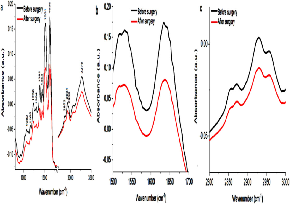
Mean 
Curve-fitting analysis of the amide I profile of blood collected from patients before (a) and after (b) surgery. Mean 
PCA analysis of blood collected from patients suffering from venous disease, which was before (black dot) and after (red dot) surgery. PCA analysis for IR ranges: 800 cm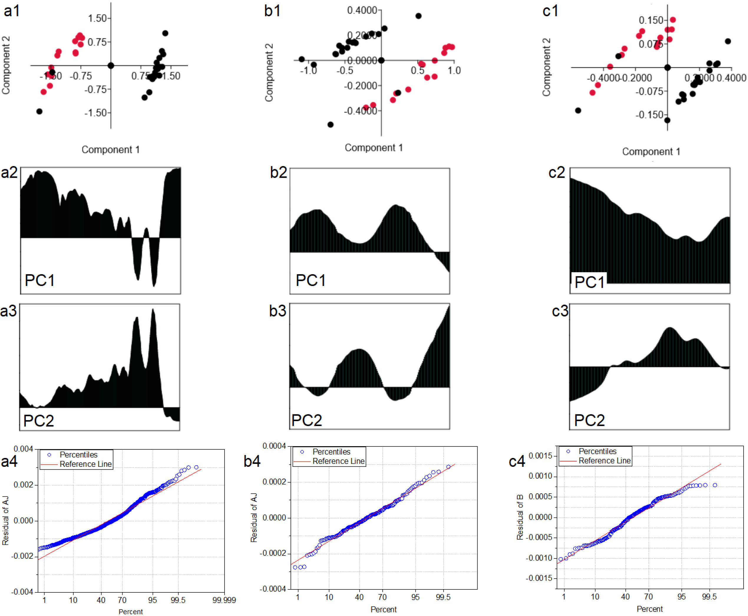
Figure 1a illustrates FTIR spectra of human blood collected from patients before (black spectrum) and after (red spectrum) surgery. In these spectra peaks corresponding to functional group building proteins, lipids, carbohydrates, were marked. Peaks at 1082 cm
FTIR spectra contain a lot of data, to be able to show, that using the FTIR technique it is possible to assess the blood of people suffering from venous disease, the PCA analysis was performed. The analysis performed for three different spectral ranges will show whether it is possible to differentiate by FTIR spectroscopy the blood of patients before and after surgery (Fig. 4). We decided to make PCA analysis for three separate IR regions: fingerprint, amide II with amide I, and region characteristic for CH lipids vibrations. Consequently, we choose the IR range of chemical compound, which the balance changes during venous disease [40].
PCA analysis of all analyzed IR ranges (Fig. 4a1–c1, respectively) showed, that it is possible to distinguish blood collected from patients suffering from venous disease, which was before and after surgery. For IR range 800 cm
To obtain information about which FTIR signals can be correlated with laboratory conditions results, the Pearson correlation test was performed (Table 4). In this section, only the correlation between analyzed features with the highest p-value will be described, while all p values for all analyzed factors were placed in Table 4. From correlation test shows that the value of WBC correlates with absorbance at 2858 cm-1 wavenumber. There are works, where authors showed a correlation between lipids and WBC levels [41]. Tolani et al. showed a significant relationship between elevated high-density lipoprotein (HDL) cholesterol levels and decreased monocyte numbers [42]. Other authors found significant positive associations between triglyceride levels and lymphocyte, neutrophil, monocyte, and basophil count as well as sex-dependent inverse relationships between HDL cholesterol levels and leukocyte counts [44]. Increased triglyceride and low-density lipoprotein (LDL) cholesterol levels were associated with increased total WBC, monocyte, lymphocyte, and neutrophil counts and increased levels are associated with increased lymphocyte number [44, 45]. Moreover, the correlation between NEU and OH vibrations, and between HCT and 1082 cm
Pearson correlation test for a medical condition (blue marked) and FTIR results (yellow marked) with bold the highest

In this study, the objective was to examine the possible changes in the blood samples taken before and after the operation of the chemical substance (CA) used to close the saphenous vein with the FTIR spectroscopic method. By detecting the changes and molecular compositions at the molecular level, it is aimed to determine possible changes in the secondary structure of proteins and to obtain a metabolic profile. Thus, it is aimed to obtain information about the course of treatment and the effect of the chemical used. In summary, for the first time, the present study revealed several profound contextual, structural, and functional changes in biomolecules such as proteins and lipids of patients who suffered from C2 to C4 grade venous insufficiency who treated by endo-venous cyanoacrylate. We used infrared spectroscopy to investigate variations. Additional infrared coupled chemometric analysis such as PCA, PLS, Curve-fitting, and correlation analysis enabled us to discriminate before and after surgery group as a new approach. Our results presented, PCA of all analyzed IR range showed, that it is possible to distinguish blood collected from patients suffering from venous disease, before and after surgery in the case of amide II, amide I region, and CH lipid vibrations range in different sensitivity and accuracy. Hence, FTIR spectroscopy and curve-fitting analysis of amide I profile successfully applied to identify qualitative and quantitative changes of protein structure in blood sera of the groups. Interestingly, in the range corresponding to CH vibrations, the value of absorbance was similar in both groups because of risk factors known for chronic venous disease. Another novelty of our study is to find a correlation between laboratory indexes of peripheral blood cell counts and spectroscopic data. It is verified that venous insufficiency blood sample parameters such as WBC, NEU, and PLT have absorbance in the wavenumber of FTIR spectroscopy in addition to changes in biomolecules.
Based on the data we have obtained as a result of this study, future studies are planned. Among the studies planned to be carried out, primarily oxidant/antioxidant enzymes and involved in these enzymes structure in quantities of the elements will be investigated. Considering these data, it is aimed to discuss whether the changes that may occur are due to the cyanoacrylate used for ablation. It is planned to investigate the effectiveness of such chemical substances and to examine the health effects of these substances and to present new suggestions.
Data availability statement
The raw/processed data employed to obtain these findings are available from the corresponding author upon reasonable request.
Funding
This work did not receive any specific grant from funding agencies in the public, commercial, or not-for-profit sectors.
Footnotes
Conflict of interest
The authors declare that there are no known competing interests.