
Abstract
BACKGROUND:
The similar elastic modulus of resin-matrix ceramics to dentin has resulted in their recent widespread application clinically. Nevertheless, the bacterial environment of oral cavity can degrade the resin composite.
OBJECTIVE:
The objective was to analyse the effect of S. mutans and its fluoride-resistant strains on the adhesion of three CAD/CAM ceramics.
METHODS:
S. mutans UA159 (UA) was identified, and its fluoride-resistant strain (FR) was induced. For crack observation, three kinds of CAD/CAM ceramics (IPS Empress, Lava Ultimate and Vita Enamic) were bonded with tooth complex (enamel, dentin and flowable resin) through adhesive. For micro-tensile test, ceramics were bonded with flowable resin, and cut into strip test pieces. Then specimens were immersed into the UA, FR and the control solution (BHI) separately for 14 d. Ceramic-adhesive interface and adhesive-tooth complex interface were observed and analyzed through electron microscope and stereomicroscope. Micro-tensile test was conducted.
RESULTS:
Specimens in bacterial solutions had more cracks and comparatively weaker micro-tensile strength than those in BHI. In ceramic-adhesive interface, Lava Ultimate produced the most cracks. In adhesive-tooth complex interface, adhesive-dentin produced the most cracks. Meanwhile, IPS Empress had the strongest micro-tensile strength when bonded with resin.
CONCLUSIONS:
S. mutans and its fluoride resistant strain can cause cracks in the bonding of ceramics and dental tissue, especially resin-matrix ceramic and dentin, and weaken the bonding strength between ceramics and resin.
Introduction
Ceramic prosthodontic materials have gradually played a main role of fixed prosthesis because of their high aesthetics and strong biocompatibility [1]. Especially, ceramics made by CAD/CAM chair-side repair technology are widely favored due to the short treatment cycle and convenience. Among them, glass ceramic has dominated during the past 30 years, with high translucency and superior mechanical properties. However, according to the long-term follow-up, the elastic modulus between glass ceramic and teeth is quite different, so teeth have possibility of fracture [2]. In order to solve this problem, scholars try to combine resin with glass ceramic to form a new composite material, resin-matrix ceramic. This composite has the advantages of both ceramic and resin, and its elastic modulus is similar to that of dentin. The representatives of resin-matrix ceramics are Lava Ultimate (3M Espe, USA) and Vita Enamic (Vita Zahnfabrik, Germany). They have different structures. The former is to add nano-sized glass-ceramic filler into the uniform resin matrix, while the latter is to infiltrate the liquid resin into the glass-ceramic mesh frame at high temperature [3].
CAD/CAM restorations are commonly used in full crowns, inlays, onlays, veneers, etc. For Class II cavities, onlay is the best choice. In order to have a good neck closure, the neck margin is usually required to be supragingival. But for patients whose damaged dental tissues are subgingival due to deep caries, we usually apply flowable resin to the subgingival tissue to reconstruct a supragingival margin, which can form a good bond with the CAD/CAM onlay, this technique is referred to as Cervical Margin Relocation, CMR [4].
In terms of the Class II cavity, there is a long marginal line between teeth and prosthesis. They are under the complex environment of saliva and oral cavity for a long time, which may lead to repair failure due to many factors. One of these factors is bacteria. In addition to the unstable temperature in the oral environment, the bacterial environment is also extremely complex. Streptococcus mutans (S. mutans) is the dominant in saliva, which exists in the normal flora and has strong acid production capacity and acid resistance compared with other bacteria. Thus, S. mutans can lead to teeth demineralization and it is the main cariogenic bacteria. In recent years, researchers have found that cariogenic bacteria such as S. mutans can not only demineralize tooth hard tissue, but also degrade composite resin. This is because they contain a special esterase, which can act on the ester bond of the resin and degrade the resin matrix into macromolecular monomer including BisGMA, TEGDMA, UDMA and small monomer, Bis-HPPP. It is called “biodegradation”. Some scholars put the composite resin into the environment of S. mutans UA159, the monomer bis-HPPP [5] can be detected, and the erosion of the resin surface can be observed [6].
As the fact that the main components of resin-matrix ceramic, adhesive, and flowable resin for CMR are all resins, we speculate that they may also be affected by S. mutansin the long-term bonding of Class II cavity, but few researches have been done so far.
Another issue that needs attention is that with the development of science and technology, people gradually realize that fluoride can prevent caries. Correspondingly, the use of fluoride toothpaste and fluoride mouthwash is also increasing year by year, and some regions even add fluoride into running water. Under such long-term local high-concentration fluoride use conditions, the fluoride-resistant strains of some bacteria can selectively grow. Especially the fluorine-resistant strains of S. mutans have been isolated from the oral cavity. Some biochemical properties of the fluoride-resistant strains are different from those of the wild strains, such as higher acid production and acid resistance [7]. However, it has not been studied whether the biodegradability of fluorine-resistant strains has changed and whether they will do harm to resin-matrix ceramic and its bonding to teeth. If so, maybe we need to pay more attention to the use of fluoride toothpaste after ceramic bonding.
In the past, most of the research on the bonding of ceramic to teeth mainly focused on comparing different ceramic materials, adhesives, bonding methods, surface treatments. Few studies are about the influence of bacterial environment. Therefore, we took S. mutans and its fluoride-resistant strains as influence conditions.
The former part of this experiment focused on observation of the bonding interface, because the bonding interface can more intuitively reflect the effect of bacteria on each part. The biggest difference between bonding and loss of bonding is whether the bonding interface is tightly combined. If there are cracks between the interfaces, it means that the bonding is lost. Therefore, in this experiment we calculated the proportion of cracks on each bonding interface. Then, in the latter part, it was planned to study the micro tensile strength after the adhesion of ceramic and flowable resin, which could further reflect the effect of bacteria on adhesion by mechanical parameters.
Methods
Strain identification
Bacteria frozen at
Bacteria were dissolved in 50
Prime sequence for PCR
Prime sequence for PCR
Inoculate S. mutans on BHI medium containing NaF 50
Observation of cracks in the bonding interface of ceramic- adhesive-tooth complex
Preparation of ceramic-adhesive-tooth complex specimens
The isolated molars which stored in thymol solution at 10
Preparation of ceramic-adhesive-tooth complex specimens. A. Low-speed cutting machine. B. Finished specimens. Ceramics were bonded with tooth complex (enamel, dentin and flowable resin). C. Schematic diagram of specimen making. D. Flow chart and grouping.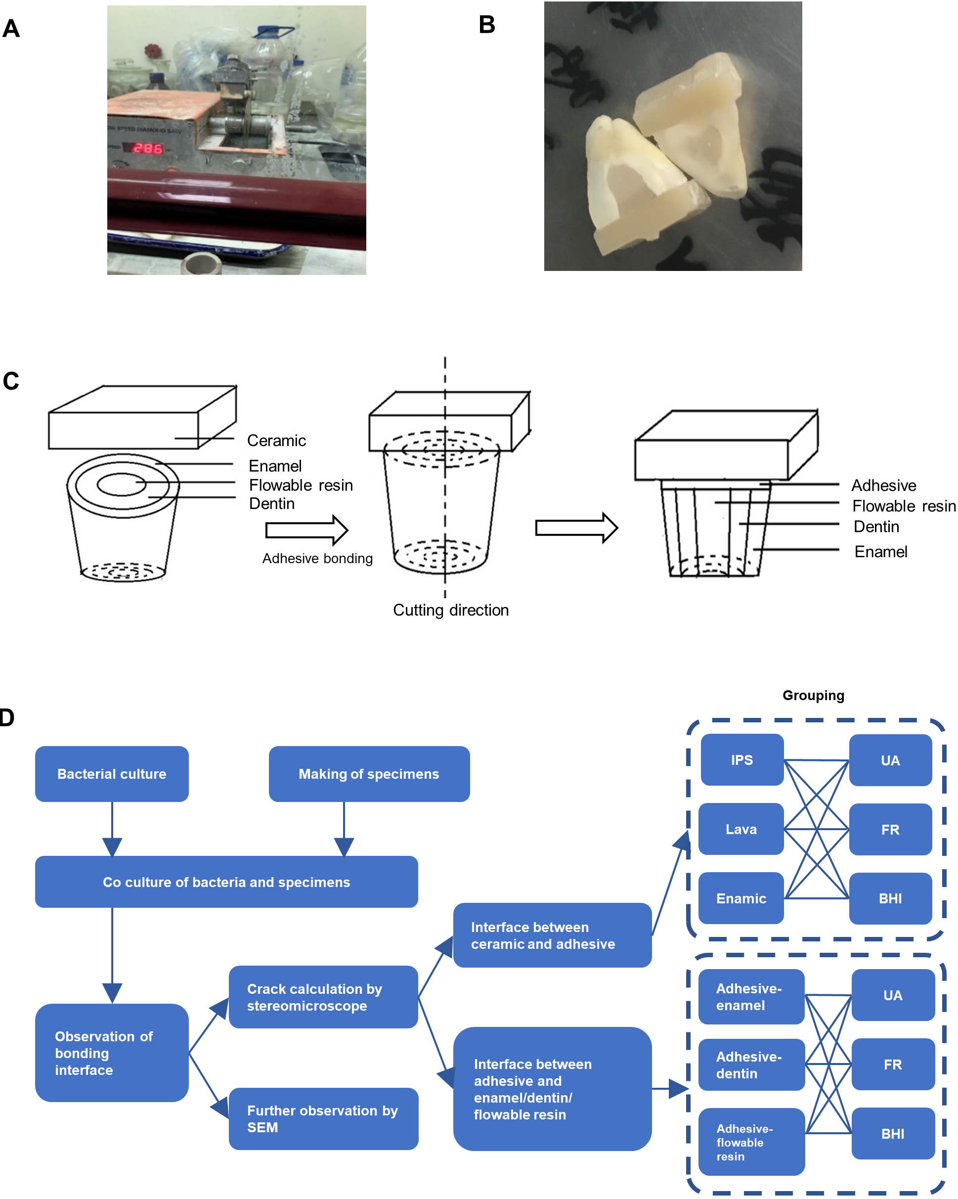
The cylindrical bur SF-13 was used to drill a hollow cylinder with a diameter of 3 mm downward at the center of the cross section. Dentin debris was removed by running water. The self-etching adhesive Single Bond (3M ESPE, USA) was applied to the cavity wall of the hollow cylinder, light-cured for 10 s, then the flowable resin, Tetric N-Flow (Ivoclar Vivadent, Liechtenstein) was evenly extruded into the cavity to ensure that the resin was flush with the coronal cross-section, and light-cured for 20 s. Then tooth complexes were finished. Cut the three kinds of ceramic blocks into cuboids whose size matches the cross section of the molars.
Cross sections of teeth were treated by 35% phosphoric acid for 30 s, rinsed with running water, air-dried. Then dentin treatment agent Syntac Primer, dentin sealer Syntac Adhesive, and enamel adhesive Heliobond (Ivoclar-Vivadent, Liechtenstein) were used in turn.
The bonding surfaces of the three types of ceramic blocks were polished with silicon carbide sandpaper. After rinsing with clean water, they were coated with hydrofluoric acid for 60 s. Coupling agent Monobond N was continued to react for 60 s, and then continued to dry.
Adhesive was applied to the bonding surface of the molars to bond with the ceramic block. Finally the bonded specimens were light-cured in all directions for 20 s. The bonded specimens were placed in distilled water at 37
Next, parallel to the long axis of the tooth and along the diameter of the cross section of the molar, the specimens were cut slowly by the low-speed cutting machine to expose the bonding surface of tooth complex and ceramic (Fig. 1B). Check them with a stereo microscope, and discard the specimens with cracks. The schematic diagram of the experiment was shown in the figure (Fig. 1C).
The specimens were disinfected with UV light for 4 h, soaked in 75% ethanol for 24 h, and washed with a large amount of sterile distilled water.
Adjust the absorbance of S. mutans UA159 solution (UA) and fluoride-resistant strain solution (FR) to A
Observation of cracks under a stereo microscope
After 14 days, the specimens were taken out and placed in sterile distilled water for 10 min of ultrasonic vibration. Under a stereo microscope, the bonding interfaces were observed with a magnification of 20 times. Two interfaces were observed: a. ceramic-adhesive interface, b. adhesive-tooth complex (enamel/dentin/flowable resin) interface. Save the photos, and use Image-Pro Plus 4.0 software to calculate the percentage of crack length to the total length of bonding surface on each interface.
Observation of ceramic-adhesive interface by scanning electron microscope (SEM) magnified 1000 times
The specimens were dehydrated in ethanol with gradient concentration, sprayed with gold. The bonding interface was observed by SEM under 20 kV accelerating voltage. The experimental flow chart is shown in Fig. 1D.
Interface observation and micro-tensile test of the direct bonding between ceramic and resin with adhesive
Preparation of ceramic-resin specimens and co-culture with bacteria
The three kinds of ceramic blocks were polished with silicon carbide sandpaper, and the adhesive surface was coated with hydrofluoric acid for 60 s, rinsed with running water, and dried. A double-sided tape with a 8*8 mm square hole was glued on the bonding surface to limit the bonding area and the thickness of the adhesive. After evenly adding the dual-curing resin adhesive into the hole, light-cure each hole for 20 s. Place a polyethylene mold with a cross section of 8*8 mm and a height of 4 mm facing the square hole, extrude the flowable resin into the mold and light-cure it. Store in normal saline at 37
Process of micro-tensile test. A. Finished strip test pieces by a low-speed cutting machine. B. Test pieces glued on a mold. C. The mold fixed on the universal material testing machine.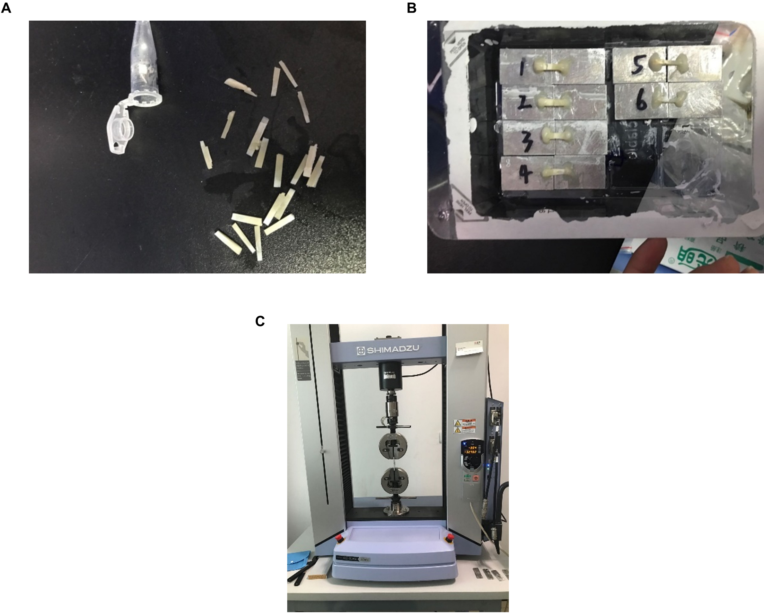
Bonded complexes were cut into 1*1*8 mm strip test pieces by a low-speed cutting machine, whose saw blade was perpendicular to the bonding surface (Fig. 2A). Test pieces with cracks in bonding interfaces were abandoned under a stereo microscope. The test pieces were co-cultured with UA, FR, and BHI respectively, under the same conditions as outlined above.
After 14 days, test pieces were taken out, and the bonding interface was observed by SEM magnified 200 times.
Micro-tensile test
A test piece was glued on a mold (Fig. 2B), and then fixed on the universal material testing machine (SHIMADZU, Japan). Note that the direction of the test piece should be parallel to the loading direction of the force (Fig. 2C). The loading speed was 1 mm/min. The maximum force value (F) of facture was recorded, and the fracture surface area (S) was measured with a micrometer. Micro tensile strength
Result
Identification of S. mutans UA159 and induction of its fluoride resistant strains
The colony form of S. mutansua159 is round, light yellow and opaque, and the surface is slightly rough, the boundary is irregular (Fig. 3A).
Identification of S. mutans UA159 and induction of its fluoride resistant strains. A. The colony of S. mutans UA159 is round and light yellow. B. The sequencing result. C. The phylogenetic tree of 16sRNA. The genetic distance between the sample and S. mutans is the closest. D. From Genebank Blast, the homology with S. mutans UA159 is 100%. E. Colonies of fluoride resistant strains are round, slightly larger than the wild strain.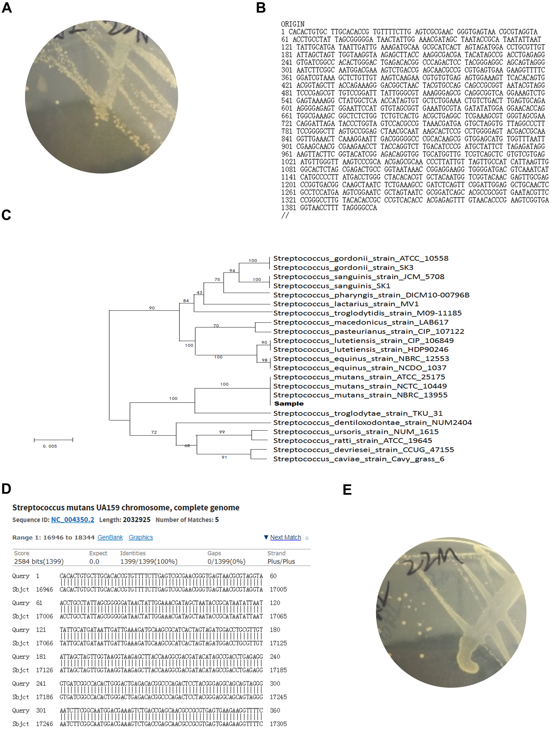
Figure 3B shows the sequencing result and the phylogenetic tree of 16sRNA.
It can be seen that the genetic distance between the sample and S. mutans is the closest (Fig. 3C). Through further sequence alignment with Genebank Blast, it is found that the homology with S. mutans UA159 is 100% (Fig. 3D), which further proves that the recovered strain in this experiment was S. mutans UA159.
After continuous passage, strains with stable morphology were screened out by BHI medium with NaF. Colonies of fluoride resistant strains are round, slightly larger than the wild strain (Fig. 3E).
Observation of the bonding interface. A. Under the stereomicroscope, two interfaces were observed. The upper, ceramic-adhesive interface and the lower, adhesive-tooth complex interface. B. The adhesive-tooth complex interface. C. Percentage of cracks in ceramic-adhesive interface. D. Percentage of cracks in the adhesive-tooth complex interface. E. Observation of ceramic-adhesive interface under SEM magnified 1000 times.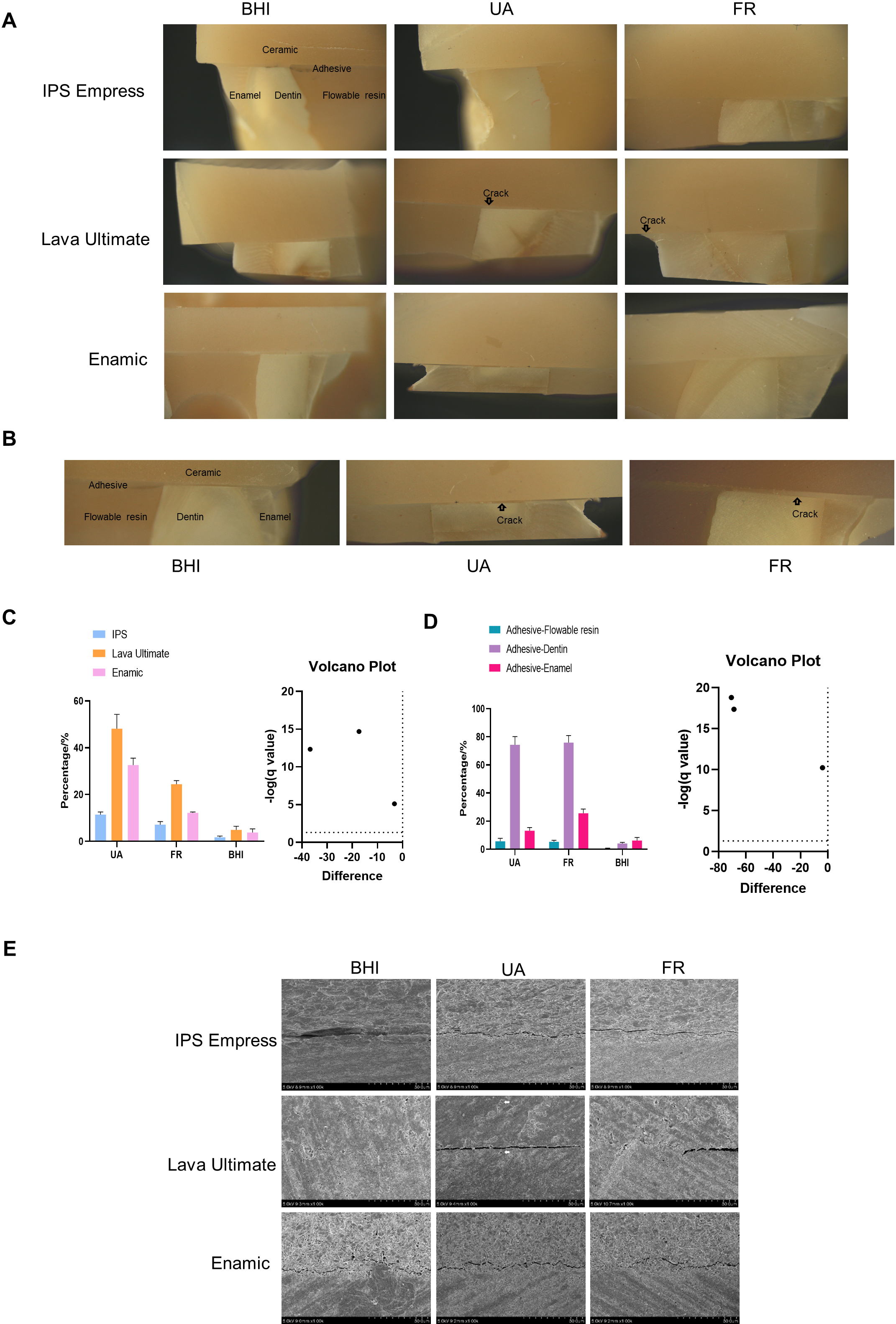
Figure 4A shows that under the stereomicroscope, the three ceramics are bonded to tooth complexes through adhesive. We mainly observe two interfaces: the upper, ceramic-adhesive interface and the lower, adhesive-tooth complex interface. Figure 4B further shows the cracks in the lower interface.
Figure 4C shows the cracks in ceramic-adhesive interface. It can be seen that compared with the control group, UA and FR cause significantly more cracks between ceramic and adhesive, and the influence of wild strain UA is greater than that of fluoride resistant strain. Compared the three kinds of ceramics with each other, cracks between resin-matrix ceramic and adhesive are higher than those between glass ceramic IPS Empress CAD and adhesive. Lava Ultimate-adhesive interface has the most cracks.
Figure 4D shows the cracks in the adhesive-tooth complex interface. Compared with the control group, the cracks of the specimens immersed in bacterial solution increased significantly, but there was no significant difference between FR group and UA group.
Among adhesive-enamel interface, adhesive-dentin interface and adhesive-flowable resin interface in the bacterial solution (UA and FR groups), cracks in adhesive-flowable resin interface are the lowest, while cracks in adhesive-dentin interface occur most, cracks in adhesive-enamel interface are in the middle, indicating that both UA and FR can cause cracks. Dentin is the most fragile part when bonding with adhesive in S. mutans environment.
Observation of ceramic-adhesive interface under SEM magnified 1000 times
It can be seen from Fig. 4E that glass ceramic impress IPS is closely bonded with adhesive, and there are fewer cracks after soaking in bacterial solution compared with the other two resin-matrix ceramics. In addition, the bonding surface of glass-ceramic is wavy under the electron microscope, forming a lock like structure with the adhesive, but the one of resin-matrix ceramic is relatively flat.
Irregular leucite particles can be seen in the ceramic matrix of glass ceramic, and there is no obvious crack even if there is a bacterial condition. The structure of Lava Ultimate shows that dispersed fillers are distributed in a relatively uniform resin matrix. After soaking in bacterial solution, small cracks can be seen between the resin matrix, as shown by the arrow. In the meanwhile, the visual field of Vita Enamic is relatively messy. Resin matrix is distributed between the brighter glass ceramic skeleton, and there are fewer cracks after being soaked in bacterial solution compared with Lava Ultimate. A small amount of fillers can be seen in the cracks between the two resin-matrix ceramic and adhesive, especially Lava Ultimate. There are many cracks in the adhesive part of each group.
Interface observation and mechanical testing of ceramic bonded with flowable resin by adhesive. A. Ceramic-adhesive-flowable resin interface under SEM magnified 200 times. CE: ceramic, LC: adhesive, FR: flowable resin. B. Micro tensile test.
Figure 5A shows that the layers of 1a, 2a and 3a in the control group are closely bonded without large cracks. However, specimens immersed in bacterial solution UA and FR have large and irregular cracks in the interface between ceramic and adhesive, and even the ceramic and adhesive are completely separated. Moreover, cracks of glass-ceramic group (IPS group) are significantly fewer than those of resin-matrix ceramic (Lava Ultimate and Vita Enamic groups) in ceramic-adhesive interface. The interface between adhesive and flowable resin is still tightly bonded without cracks.
Micro tensile test
Regardless of the type of ceramics, the micro tensile strength of UA and FR groups is lower than that of the control group, but there is no significant difference between UA and FR groups, indicating that S. mutans UA159 and its fluorine resistant strain weaken the bonding strength, but the degree of weakening is similar. In addition, in the control group, the bonding strength of glass ceramic IPS Empress CAD is significantly greater than that of the two resin-matrix ceramics, but there is no significant difference in the tensile strength between Enamic and Lava Ultimate. The strength of IPS Empress in UA and FR solution is slightly higher than that of the other two ceramics in UA and FR (Fig. 5B).
Discussion
Effect of S. mutans on ceramic-adhesive interface
Resin-matrix ceramic is a kind of composite material rising in recent years. It combines the advantages of ceramic’s high strength and resin’s high ductility. Lava Ultimate and Vita Enamic, two products widely used in the market, are different from ordinary resins, because they are cured at high temperature and high pressure instead of traditional light and heat curing, which can effectively reduce the release of monomers. A study shows no monomer release was detected after the two products were soaked in acetone for 7 days [8]. However, the oral environment is very complicated and there are a large number of bacteria, which may affect the adhesion of the prosthesis. Bourbia et al. added resin into a bacterial solution of S. mutans UA159 for 14 days and found that resin monomer bis-HPPP could be detected, and showed obvious signs of surface erosion [5, 6]. The experimental results of Wang et al. also showed that S. mutans could weaken the bonding strength between resin and dentin [9]. Further studies show that esterase SMU_118 is an intracellular protein in S. mutans, which is expressed under cariogenic acidic conditions (ph-5.5) [6]. It can hydrolyze methacrylate [10], and plays a major role in biodegradation. It can act on the fragile ester bond of the resin and degrade the resin matrix into BisGMA, TEGDMA and UDMA, and their small monomers which will lead to cracks in the resin and affect the quality of bonding. These monomers can be detected by liquid chromatography, mass spectrometry and UV absorption [11]. In contrast, the polymer degradation activity of SMU_C gene knockout strain of S. mutansUA159 decreases significantly [12]. S. mutans UA159 was selected as the experimental strain because it has the strongest esterase activity with p-nitrophenol butyrate (p-NPB) as the substrate among all strains of S. mutans. This activity has been proved to be related to the degradation of composite resin [13].
Whether S. mutans has an effect on resin-ceramic composites has not been fully studied yet. Therefore, in this experiment, two typical composite ceramics on the market, Lava Ultimate and Vita Enamic, are used as the research objects, while the glass ceramics Empress CAD is equivalent to a comparative study. Although these two composite ceramics are composites of ceramics and resins, their structures are completely different. The former has a glass-ceramic filler embedded in a uniform resin matrix, while the latter has a mesh-like glass-ceramic frame, and the liquid resin penetrates into the mesh-like frame at high temperature. Because the latter has a relatively stable framework, and previous experiments have shown that the latter has slightly stronger mechanical properties than the former [14], it was assumed that the two have different resistance to cariogenic bacteria in the early stage of this experiment. The experimental results show that there are more cracks observed between the Lava Ultimate resin matrix under SEM, while there are very few cracks in the Vita Enamic, and the glass ceramic basically do not have cracks. It shows that S. mutans can affect resin-matrix ceramic, especailly Lava Ultimate, probably because the structure of Lava Ultimate is not as stable as Vita Enamic, a polymer infiltrated ceramic network (PICN) material. PICN has a framework in which resin and glass-ceramic are cross-linked. However, the micro-tensile test in this experiment showed that there was no significant difference in the micro-tensile strength between the two, probably because the generation of cracks was not enough to affect the effect of force on the ceramic bonding.
The adhesive used in this experiment is the double curing adhesive resin VariolinkN, which means the curing process requires both long enough light and certain chemical curing time. Therefore, the bonded specimens were stored in normal saline at 37
Variolink N is mainly composed of bisphenol A, glycidyl methacrylate (Bis-GMA) and polyurethane dimethacrylate, both of which belong to methacrylate. Therefore, as mentioned above, it can be biodegraded in the environment of S. mutans. In this experiment, cracks between adhesive were observed, indicating that S. mutans can destroy the polymer structure in adhesive, which is consistent with the conclusions drawn by previous scholars. This suggests that the bonding edge line of prosthesis should be minimized, because the long edge line will expose more bonding interfaces to the oral bacterial environment. We get the hint that when selecting the prosthesis, we must consider the bonding surface factor and try to reduce the percentage of the adhesive interface directly exposed to the oral cavity. If we have to adopt the design of long edge line, we should pay attention to the bonding process, in which the prosthesis should be continuously pressurized for enough time, patients can chew food only after the adhesive has solidified, and the excess adhesive must be removed. What’s more, the ability of total acid etching system to resist biodegradation is stronger than that of the acid etching system [15].
Experiments show that the percentage of cracks between ceramic and adhesive is related to ceramic materials, and cracks between glass ceramic IPS Empress CAD and adhesive are the least. Under SEM, it is also observed that compared with resin-matrix ceramic, the acid etching bonding surface of glass ceramic and adhesive form a lock like structure, the adhesive in the flowable state penetrates into the pits produced by hydrofluoric acid etching. Since there is such a lock like structure, so that the adhesive is not easy to separate from the ceramic body, which may explain why glass ceramic is less affected by S. mutans. In addition, the results of micro tensile test also show that the bonding strength between glass ceramics and flowable resin is stronger than that of resin-matrix ceramic, which also provides a certain basis for this conclusion. On the contrary, hydrofluoric acid cannot produce enough pits on the surface of resin-matrix ceramics, and the adhesive cannot form a lock like structure, which will be easier to separate from the ceramics.
S. mutans and adhesive-tooth complex interface
Pulp chamber retention is normally needed after the onlay restoration of posterior teeth finished with perfect root canal treatment. We usually use flowable resin to form a flat bottom in pulp chamber, which may help with the bonding of ceramic. In addition, if the tooth tissue is subgingival in Class II cavity, the flowable resin should be used to restore the gingival wall to supragingival position, which is called Cervical margin relocation, also known as gingival wall lifting. In these two cases, enamel, dentin and flowable resin are bonded to the ceramic through the adhesive, and then there are interfaces of adhesive-enamel, adhesive-dentin and adhesive-flowable resin which may contact with S. mutans in the oral cavity. The purpose of this experiment is to observe whether these three interfaces will be affected by S. mutans. Compared with the method of bonding enamel, dentin and flowable resin with ceramic respectively, this experiment is simpler and more convenient. In one specimen, three interfaces bonded with one ceramic can be observed at the same time, which reduces cumbersome steps of making specimens separately. What’s more, the interfaces can be controlled in the same bacterial environment, other influencing factors can be ignored. In addition, the design also simulates the interface directly exposed in the oral cavity during onlay bonding, which is closer to the physiological environment.
Total etching is the most common way for enamel bonding. Its principle is that phosphoric acid can cause micro pits on hydroxyapatite, thus adhesive can form a lock-like structure with enamel. In the meantime, the treatment of dentin can be either total etching or self-etching. The principle of the former is to demineralize the dentin surface and expose the collagen network structure, and then use pretreatment agent to facilitate the infiltration of resin into the network; the principle of the latter is to form a smear layer [16]. In this experiment, the total etching adhesive is used. It was shown that its bonding effect is stronger than that of self-etching adhesive [17], but the total etching steps are complex, and each step needs to be strictly moisture-proof. After phosphoric acid etching, the locking effect of enamel produces strong mechanical retention, but the demineralized dentin collagen fiber is easy to collapse, which is not conducive to bonding. Therefore, the bonding between adhesive and enamel is stronger than that between adhesive and dentin, which can explain that cracks in adhesive-enamel interface is fewer than dentin.
The flowable resin used in this experiment is TetricN-Flow, whose component is similar to the adhesive, Variolink N double curing resin adhesive, so the two are bonded most closely, which can explain that the cracks between the two are the least after soaking in bacterial solution.
S. mutans can not only degrade the adhesive through esterase, resulting in cracks between the adhesive and tooth tissue, but also produce hydrolase and bacterial collagenase. The content of organic in dentin can reach 30%, of which collagen accounts for the largest proportion. It is very likely to be affected by bacterial collagenase when exposed to a high concentration of bacterial environment.
Micro tensile test
The effect of a bonding system can be evaluated by bonding strength, which is often measured by tensile test and shear test. However, many studies have found that the fracture type of shear test is closer to cohesive failure (resin internal failure), rather than interface failure, which is more common in clinic [18]. The fracture type of tensile test is the interface failure, which is more conducive to evaluate the bonding strength of intraoral prosthesis. However, the results of tensile and shear tests are easily affected by specimen’s geometry and uneven stress distribution during force application, so micro tensile test is introduced. This test requires the bonding area to be close to 1 mm
However, the micro tensile test has high requirements for the test piece. Therefore, the test is carried out only after the strip test piece without cracks is selected under the stereomicroscope.
The experimental results show that the bonding strength of glass ceramic Empress IPS is the largest, which is also consistent with the fewest cracks in ceramic-adhesive interface observed. Moreover, the bonding strength of glass-ceramic is still stronger than that of the other two resin-matrix ceramic even if it is under the environment of S. mutans. This suggests that traditional glass ceramics cannot be completely replaced by new emerging materials, and we should pay special attention to the removal of plaque when applying resin-matrix ceramics.
S. mutans and its fluorine resistant strain
Fluoride can achieve bacteriostatic effect by affecting the acid production, acid resistance and adhesion of bacteria. However, if fluoride preparations are used at specific sites for a long time, it may induce the growth of fluoride resistant strains, especially the fluoride resistant strains of S. mutans, which have stronger acid production and acid tolerance than their wild strains.
In this experiment, both kinds of bacteria can weaken the bonding strength of ceramic blocks and cause cracks, and there is little difference between the two. However, at the ceramic-adhesive interface, the wild strain cause more cracks than its fluorine resistant strain, which may be due to the change of esterase synthesis of fluorine resistant strains, the hypothesis needs to be verified by further experiments. This result also suggests that the use of fluoride such as fluoride toothpaste will not interfere too much with the bonding of resin based restorations at the bacterial level.
The results of this experiment can suggest that S. mutans, the most cariogenic bacterium in the oral cavity, can affect the bonding between ceramic and tooth tissue. In clinical practice, it is necessary to strictly control the bonding process to ensure moisture isolation and tight edges. In daily maintenance, it is necessary to ensure the cleanness of prosthesis and remove plaque in time.
Moreover, this experiment needs to further study whether the biodegradation of S. mutans occurs at the bonding interface. Liquid chromatography-mass spectrometry (LC-MS), which is commonly used in metabonomics, can be used to detect whether there are free monomers in composite ceramics.
In addition, it is necessary to further detect whether the gene encoding of the esterase SMU_C in the fluoride resistant strain of S. mutans mutates, whether the expression of SMU_C is changed under the condition of fluorine or no fluorine, so as to more accurately explain whether the biodegradability of the fluoride resistant strain of S. mutans changes.
Conclusion
S. mutans and its fluoride resistant strain can cause cracks in the bonding of ceramics and dental tissue, especially resin-matrix ceramic and dentin, and weaken the bonding strength between ceramics and resin.
Footnotes
Acknowledgments
Xinwei Guo and Hongyan Zhao contributed equally to this work.
Conflict of interest
The authors declare that they have no conflict of interest.