
Abstract
BACKGROUND:
The dielectric properties of tissues are very important physical factors for the investigation and application of bio-electromagnetism. However, the size of the active sample tissue is usually limited in actual measurement, making it difficult to meet the requirements of the existing high-frequency measurement methods, thus influencing the measurement results.
OBJECTIVE:
The present study aimed to systematically investigate the various factors influencing the effective measurement area of the open-ended coaxial probe, including the design size of the probe and the dielectric properties of the object to be measured.
METHODS:
The simplified material mixing model, in which several types of materials were set as the material under test (MUT) and the perfect conductor (PEC) was set as the specific material, was used in the simulation to study the effective measurement area of eight types of probes with different sizes for the dielectric measurement of different MUTs. Different concentrations of NaCl solutions and three types of coaxial probes were used in the actual measurement to verify the simulation results.
RESULTS:
According to the simulation results, the effective measurement area, especially the effective measurement radius, was closely related to the outer conductor radius of the probe. The effective measurement area of the probe decreased when the outer conductor radius of the probe reduced. Moreover, the change in the effective measurement area of the probe was independent of the MUT when the cross-sectional size of the probe was smaller than a certain threshold value. The experimental results also confirmed this conclusion.
CONCLUSION:
According to the research results, the independent variable dimension could be effectively reduced and the modeling difficulty was reduced when the analysis model of the effective measurement area of the probe was established.
Keywords
Introduction
Dielectric properties of biologically active tissues are related to not only the microstructure of the tissues [1, 2] but also various physiological activities inside the tissues [3]. Therefore, the dielectric property is an important factor for biophysical research on the impact of multiple electromagnetic radiation environment on organisms. It also has important applications in the study of biological electromagnetic effects, research and development of medical and health equipment, effectiveness evaluation of new military weapons, manned space engineering, and other fields. Since the beginning of the last century, people have carried out measurement research on the dielectric properties of tissues [4, 5, 6, 7]. At present, relevant technologies for measuring the dielectric properties of bioactive tissues in the middle and low frequency bands (10 KHz to 100 MHz) have been established [8, 9], but the high-frequency bands still have unresolved issues.
The main method for measuring the dielectric properties of high-frequency bands is the open-ended coaxial reflection method [6]. The dielectric properties of the tissue at the end of the probe were calculated by measuring the ratio of absorption and reflection of electromagnetic energy to biological tissue at the end of the probe and the change in the phase. The use of this method was mainly based on two assumptions: first, the measured tissue should be single and homogeneous. Second, the size of the measured tissue should be large enough to avoid the impact of tissue boundaries on the measurement results [6]. However, in the actual measurement, the size of active samples collected clinically for measurement was usually limited to keep the samples homogenous, and the tissue boundary had a significant impact on the measurement results [10, 11]. As a result, the actual measurement results were the equivalent dielectric properties of multiple substances mixed in the probe measurement area, rather than the dielectric properties of the target tissue itself, which is the main problem faced by the current measurement of dielectric properties of active tissues in the high-frequency band [12].
When measuring the dielectric properties of tissues in vitro using the open-ended coaxial probe, the sample tissue size should be larger than the effective measurement area of the coaxial probe to avoid the influence of the tissue boundary. As a result, the determination of the measurement area of the probe is the primary prerequisite for the accurate measurement of high-frequency dielectric properties. Hagness believed that the size of the measured area varied with the design size of the probe, indicating that if the size of the probe was reduced, the measurement area would decrease accordingly [13]. However, Gabriel’s study showed that the design size was not the only factor affecting the measurement area. The working frequency and the dielectric properties of the measured object might affect the measurement area, so the probe size required to measure different biological tissues might also be different [14]. As no analytical model determining the measurement area is available, many research groups mainly determined the measurement area of their respective coaxial probes based on the actual measurement. Hagness et al. explored the measurement region of their 2.2-mm and 3.58-mm coaxial probes in the 1–20 GHz band based on the dielectric properties of salt solution. By comparing the measurement results of the probes at different positions in the salt solution, the measurement depth and breadth (the transverse measurement size of the probes) of the two probes were, respectively, 1.5 mm/5 mm and 3 mm/11 mm [13]. Meaney et al. compared the measurement results of the probe at different positions of the double-layer mixed substance composed of salt solution and poly tetra fluoroethylene (PTFE), and studied the measurement depth of the 2.2-mm coaxial probe at the 300-KHz to 8.5-GHz frequency band at 0.2–0.5 mm [15]. In the subsequent study, the measurement depths of several probes of different sizes were compared [16]. However, the experimental results were different from those of Hagness in terms of the depth measured by the probe. Porter’s team investigated the measurement depth and lateral size of a 2.2-mm probe in the 300-MHz to 8.5-GHz band based on simulation and verified it with a two-layer mixture of rubber, salt solution, and part of pig tissue. The measurement area of the probe was mainly affected by the surface material of the measured sample, but the probe measurement depth changed with the different frequencies and the measured materials [17, 18, 19]. Yonghong explored the induction range of the coaxial probe based on the layered model composed of various materials, such as ethanol, deionized water, and salt solution, and believed that it was affected by both the measurement parameters and the dielectric properties of the measured object, among which the measurement depth was greatly affected [20].
Most of the current researches on the probe measurement area were need based and explored only the measurement area of the probe with a specific size in the measurement object by experimental methods. Once the probe size or the measurement object changed, it could not be concluded whether the measurement area would change. Therefore, the conclusions of the existing studies are of limited use in practical measurement. A general analysis method or model for the effective measurement area of the probe is required. Therefore, this study systematically investigated the various factors influencing the effective measurement area of the open-ended coaxial probe. The design size of the probe and the dielectric properties of the object to be measured were discussed based on the simulation and measurement in the 100-MHz to the 3-GHz band to establish the analysis and calculation model of the effective measurement area of the coaxial probe.
Methods
Analysis method for measuring the area of the open-ended coaxial probe
The probe terminal was attached to the surface of the tissue and measured the reflection coefficient
In Eq. (1),
According to the previous research conclusion [21], the transverse sectional size of the measurement area of the coaxial probe in the tissue gradually decreased with the increase in the measurement depth. Therefore, the effective measurement area as a whole presented an approximate inverted conical shape, as shown in Fig. 1. The measurement radius
Schematic diagram of the effective measurement area of the probe.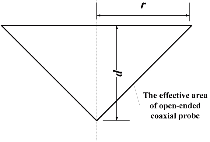
The equivalent input admittance of
In Equ. (2.1),
According to the analysis method of the measurement area of the probe, the effective measurement area parameters
Schematic diagram of simulation model structure: (a) 
As shown in Fig. 2, the object measured in the simulation was mainly composed of two parts: one was the target material under test (MUT), and the other was the specific material. The perfect conductor (PEC) was adopted to highlight the difference. In Fig. 2a, the transverse section size of PEC was consistent with that of the MUT. In the depth direction, the effective measurement depth
The simulation working frequency was set as 100 MHz to 3 GHz. The dielectric parameters of the MUT were set directly according to the research needs. In the simulation study, when the MUT was identified as the biological tissue, its dielectric properties could be analyzed and calculated using Gabriel’s fourth-order Cole–Cole equation, as shown in Eq. (3), and imported into the model [4]:
where
Coaxial probes with an open terminal in different design sizes
Coaxial probes with an open terminal in different design sizes
The main factors affecting the effective measurement area of the probe were the design size of the probe and the dielectric properties of the measured materials. The probe design size mainly included the probe inner radius
Dielectric properties of different measurement objects
Three types of coaxial probes with different sectional sizes were used to test and verify the effective measurement area of the probe. The design sizes of the three probes were as follows: probe 1 (PTFE filled, internal and external diameter 0.29/1.19 mm), probe 2 (PTFE filled, internal and external diameter 0.53/2.15 mm), and probe 3 (PTFE filled, internal and external diameter 1.2/4.5 mm). The vector network analyzer NanovNA-F V2, which had its own open circuit, short circuit, and matching load accessories for calibration was used for measuring the frequency range of 100 MHz to 3 GHz. The NaCl solutions at different concentrations (0.005M, 0.01M, 0.03M, 0.05M, and 0.15M) were used as the MUT.
According to the simulation settings in Fig. 2, the 100 ml beaker with the diameter 5.7 cm and height 7.3 cm containing NaCl solution was placed on a metal base to form a longitudinal layered measured object model. The probe terminal was placed at the bottom of the beaker for initial measurement. The probe terminal position was continuously raised to observe and record the measurement results and the probe terminal height, and the effective measurement depth
Schematic diagram of simulation model structure: (a) probe measurement depth measurement setting (the NaCl solution was 4.8 cm in diameter and 5.0 cm in height), (b) probe measurement breadth measurement setting, (c) the measurement system combination.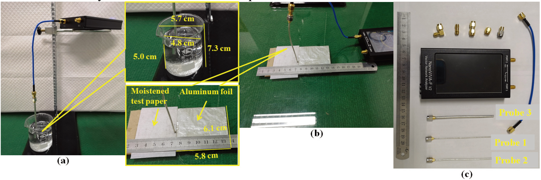
Study of the probes featuring different openings of the outer conductor b
The radius of the outer conductor
Comparing Fig. 4a, c, and e, it was seen that the effective measuring radius
Real part of the measurement sensitivity 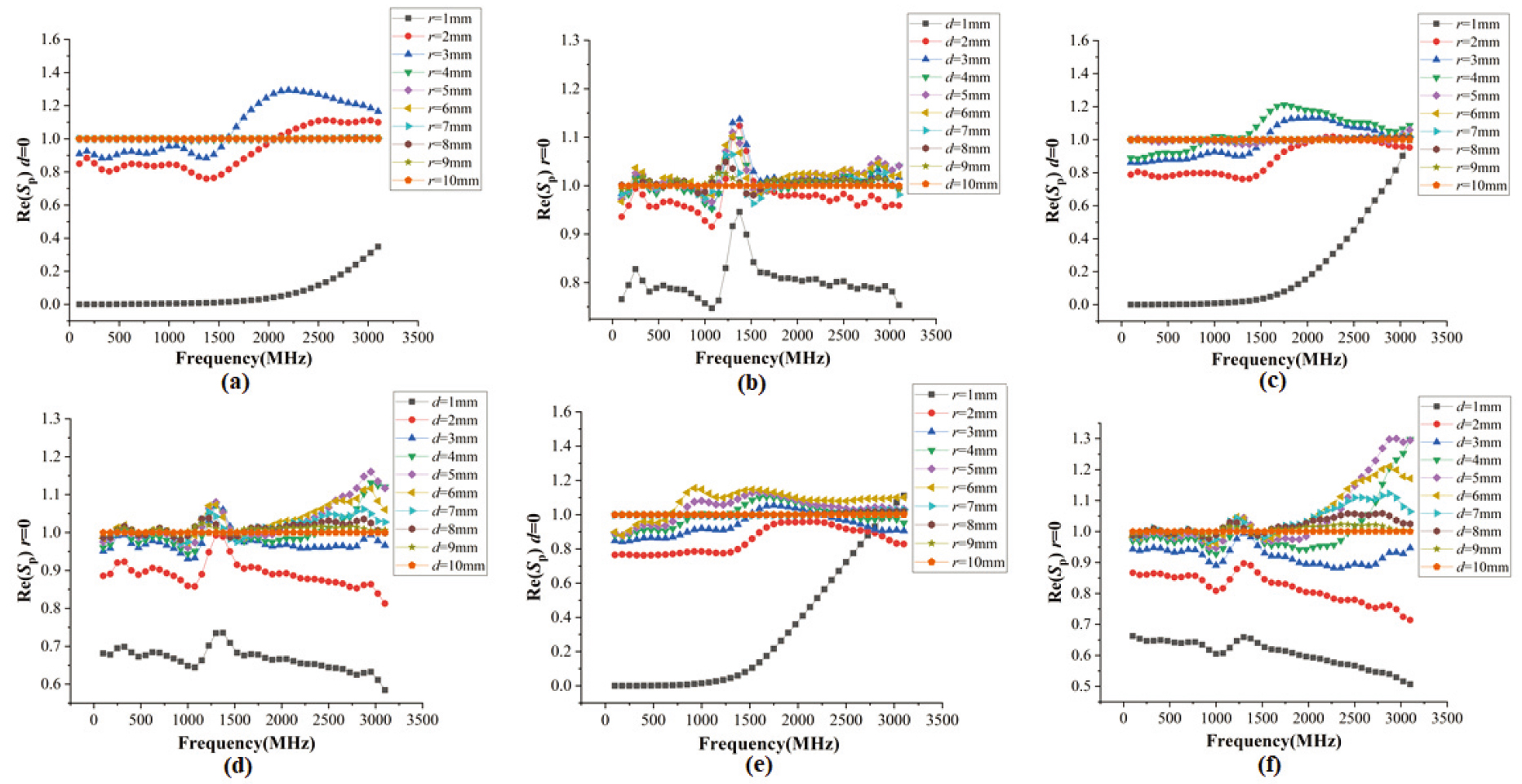
The radius of the inner conductor
Real part of the measurement sensitivity 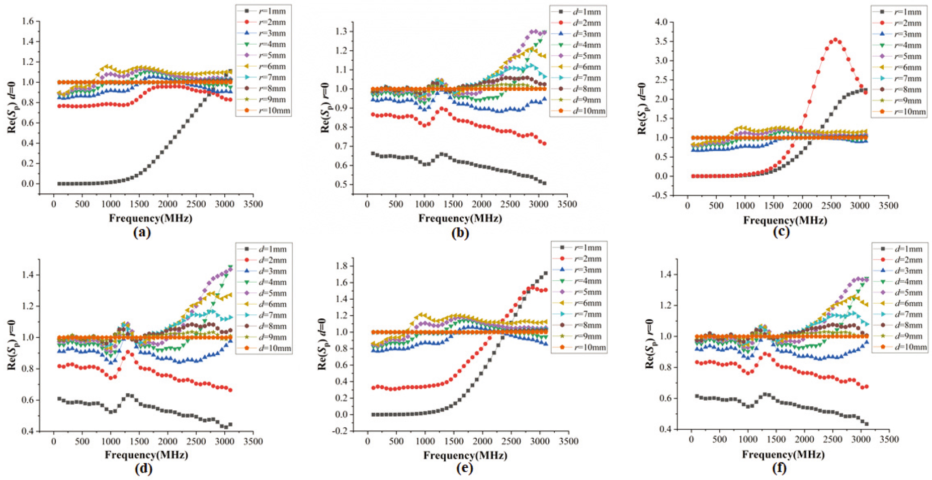
In the previous section, the main factors influencing the dimension of the probe were examined for the dielectric measurement. However, the characteristic impedance
On the contrary, Fig. 6 shows that the increase in the inner and outer radius of the probe could significantly improve the effective measuring radius
Study of the probes with different MUTs
The effect of dielectric properties on the effective measurement area of the probe was discussed in this section. The type A, type G, and type H probes in Table 1 and different measurement objects in Table 2 were used for the simulation. Table 3 shows the results of simulation.
Effective measurement area parameters of the probe under different MUTs
Effective measurement area parameters of the probe under different MUTs
Real part of the measurement sensitivity 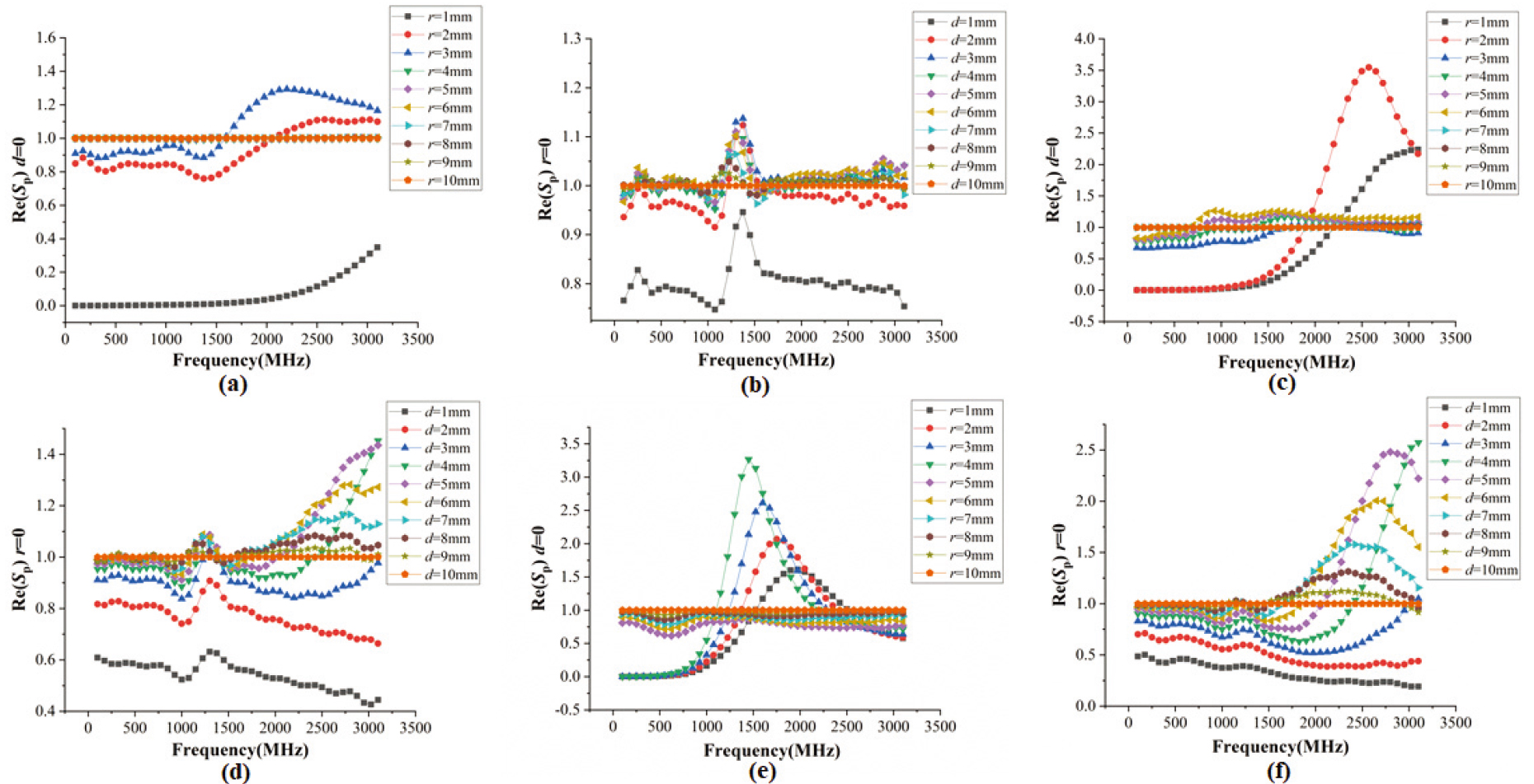
As shown in Table 3, the effective measurement radius
Figure 7 shows the real part of the measurement sensitivity
Effective measurement area parameters of the probe for different NaCl solutions
Effective measurement area parameters of the probe for different NaCl solutions
Real part of the measurement sensitivity 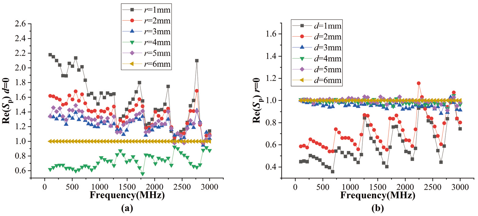
As shown in Fig. 7, when the probe 3 was used for measuring 0.03M NaCl solution, the effective measuring radius
The measurement of dielectric properties of biological tissues has always been a concern for the researchers. The final research goal of this study was to explore a method to accurately measure the dielectric properties of biological tissues based on the principle of coaxial probe measurement. In this study, we focused on the methodologies for in vitro measurement of the effective area with coaxial probes.
Although the structure of biological tissue is complex, the homogeneity of the samples can be ensured by controlling the measurement of the sample size in vitro. As a result, the tissue samples measured in vitro using the open-ended coaxial probe in this study were considered to be homogeneous. However, the tissue boundary had a significant impact on the measurement results when the size of the samples was smaller than that of the probe measurement area. At this time, the actual measurement results were the equivalent dielectric properties of the target tissue mixed with the air in the probe measurement area, rather than the dielectric properties of the target tissue itself. Therefore, the measurement area of the probe in in vitro measurement was investigated in this study for the analysis and modeling to determine the measured sample size required to ensure that the measured tissue size was larger than the probe measurement area.
According to the results of simulation and experimental tests in the previous section, the increase in the outer conductor radius
The characteristic impedance
where
Figures 4–7 show that the measurement sensitivity
The effective measurement area of the probe was firstly studied based on simulation in this paper. Then the simulation results were verified by actual measurement. As a result, a discussion of the metrological aspects of measurement is very important for actual measurement [22, 23]. The room temperature was constant at 26
In this study, the key factors influencing the effective measurement area of the open-ended coaxial probe, including the probe design dimension and the dielectric properties of the MUT, were studied in the frequency ranging from 100 MHz to 3 GHz based on simulation and measurement. The simplified material mixing model, in which several types of materials were set as the MUT and the PEC was set to be the specific material, was used in the simulation to study the effective measurement area of eight types of probes of different sizes for the dielectric measurement of different MUTs. The NaCl solutions with different concentrations and three types of coaxial probes were used in the actual measurement to verify the simulation results. According to the results, the effective measurement area, especially the effective measurement radius, was closely related to the outer conductor radius of the probe. The effective measurement area of the probe decreased when the outer conductor radius of the probe was reduced. Further, the change in the effective measurement area of the probe was independent of the MUT when the cross-sectional size of the probe was smaller than a certain threshold value. As a result, the change in the effective measurement area of the probe whose cross-sectional size was smaller than 2.15 mm was independent of the MUT in the frequency ranging from 100 MHz to 3 GHz according to the results in this paper. This conclusion is very important to establish the analysis model of the effective measurement are of the open-ended coaxial probe. The independent variable dimension can be effectively reduced and the modeling difficulty can be reduced when the analysis model of the effective measurement area of the probe is established.
Footnotes
Acknowledgments
This research was supported by the National Key R&D Program of China (2019YFC0119103) and the NSFC (31900978).
Conflict of interest
None to report.