



Abstract
BACKGROUND:
The femoral neck system (FNS) has been considered as a novel strategy for femoral neck fracture. The diversity of internal fixation creates difficulties in choosing an effective option for Pauwels III type femoral neck fractures. Therefore, it is significant to investigate the biomechanical effects of FNS versus conventional approaches on bones.
OBJECTIVE:
To evaluate the biomechanical characteristics of FNS versus cannulated screws coupled with medial plate (CSS
METHODS:
Through three-dimensional computer software (Minics, Geomagic – Warp), the proximal femur model was rebuilt. Based on the present clinical characteristics, models of internal fixation were reconstructed in SolidWorks, including cannulated screws (CSS), medial plate (MP) and FNS. After parameter setting and meshing, boundary conditions and loads were set up for the final mechanical calculation in Ansys Software. Under identical experimental conditions, such as the same Pauwels angle and force loading, the peak values of displacement, shear stress and equivalent (von Mises) stress were recorded.
RESULTS:
This study showed that the displacement of the models was CSS, CSS
CONCLUSION:
CSS
Introduction
Globally, the tendency of femoral neck fractures (FNFs) has increased continuously due to increased average age and osteoporosis, which accounts for approximately 50% of hip fractures [1]. By 2050, the cases of FNFs are estimated to be 6.7 million annually [2]. Of particular note is the morbidity and mortality of FNFs, especially in the developed countries [3]. FNFs can be divided into three types in compliance with the angle between the fracture face and the horizontal plane [4]. Of these fracture patterns, Pauwels type III is considered to be extremely unstable, which causes clinical nonunion and internal plant failure [5]. Therefore, for patients aged 65 and younger, the application for effective internal fixations has become a clinical research priority recently.
While the best treatment of FNFs remains controversial. Cannulated screws (CSS) and dynamic hip screw (DHS) are widespread adoptions to stabilize FNFs [6, 7, 8]. DHS and CSS have significantly similar complications, including non-union and revision rate, but CSS has superior to DHS on avascular necrosis [9]. It is universally acknowledged that fixation by CSS has been a standard method for stable and undisplaced femoral neck fracture [10]. However, for unstable fractures (Pauwels Type III), this strategy is prone to failure due to the shear forces mentioned above. To address this issue, researchers have successfully optimized fixation techniques by using a medial buttress plate (MP). Some finite element experiments have shown that this auxiliary technique has the effect of reducing the torsional force and shear stress while maintaining the axial compressive stress [11, 12, 13]. The recently applied implant Femoral Neck System (FNS) (DePuy Synthes, Zuchwil, Switzerland) can provide a minimally invasive surgical while allowing a reduced implant footprint. By offering angular stability and intraoperative compression, FNS has been considered as a novel strategy for femoral neck fracture (Fig. 1). In addition, based on excellent biomechanics behaviour, the stability of FNS has been proved to be comparable to DHS with anti-rotation screw and superior to cannulated screws [14, 15].
Representative X-ray image of one clinical case who was treated for vertical femoral neck fracture using Femoral Neck System.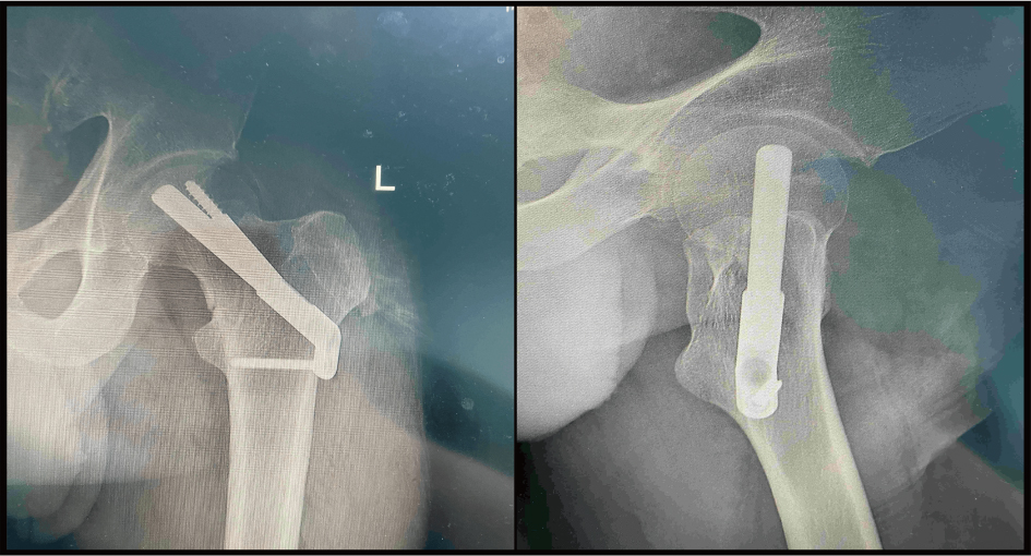
Some studies have shown the advantages of both CSS
Building a normal proximal femur model
The volunteer we recruited (40-year-old, female, 60 Kg, 160 cm) did not have any hip and systemic disease. Written informed consent was acquired before the examination. The normal proximal femur of the volunteer was scanned using a computed tomography scanner (Philips, Netherlands, 128 spiral). A series of images (Slice Thk 0.5 mm) were exported and saved as Digital Imaging and Communications in Medicine (DICOM) format. These images were imported to Mimics 20. After predefined threshold, region growing, mask modifying and preliminary borders smoothing, the project was outputted in STL format. This model was imported into Geomagic – Wrap 2017 software for advanced smoothing, adjusting polygon mesh and fitting surface and also integrated into the SolidWorks 2017 software. The three-dimensional (3-D) model of proximal femoral bone was established by Boolean operation.
Modelling of FNFs and internal fixation
By using the part pattern, the model was divided into two parts, and the angle between the fracture face and horizontal plane was 60 (Fig. 2a). Based on the present clinical characteristics, models of internal fixation were reconstructed through schematic drawing function. The medial reconstructive plate was modeled utilizing equidistant offset surface method, and the three CSS (7.3 mm) were arranged in an inverted triangle. Under assembly pattern, models of FNFs and internal fixations were combined into one part, which could be edited directly (Fig. 3). In the end, all parts were detected by interference to evaluate their process quality, then were imported into Ansys 17.0 software.
Details of proximal femoral bone model: Pauwels type III (a); Uneven meshes of femur model (b); load and muscle forces (c).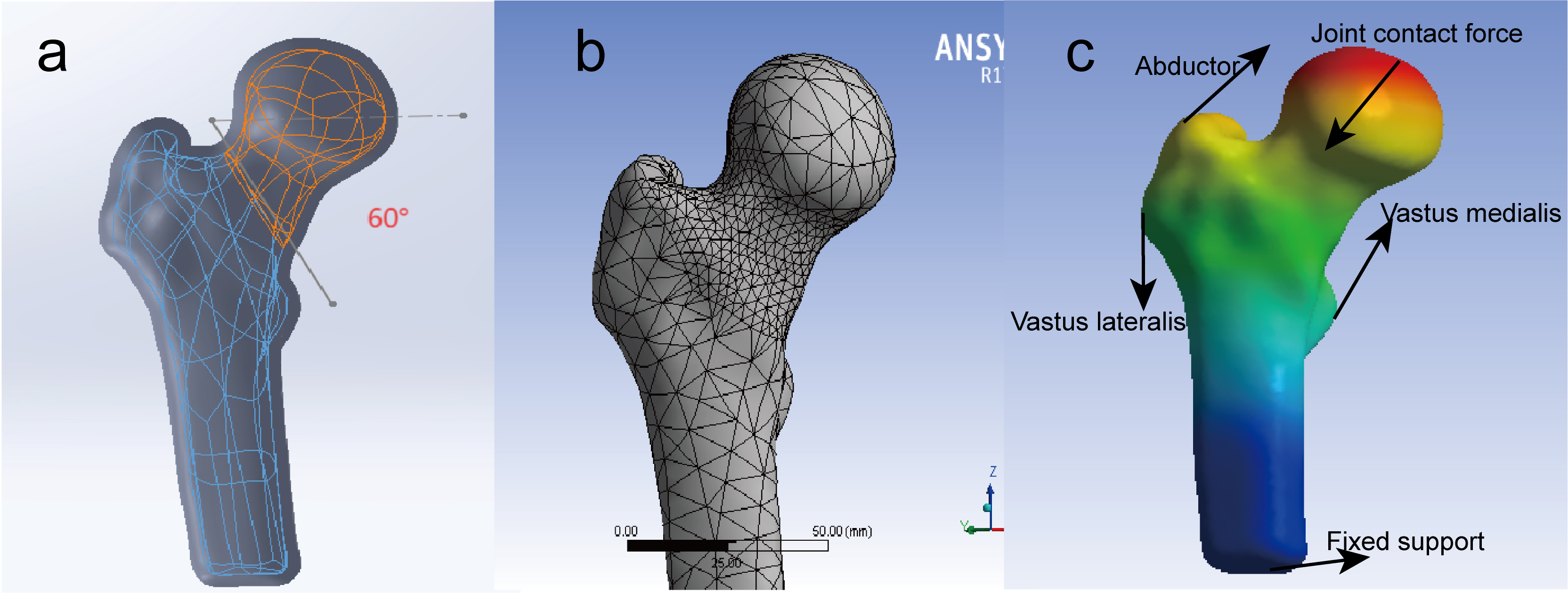
According to the previously set material property parameters [11, 18], the model including cortical bone, cancellous bone, and internal fixations was assigned to different elastic modulus and Poisson’s ratio (Table 1). The connection between the fracture surface was set to friction, and the coefficient of friction was 0.46 [19]. The relationship between the internal fixations and bone was set as binding relation.
Material properties of models in this study
Material properties of models in this study
Details of the modeling: CSS(a); CSS
To improve the calculation accuracy, a mixed mesh method was adopted to build meshes of the femoral neck, and different mesh types were established (Fig. 2b). This method allowed for more nodes and elements, which resulted in higher accuracy of biomechanical analysis. The minimum effective size of a four-node tetrahedron body element was 1.5 mm, which was used for bone and all kinds of internal fixations.
We created a simplified model in which the various materials were recognized to be homogeneous, isotropic linear. An 1800 N load corresponding to 3 times body weight [20, 21] was uniformly applied to the weight-bearing region of the femoral head along the specific direction. The load and muscle forces were simplified [12] (Fig. 2c).
Evaluation criteria
First, the statistics of all reconstructed models were recorded, including their nodes and elements. Second, the displacement, shear stress and equivalent (von Mises) stress of different internal fixations were observed.
Details of displacement: CSS(a); CSS
Schematic diagram of shear stress of internal fixations: CSS(a); CSS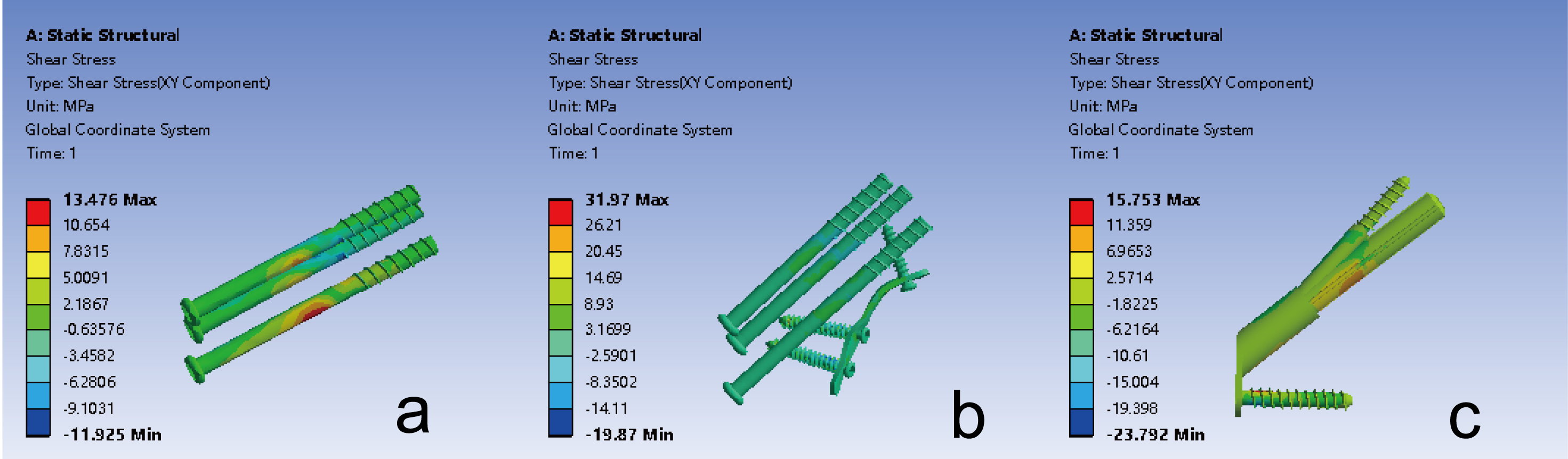
Maximal displacement of femur and internal fixators
From the vector display, the displacement of models was the same direction as the force. Meanwhile, this study showed roughly the same displacement direction in FNS and CSS
Peak value and distribution of shear stress
As shown in Fig. 5, the maximum shear stress occurred at the contact surface between the internal fixation and the fracture. The shear stress of CSS was located in the middle of the distal screw; the maximum value was 13.47 Mpa. The principal shear stress of CSS
The interfragmentary shear stress of CSS, CSS
Peak value and distribution of equivalent (von Mises) stress
The distribution map of equivalent (von Mises) stress was showed in Fig. 6. The stress was scattered at the femoral medial cortical bone. However, the peak value of equivalent stress was located in the internal fixator. The maximum equivalent stress of CSS was concentrated on the middle of three screws; the peak value was 52.01 Mpa. The maximum equivalent stress of CSS
Schematic diagram of equivalent stress of internal fixations: CSS(a); CSS
In the treatment of FNFs, implant choice has been one of the most critical yet contentious questions. A meta-analyze showed the incidence of implant failure was 9.7% [22], which leading to a high rate of reoperation. It is significant to choose most effective internal fixation approach to treat unstable fractures (Pauwels Type III). A previous FEA evaluated the biomechanical stability of cannulated screws with different configurations [23]. The author considered inverted triangle configuration as the recommended choice. However, it is not particularly appropriate for unstable fractures because of vertical shear force. To address this issue, a medial buttress plate was recommended to assist in the operation, affording increased resistance of shear stress [24]. As an improvement in classic minimally invasive surgery, CSS
Accordingly, we would like to analyze the biomechanical properties of CSS and FNS by the finite element method [29] in order to provide help for clinical selection. Compared to traditional biomechanical analysis, the finite element analysis can provide more convenient conditions, such as various loading methods, multivariate test indexes and relatively low research costs. At present, the FEA of FNFs mainly focuses on the comparison of internal fixation methods of different types to optimize surgical approaches. In our study, models with the same Pauwels angle were rebuilt through finite element analysis, which enhanced the accuracy and repeatability of experiments. A more elaborate model of FNS was rebuilt with high amount of details. Meanwhile, the shear stress was recorded for the first time to evaluate the stability of models.
In this finite element analysis, we evaluated the biomechanical characteristics of FNS and CSS
Our results showed that the internal fixation shear stress of FNS increased by 16% compared to CSS, while the shear force of MP increased by 137%. It indicates that the medial plate also continues to be subjected to more shear forces as time increases, which raises the risk of internal fixation failure. From the results, the CSS
However, this study has some limitations. First, the simplified finite element models were reconstructed without muscles, joint capsules and ligaments. Therefore, the accuracy of the experimental results was slightly lower than the actual results, but it could reflect the trend of change. Second, this study was designed to simulate the immediate mechanical environment after the operation. After all, it’s hard to evaluate the force variation in the healing process of femoral neck fracture. Third, different bone mass, running and other different conditions were not taken into consideration. Thus, further biomechanical tests and clinical assessments need to be performed.
Conclusions
CSS
Ethics approval and consent to participate
This study was approved by the Medical Ethics Committee of the Baoding No.1 Central Hospital ([2020]038). All methods were carried out in accordance with relevant guidelines and regulations. The volunteer agreed to the trial protocol and her informed consent was obtained.
Availability of data and materials
The datasets used and/or analysed during the current study are available from the corresponding author upon reasonable request.
Funding
The study was financially supported by the Baoding City Science and Technology Plan Project (2041ZF103).
Author contributions
CN: Conceptualization, Methodology, Software, Writing-review & editing. YL: Conceptualization, Methodology, Software, Editing. YL: Visualization, Investigation. LM: Software, Validation. ZM: Supervision, Writing – review & editing. All authors read and approved the final manuscript. CN and YL contributed equally to this work and should be considered co-first authors.
Footnotes
Acknowledgments
Not applicable.
Conflict of interest
The authors declare that they have no competing interests.