



Abstract
BACKGROUND:
At present, studies on MircoRNA-22-3p (miR-22-3p) in lung adenocarcinoma use a single method, lack multi-center validation and multi-method validation, and there is no big data concept to predict and validate target genes.
OBJECTIVE:
To investigate the expression, potential targets and clinicopathological significance of miR-22-3p in lung adenocarcinoma (LUAD) tissues.
METHODS:
LUAD formalin-fixed paraffin-embedded (FFPE) tumors and adjacent normal lung tissues were collected for real-time quantitative polymerase chain reaction (RT-qPCR). Collect miR-22-3p in LUAD and non-cancer lung tissue from high-throughput datasets, standardized mean difference (SMD) and area under the curve (AUC) of the comprehensive receiver operating curve (summary receiver operating characteristic cure, sROC curve) were calculated. Cell function experiments on A549 cells transfected with LV-hsa-miR-22-3p. Target genes were predicted by the miRwalk2.0 website and the resulting target genes were subjected to Gene Ontology (GO) pathway enrichment analysis and constructed to protein-protein interaction network. Finally, the protein expression level of the key gene TP53 was validated by searching The Human Protein Atlas (THPA) database to incorporate TP53 immunohistochemical results in LUAD.
RESULTS:
RT-qPCR result from 41 pairs of LUAD and adjacent lung tissues showed that miR-22-3p was downregulated in LUAD (AUC
CONCLUSION:
Overexpression of miR-22-3p may inhibit LUAD cell proliferation, migration and invasion through TP53, and promote cell apoptosis.
Introduction
Primary lung cancer is the deadliest cancer in the world [1, 2]. As the most common histological type of lung cancer, lung adenocarcinoma (LUAD) has become more prevalent over the past two decades [3]. Due to the development of precision medicine and non-small-cell lung cancer (NSCLC) therapy for genetic changes, many microRNAs (miRNAs) can be used as ideal markers for early diagnosis and prognosis prediction of lung cancer [4, 5], and are also potential therapeutic targets for lung cancer [6]. However, resistance to current targeted therapies is inevitable, and immune-based therapies still lack robust predictive biomarkers [7]. Therefore, it is still imperative to find effective therapeutic targets for LUAD patients.
MiRNAs are non-coding RNAs that are thought to play crucial roles in human physiology and lesion motility. Benefiting from high-throughput sequencing technologies, more and more non-coding RNAs are being discovered and defined as key regulators of tumorigenesis, aging and immune defense [8]. Dysregulated miRNAs cause various human diseases, including miR-22-3p. Previous studies have shown that upregulated miR-22-3p in extracellular vesicles (EVs) derived from human adipose tissue stem cells (AT-MSCs) can regulate osteogenesis [9]. The expression levels of miR-22-3p in serum may predict breast cancer at an early stage [10]. MiR-22-3p directly targets TCF7L2 in osteosarcoma and ENO1 in glioblastoma [11, 12]. Therefore, detecting the expression levels of miR-22-3p helps us to predict the risk of disease, diagnose disease at an early stage and evaluate the effect of treatment.
However, in the current studies of miR-22-3p, it has been found that the expression of miR-22-3p in LUAD is lower than that in normal tissue by PCR detection of clinically collected lung tissue [13, 14]. There are also studies using NSCLC lung tissue or blood PCR to learn that the expression of miR-22-3p in NSCLC is lower than that in normal lung tissue [15]. Some studies have verified the expression of miR-22-3p by cell function experiments [16]. Another study has concluded that the expression level of miR-22-3p is related to the prognosis of patients by extracting miRNA from patients’ blood and analyzing clinical information [17]. These research methods are relatively simple, lack multi-center validation and multi-method validation, and none of them have big data concepts to predict and validate target genes. Therefore, this study comprehensively analyzed an online database integrating in-house real-time quantitative polymerase chain reaction (RT-qPCR) data to validate the expression pattern of miR-22-3p, overexpressed miR-22-3p. In vitro experiments were carried out to detect the cell function after Lv-has-mir-22-3p lentiviral vector transfection, and computational biology was performed to predict the potential regulatory mechanism of miR-22-3p in LUAD.
Materials and methods
Clinical samples
41 pairs of formalin-fixed paraffin-embedded (FFPE) tumors and adjacent normal lung tissues were collected from LUAD patients from January 2014 to February 2016 from the First Affiliated Hospital of Guangxi Medical University. All involved patients received no treatment before surgery and were diagnosed postoperatively by two pathologists. All experiments were approved by the Medical Ethics Committee of the First Affiliated Hospital of Guangxi Medical University.
RT-qPCR
The extraction, reversion transcribed into cDNA and correction of total RNA were processed according to previous study [18]. RT-qPCR assay was performed on the Applied Biosystems PCR 7900. The expression of miR-22-3p was detected by miRcute Plus miRNA qPCR Kit (SYBR Green) (TIANGEN, Beijing, China). The expression of miR-22-3p was calculated by formula 2-
Forward GGCTGAGCCGCAGTAGTTCT, Reverse GTGCAGGGTCCGAGGT; and the primer sequence of the endogenous control RNU6B was as follows: Forward CTCGCTTCGGCAGCACA, Reverse GTGCAGGGTCCGAGGT.
Data source and processing of miR-22-3p
Microarray datasets containing miR-22-3p expression were included by searching public databases such as ArrayExpress, Gene Expression Omnibus (GEO), Oncomine, and The Cancer Genome Atlas (TCGA). The inclusion criteria for the dataset were: (1) the study subjects were humans; (2) included miR-22-3p expression data. The exclusion criteria are: (1) duplicate or incomplete sample data; (2) the number of samples in the LUAD group and the control group in the combined data set is less than 3 in any one.
Log
Cell culture, transfection and functional experiments
The LUAD cell line A549 was purchased from the Shanghai Cell Bank of the Chinese Academy of Sciences. A549 cells were cultured in RPMI 1640 medium containing 10% fetal bovine serum (FBS) and 1% penicillin/streptomycin (Hyclone, Logan, UT) and maintained at 5% carbon dioxide, 37.0
Prediction of candidate target genes
“Limma” package from R software was used to screen for differentially expressed genes in microarray and sequencing data from LUAD. Compared with normal lung tissue, genes with SMD
Analysis of potential molecular mechanisms of candidate target genes
“Clusterprofiler” is a powerful R package [23] that supports enrichment analysis of Gene Ontology (GO). GO enrichment analysis included biological process (BP), cellular component (CC) and molecular function (MF), mainly showing the biological pathways involved in candidate target genes. Protein-protein interaction (PPI) network analysis can study the molecular mechanism of LUAD from a systems perspective, which is helpful to discover the hub of miR-22-3p. We input candidate target genes into the STRING (V11.0) website (
Data source and processing of TP53
The PPI network constructed from the candidate target genes showed that TP53 was a key gene of the miR-22-3p pathway (see 3.3.2 for details). TP53 multi-center sample information collection and data processing methods are the same as the above (see 2.3). The protein expression level of TP53 was verified by searching The Human Protein Atlas (THPA) database to incorporate TP53 immunohistochemical results in LUAD.
Statistical methods
Wilcoxon rank sum test and SMD were used to determine differences in gene expression between the LUAD and control groups. A
All the experiments were independently conducted at least three times. Experiment data were analyzed by SPSS 22.0 statistical software package. Normal distribution data were presented as Mean
All calculation and visualization steps of data from public databases were performed in R software (v4.1.0) except that the integrated receiver operating characteristic curve was drawn in Stata 15.0.
Results
Expression of miR-22-3p in LUAD
RT-qPCR of samples from our hospital
In the 41 pairs of lung tissues collected in our hospital, the expression of miR-22-3p in LUAD showed a downward trend compared with the adjacent normal tissues (Fig. 1A). In addition, miR-22-3p expression levels were not significantly different from clinicopathological parameters (
MiRNA-sequencing and microarray data
Basic information about the microarray chips included in this study
Basic information about the microarray chips included in this study
N1: Number tumor; M1: Mean tumor; SD1: Standard deviation tumor; N2: Number control; M2: Mean control; SD2: Standard deviation control.
Analysis of RT-qPCR data indicates that miR-22-3p expression was down-regulated in LUAD. A. violin diagram. B. ROC chart. LUAD: lung adenocarcinoma. AUC: area under the curve. 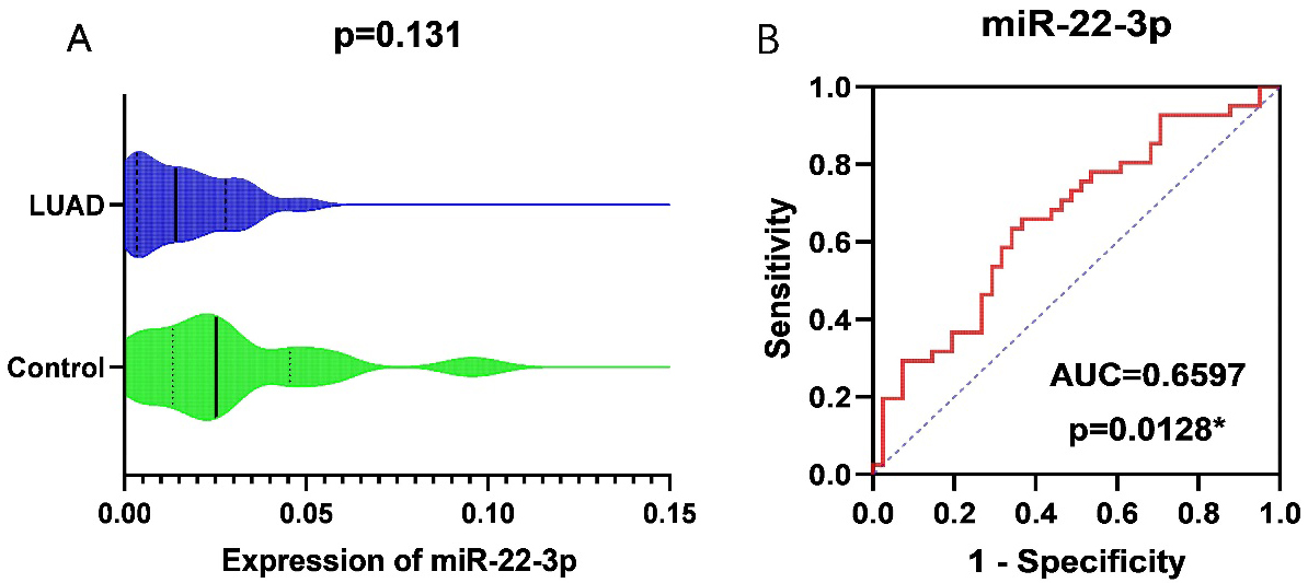
Integrated analysis of RT-qPCR and all microarray chips of miR-22-3p got from public databases. A. Standardized mean difference (SMD) forest map. B. Funnel chart. C. Summary receiver operating curve (sROC) curve.
The data included in this study were shown in Table 1. We obtained RNA-sequencing data for 521 LUAD tissues and 46 normal lung tissues from the TCGA database which composed with very few Asian patients. Compared with the normal lung tissues, miR-22-3p was down-regulated in LUAD tissues (
LV-miR-22-3p transfection
To explore the function of miR-22-3p in LUAD, A549 cell lines were transfected with LV-miR-22-3p and no-load virus, respectively, and a blank control group was set. The interference efficiency was verified by RT-qPCR. The results showed that A549 was successfully transfected into LV-miR-22-3p, and compared with the control group transfected with no-load virus, the relative expression of LV-miR-22-3p in lung cancer cells was significantly higher decreased (
Over expression efficiency of LV-miR-22 in lung cancer cell
Over expression efficiency of LV-miR-22 in lung cancer cell
In this study, the effect of miR-22-3p on the proliferation of lung cancer cell A549 was detected by MTS kit. The established experimental group and control group were the same as the above experiments. It can be concluded that the cell proliferation ability of the experimental group added with miR-22-3p inhibitor was lower than that of the control group on the 5th day, and was stronger than that of the control group on the tenth day, but there was no significant difference. Compared with the control group, the cell proliferation ability of the experimental group of the mock gradually decreased on the fifth day and the tenth day, and the difference was statistically significant (Fig. 3, Table 3).
The effect of transfection of miR-22-3p on the proliferation of A549 cells was detected by MTS
The effect of transfection of miR-22-3p on the proliferation of A549 cells was detected by MTS
CCK-8 kit was used to detect the effect of transfected LV-miR-22 on the proliferation of A549 cells
To further validate the ability of transfected miR-22-3p to inhibit cell proliferation, CCK-8 cell proliferation assay was used. The growth curve of the transfected LV-miR-22-3p group was lower than that of the empty virus group. And at 60 hours, the overexpression of miR-22-3p group compared with the empty group had a significant difference in the effect on cell proliferation (
The scratch assay was used to detect the effect of overexpression of miR-22-3p on the migration ability of A549 cells in vitro. Figures 5 and 6 show that after the use of lentiviral vector to interfere with miR-22-3p expression, the in vitro migration ability of A549 cell line was consistently inhibited, and was statistically different from the corresponding empty group at 18 hours and 24 hours (Table 5).
Overexpression of miR-22-3p inhibits the migration of A549 lung cancer cells
Overexpression of miR-22-3p inhibits the migration of A549 lung cancer cells
The effect of transfection of miR-22-3p on the proliferation of A549 cells was detected by MTS (
The growth curve of A549 cells transfected with LV-miR-22 was detected by CCK-8 kit (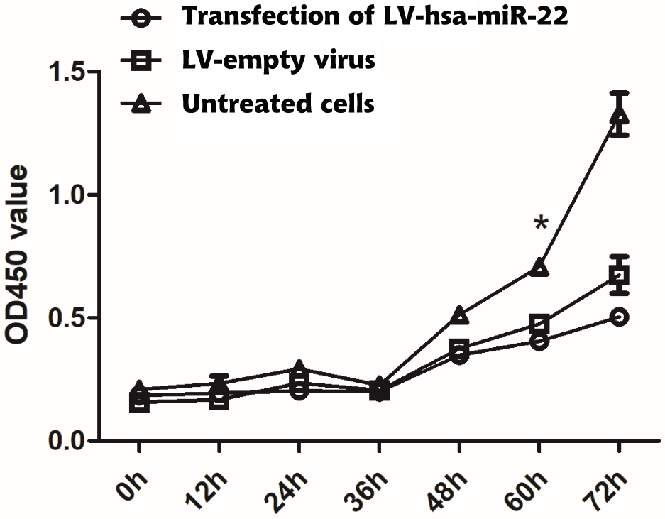
Migration ability of A549 lung cancer cells overexpressed with miR-22-3p (
Over-expression of miR-22-3p inhibits the migration of A549 lung cancer cells. A. 18 hours later no-load virus group. B. 18 hours after transfection with LV-miR-22 group. C. 24 hours after no-load virus group. D. 24 hours later, the cells were transfected into LV-miR-22 group.
The effect of over-expression of miR-22-3p on the migration of lung cancer cells was detected by Transwell assay. A549 cells were (A) transfected with LV-miR-22 and (B) without virus transfected for 24 hours and then stained with crystal violet. The number of migration cells was observed under microscope (200 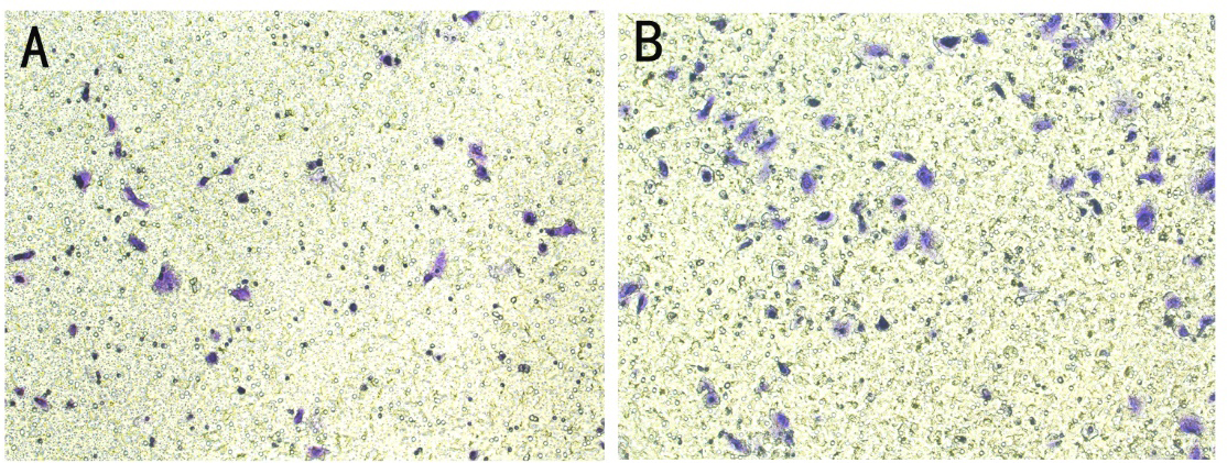
To further verify the scratch test results, Transwell assay was used to detect the effect of miR-22-3p on the migration ability of A549 cells. In the A549 cell line, the number of cells transfected with LV-miR-22-3p was less (Fig. 7), which was statistically different from the control group (
The effect of over-expression of miR-22-3p on the migration of A549 cells was detected by Transwell assay
The effect of over-expression of miR-22-3p on the invasion ability of A549 cells was detected by Matrigel Transwell assay
Caspase-3/7 kit was used to detect the effect of miR-22-3p on apoptosis of lung cancer cells
The effect of over-expression of miR-22-3p on the migration of A549 cells was detected by Matrigel transwell assay. A549 cells (A) transfected with LV-miR-22 and (B) without a virus were stained with crystal violet 48 hours. The number of invading cells was observed under microscope (200 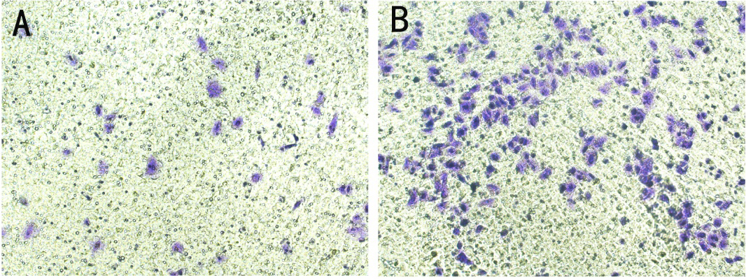
Caspase-3/7 kit was used to detect the effect of miR-22-3p on apoptosis of A549 cells.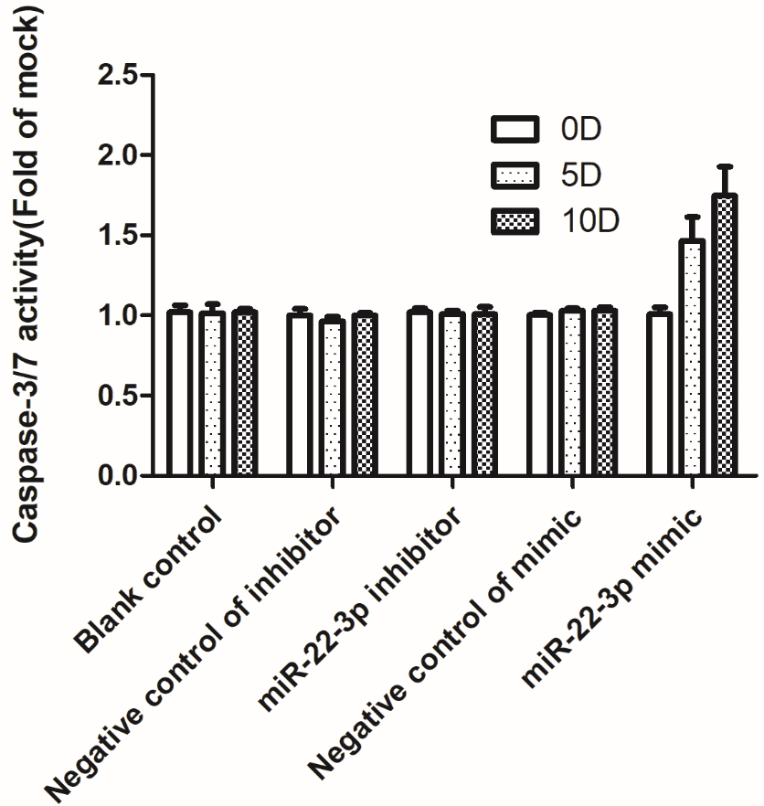
Potential mechanism of miR-22-3p in lung adenocarcinoma. A. The Venn diagram of predicted target genes and DEGs. B. Bar chart of candidate target genes enriched in cellular component.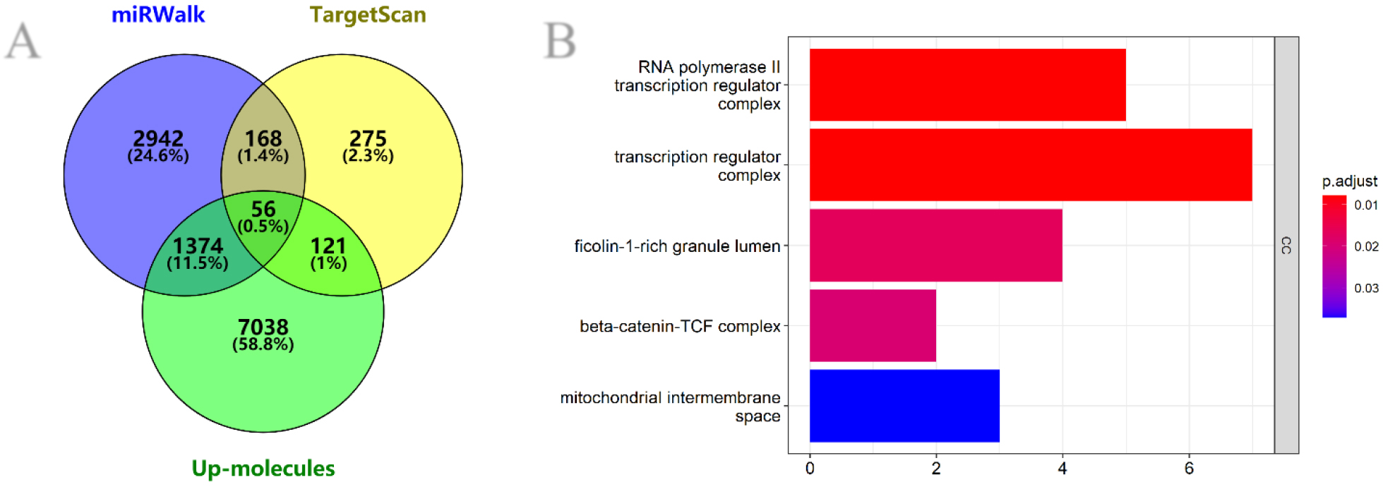
PPI analysis of candidate target genes in the network that interact most with other genes. TP53 is the determination gene within the whole PPI network.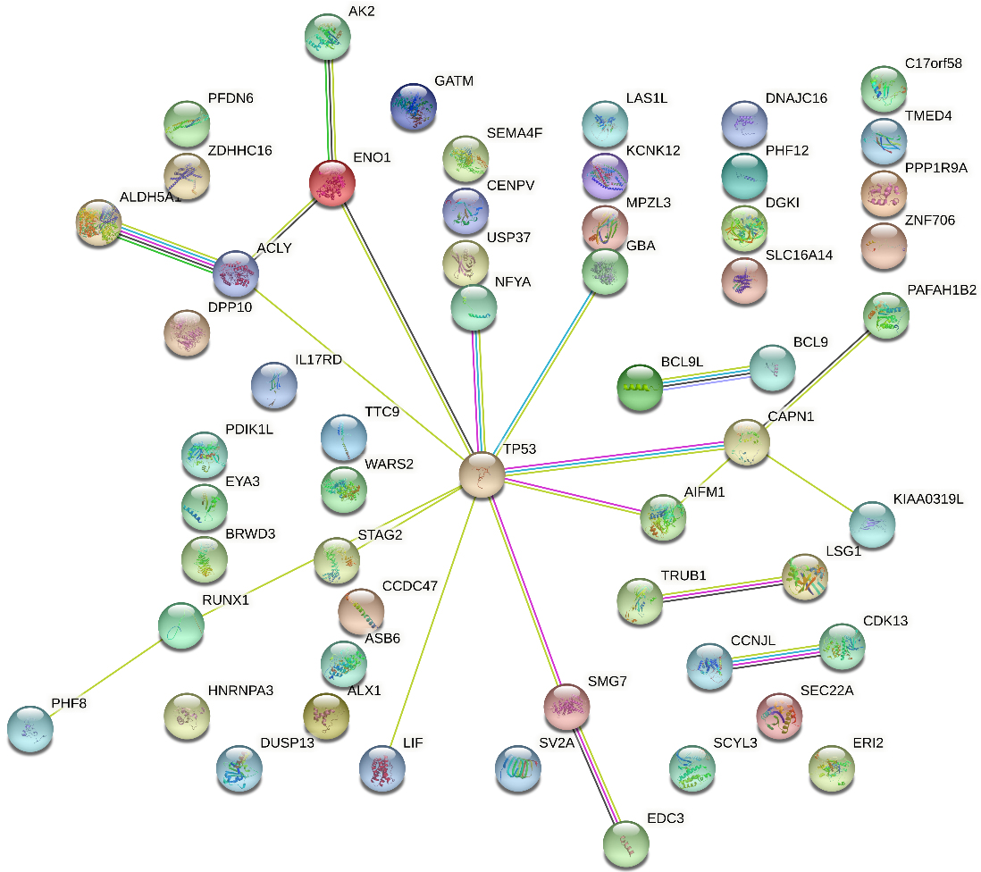
Integrated analysis of expression levels of TP53 in LUAD got from public databases. A. SMD forest map. B. Funnel chart.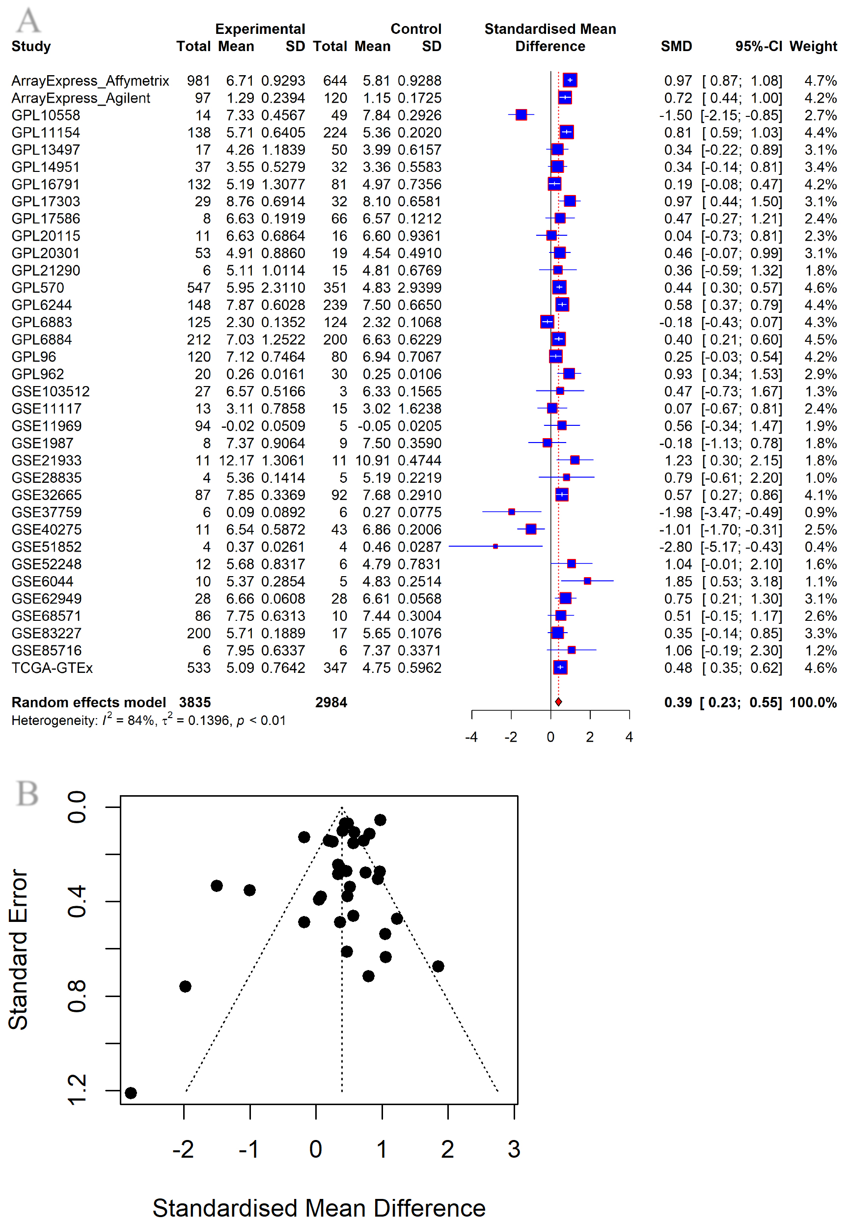
Protein expression of TP53 in LUAD and control tissues. N: Normal tissue; T: Tumor tissue.
Matrigel Transwell assay was used to verify the regulatory effect of overexpression of miR-22-3p on A549 cell invasion ability. The results showed that after A549 cells were transfected with LV-miR-22, the invasive ability of A549 cells was weakened (Fig. 8), and there was a statistical difference (
The effect of miR-22-3p on apoptosis of A549 cells was detected by Caspase-3/7 kit. After transfection of miR-22-3p inhibitors and mimics into A549 cells, apoptosis was promoted on both the fifth and tenth days, but the changes were not obvious (Fig. 9, Table 8).
Analysis of pathway and potential molecular mechanisms of miR-22-3p
Candidate target gene prediction
According to the screening criteria, 8589 up-molecules were selected from the microarray chip and sequencing dataset. 4540 related target genes were predicted on the miRwalk2.0 website. 620 relevant target genes were predicted on the TargetScan website. We intersected these ascending genes with related target genes, resulting in 56 candidate target genes (Fig. 10A).
Analysis of potential molecular mechanisms of candidate target genes
The possible mechanisms of 56 candidate target genes were analyzed using the “clusterProfiler” R package and STRING. The GO analysis results mainly focused on the RNA polymerase II transcriptional regulatory complex, the transcriptional regulatory complex, the ficolin-1-rich granule lumen, the
Integrated analysis of TP53 public database
Globally, a total of 114 high-throughput datasets were included, including 3897 LUADs and 2993 noncancerous lung tissues. After removing batch effects, 37 platforms were finally obtained. A random-effects model was further constructed to calculate SMD, and the expression level of TP53 (the gene expression value included in the 35 platform datasets) was integrated for analysis. The results showed that TP53 expression was up-regulated in the LUAD group compared with the control group (SMD
The expression of TP53 in LUAD was verified from the protein level. In this study, a total of 23 LUAD tissues and 12 control tissues were retrieved through the THPA database to obtain immunohistochemical pictures. As shown in Fig. 13, the TP53 protein expression level in the LUAD group was higher than that in the control group (
Discussion
In recent years, thanks to significant advances in molecular oncology, there have been more effective approaches to the treatment of NSCLC, improving survival for patients with advanced stages. However, the drug resistance of targeted therapy is still a problem of precision therapy [7]. As the most common NSCLC, LUAD has poor survival and high recurrence rate [2]. is the focus of current LUAD treatment research to find effective therapeutic targets and avoid drug resistance. Despite tremendous progress in lung cancer treatment, the 5-year survival rate for patients with stage remains around 55% [24]. Therefore, finding effective therapeutic targets and avoiding drug resistance is the focus of current LUAD treatment research.
In this study, the expression patterns of miR-22-3p and overexpressed miR-22-3p were verified by RT-qPCR data of our hospital and cell function detection after Lv-has-mir-22-3p lentiviral vector transfection. And computational biology was performed to predict the potential regulatory mechanism of miR-22-3p in LUAD.
Previous studies on the expression of miR-22-3 in LUAD have either single-center data or only a single method. Ma et al. collected 62 LUAD patients’ tumor tissues and adjacent normal tissues from Peking Union Medical College Hospital for PCR detection of miR-22-3p expression levels [13]. Yang et al. collected 91 LUAD lung tissues and 10 normal lung tissues from the Department of Thoracic Surgery of the University of Michigan for RT-qPCR to detect the expression levels of miR-22-3p [16]. Zhenan Xu et al. collected blood samples from 2 cases of LUAD and 2 cases of healthy people in the Second Hospital of Jilin University to detect the expression of miR-22-3p by exosome microarray [15]. Zhou et al. confirmed that ENO1 is one of the targets of miR-22-3p by RT-qPCR and western blot analysis of A539 cell line overexpressing ENO1 and miR-22-3p [14]. Ulivi et al. collected 99 LUAD blood samples and matched clinical information from Medical Oncology Department of Perugia Hospital and by the Thoracic Surgery Department of Morgagni-Pierantoni Hospital of Forlì, and performed univariate analysis after detecting miR-22-3p expression. The results showed that miR-22-3p was significantly associated with disease-free survival and overall survival of patients [17]. Dong et al. used ChipBase, LncRNAdb and StarBase to identify that miR-22-3p was regulated by DGCR5, and verified by cellular luciferase experiments [25]. However, the above studies are only validated with single-center data or a single method.
In this study, RT-qPCR was performed on 41 pairs of LUAD and matched adjacent normal tissues collected in our hospital, and the results showed that miR-22-3p was expressed lower in LUAD than in normal tissues. At the same time, by incorporating high-throughput data from around the world, 838 cases of LUAD and 494 cases of non-cancer lung tissue were comprehensively analyzed, and the results were consistent with the RT-qPCR results. We also performed functional experiments with A539 cells transfected LV-has-miR-22-3p, showing that miR-22-3p inhibited cell proliferation, migration and invasion, and promoted cell apoptosis, which was consistent with the low expression of miR-22-3p in LUAD.
In order to understand the tumor suppressor effect mechanism of miR-22-3p, this study collected miR-22-3p-related genes for gene enrichment and signaling pathway analysis, and constructed PPI network. The results showed that TP53 is a key gene of the miR-22-3p-related signaling pathways. The integrated analysis of TP53 expression levels showed that TP53 is up-regulated in LUAD. At the same time, we collected the LUAD tissue immunohistochemical data of TP53 from THPA, which also showed that the protein expressed by TP53 was increased in LUAD. These indicated that miR-22-3p was inversely correlated with TP53, suggesting that miR-22-3p might inhibit TP53 expression. Research has shown that, mutated or unregulated forms of TP53 exert novel oncogenic activities [26], further verifying that miR-22-3p inhibits the development of LUAD. Qian et al. showed that TP53 and STAG2 were associated with overall survival of NSCLC patients. It may be the manifestation of PPI interaction on LUAD in this study [27].
Conclusion
miR-22-3p is underexpressed in lung adenocarcinoma and it plays a role in inhibiting tumor proliferation, migration and invasion, and promoting apoptosis in LUAD, which may be achieved through pathways related to cell cycle and cell migration. In addition, TP53 is a key gene in these pathways and its expression is up-regulated in LUAD, suggesting that miR-22-3p inhibits TP53 expression and thus acts as a tumor suppressor. The research on miR-22-3p and its related pathways needs to be verified by more in-depth in vivo studies.
Consent for publication
All authors have read the final draft of the manuscript and agreed to its publication.
Funding
This work was supported by the Guangxi Zhuang Autonomous Region Medical Health Appropriate; Technology Development and Application Promotion Project (S2020031); Guangxi Medical High-level Key Talents Training “139” Program (2020); Guangxi Medical University Undergraduate Innovation and Entrepreneurship Training Program (X202210598227); and Future Academic Star of Guangxi Medical University (WLXSZX22112).
Data availability
The datasets generated and/or analyzed during the current study are available from the corresponding author upon reasonable request.
Footnotes
Acknowledgments
The authors greatly acknowledge Guangxi Medical University and other institutions for funding the study.
Conflict of interest
None to report.