



Abstract
BACKGROUND:
Gastric cancer (GC) is the fifth most common cancer worldwide and the third leading cause of cancer death. Due to the low rate of early diagnosis, most patients are already in the advanced stage and lose the chance of radical surgery.
OBJECTIVE:
To investigate the clinical value of computed tomography (CT) dual-energy imaging in preoperative evaluation of pathological types of gastric cancer patients.
METHODS:
121 patients with gastric cancer were selected. Dual-energy CT imaging was performed on the patients. The CT values of virtual noncontrast (VNC) images and iodine concentration of the lesion were measured, and the standardized iodine concentration ratio was calculated. The iodine concentration, iodine concentration ratio and CT values of VNC images of different pathological types were analyzed and compared.
RESULTS:
The iodine concentration and iodine concentration ratio of gastric mucinous carcinoma patients in venous phase and parenchymal phase were lower than those of gastric non-mucinous carcinoma patients, and the differences were statistically significant (
CONCLUSION:
Dual-energy CT imaging plays an important role in the preoperative evaluation of patients with gastric cancer. The pathological types of gastric cancer are different, and the iodine concentration will change accordingly. Dual-energy CT imaging can effectively evaluate the pathological types of gastric cancer and has high clinical application value.
Introduction
Gastric cancer (GC) is the fifth most common cancer worldwide and the third leading cause of cancer death [1]. The 5-year survival rate of early gastric cancer can reach more than 90%. However, due to the low rate of early diagnosis, most patients are already in the advanced stage and lose the chance of radical surgery [2]. The most commonly used imaging diagnostic methods for gastric cancer are digital fiber endoscopy, gastrointestinal angiography and traditional CT examination [3]. With the development of individualized diagnosis and treatment mode of gastric cancer, higher requirements are put forward for the refinement of imaging diagnosis [4]. At present, a large number of studies have shown that dual-energy CT increases the energy resolution and physical and chemical properties resolution on the basis of the original spatial resolution, time resolution and density resolution of conventional CT [5]. The dual-energy CT provides more diagnostic information for clinical practice. It has obvious advantages over traditional imaging techniques. Dual-energy CT, with high density resolution, high energy resolution, high physical and chemical properties of the resolution and powerful post-processing technology, realizes the CT from single parameter analysis to tissue composition analysis, from morphological diagnosis to functional diagnosis of human energy spectrum imaging. It integrates precise imaging, whole body low-dose imaging and large-scale dynamic imaging, which is a real multi-parameter imaging [6] and can find some lesions that cannot be found by conventional CT. Dual-energy CT has become a hot spot in qualitative diagnosis, staging and typing of gastric cancer [7]. The traditional diagnosis, staging and typing of gastric cancer need to rely on invasive pathological examination, but dual-energy CT, as a non-invasive impact examination, can infer the degree of tumor invasion by the different enhancement amplitude of the lesion area in different phases of the enhanced scan and the depth of the gastric wall involved in the enhanced area on the single energy image, and use the multi-parameter advantage to detect and evaluate the lymph node metastasis of gastric cancer [6].
Imaging examination plays an important role in tumor staging and clinical diagnosis, but the current doctors pay less attention to the pathological types of gastric cancer. Preoperative histopathological evaluation is associated with prognosis, surgical and comprehensive evaluation [8]. Therefore, while paying attention to the staging of gastric cancer, it is necessary to effectively evaluate the degree of differentiation and pathological types of gastric cancer. In this study, the clinical value of dual-energy CT imaging in preoperative evaluation of gastric cancer pathological type diagnosis was explored to provide a more effective theoretical basis for clinical treatment of patients. The report is as follows.
Materials and methods
General information
A total of 121 patients with gastric cancer admitted to Maanshan People’s Hospital from December 2019 to June 2022 were selected as the study subjects. Inclusion criteria: (1) patients with gastric cancer diagnosed by gastroscopy and confirmed by histopathology; (2) patients who agreed to the test items and had complete clinical data; (3) patients without targeted or chemoradiotherapy before CT examination; (4) the lesions were clearly displayed, and the patients with good gastric cavity filling in CT images; (5) patients with clear consciousness who can cooperate with imaging examination Exclusion criteria: (1) Poor image quality, patients cannot meet the requirements of measurement and diagnosis; (2) patients in pregnancy or lactation; (3) patients with contrast intolerance or CT scan contraindication.
There were 88 males and 33 females. Age 43–89 years; postoperative pathological diagnosis: 98 cases of gastric non-mucinous carcinoma, including 49 cases of poorly differentiated adenocarcinoma, 49 cases of well-differentiated adenocarcinoma; there were 23 cases of gastric mucinous carcinoma, including 10 cases of signet ring cell carcinoma and 13 cases of mucinous adenocarcinoma. All patients agreed to participate in this study. The study was approved by the ethics committee of the hospital.
Methods
Inspection method
The patients were forbidden to use heavy metal drugs 3 days before the examination, and fasted for 8
Image analysis
Firstly, the original images of arterial phase, venous phase and delayed phase of 100 keV and Sn150 keV thin-layer CT were introduced into Sigmens Syngo.via.by workstation, and Dury-Energy mode was used for image post-processing. Second, the Liver VNC mode was used to obtain the iodine map. The iodine content in arterial phase, venous phase and delayed phase was measured by ROI, and the iodine content in lesion area and abdominal aorta was measured. After the iodine concentration of each lesion was enhanced, the standard iodine concentration was defined as the iodine concentration ratio of the abdominal aorta in the same period. A total of 3 measurements were performed on the lesion, and the average value was selected. Liver VNC mode was used to get images of VNC in arterial phase, venous phase and delayed phase. During the measurement process, the artifacts, necrosis and calcification sites caused by the gas-liquid plane boundary should be avoided, and the ROI should be kept consistent in size and shape during the 3-phase measurement.
Observation indicators
1. CT values of VNC images, iodine concentration and iodine concentration ratio of gastric non-mucinous carcinoma and gastric mucinous carcinoma in arterial phase, venous phase and parenchymal phase were analyzed and compared. 2. The CT values of VNC images, iodine concentration and iodine concentration ratio of mucinous adenocarcinoma and signet ring cell carcinoma in arterial phase, venous phase and parenchymal phase were analyzed and compared. 3. The CT values of VNC images, iodine concentration and iodine concentration ratio of well-differentiated and poorly differentiated adenocarcinoma in arterial phase, venous phase and parenchymal phase were analyzed and compared.
Statistical methods
SPSS v. 23.0 statistical software was used to process the data. Measurement data were expressed as
| Category |
|
CT values of VNC images (HU) | Iodine concentration (100 |
Iodine concentration ratio | ||||||
|---|---|---|---|---|---|---|---|---|---|---|
| Arterial phase | Venous phase | Substantial phase | Arterial phase | Venous phase | Substantial phase | Arterial phase | Venous phase | Substantial phase | ||
| Gastric mucinous carcinoma | 23 | 38.2 |
39.2 |
40.2 |
12.2 |
16.9 |
18.1 |
0.2 |
0.4 |
0.6 |
| Non gastric mucinous carcinoma | 98 | 39.4 |
40.6 |
41.4 |
14.4 |
21.3 |
20.7 |
0.2 |
0.6 |
0.7 |
|
|
|
|
|
1.774 | 3.011 | 2.115 | 0 | 4.774 | 2.387 | |
|
|
|
|
|
|
|
|
|
|
|
|
Comparison of CT values of VNC images, iodine concentration and iodine concentration ratio between patients with mucinous adenocarcinoma and signet ring cell carcinoma (X
Comparison of CT values of VNC images, iodine concentration and iodine concentration ratio between moderately and poorly differentiated adenocarcinoma patients (X
mean
The iodine concentration and pathological sections of patients with gastric antrum mucinous adenocarcinoma. A–D: Male patient, 63 years old, gastric antrum mucinous adenocarcinoma, A: arterial phase iodine concentration, B: venous phase iodine concentration, C: delayed phase iodine concentration, D: pathological section.
Comparison of CT values of VNC images, iodine concentration and iodine concentration ratio between non-mucinous gastric cancer and mucinous gastric cancer patients
The iodine concentration and iodine concentration ratio in venous phase and parenchymal phase of patients with gastric mucinous carcinoma were lower than those in patients with gastric non-mucinous carcinoma, and the difference was statistically significant (
Comparison of CT values of VNC images, iodine concentration and iodine concentration ratio between mucinous adenocarcinoma and signet ring cell carcinoma
Iodine concentration and iodine concentration ratio in venous phase and parenchymal phase of patients with mucinous adenocarcinoma were lower than those of patients with signet ring cell carcinoma (
Comparison of CT values of VNC images, iodine concentration and iodine concentration ratio between well differentiated and poorly differentiated adenocarcinoma patients
The iodine concentration and iodine concentration ratio in venous phase and parenchyma phase of patients with moderately and highly differentiated adenocarcinoma were lower than those of patients with poorly differentiated adenocarcinoma, and the difference was statistically significant (
The iodine concentration and pathological section of poorly differentiated adenocarcinoma of gastric antrum. E–H: patient male, 70 years old, poorly differentiated adenocarcinoma of the gastric antrum, E: arterial phase iodine concentration, F: venous phase iodine concentration, G: delayed phase iodine concentration, H: pathological section.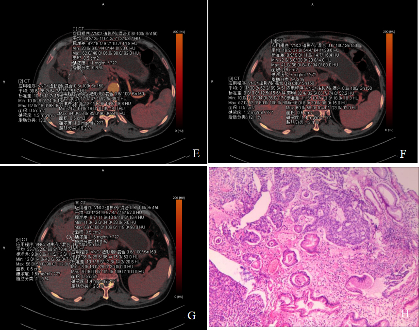
In the evaluation of tumor grading and tumor type of different tissue types, Dual-energy CT imaging has a broad prospect [9]. In this study, water-iodine-based substance was selected for quantitative analysis. The results showed that the iodine concentration and iodine concentration ratio of gastric mucinous carcinoma patients in venous phase and parenchymal phase were lower than those of gastric non-mucinous carcinoma patients, and the differences were statistically significant. There was no significant difference in CT values of VNC images in arterial phase, venous phase, parenchymal phase, iodine concentration ratio and iodine concentration in arterial phase between patients with gastric non-mucinous carcinoma and patients with gastric mucinous carcinoma. The main reasons for this result are different density of tumor blood vessels and whether the tumor is rich in mucus components. The analysis of different subtypes of gastric mucinous carcinoma showed that although both mucinous adenocarcinoma and signet ring cell carcinoma were gastric mucinous carcinoma, the iodine concentration obtained after enhanced signet ring cell carcinoma was higher than that of mucinous adenocarcinoma [10].
Different degrees of differentiation in patients with gastric non-mucinous carcinoma have different iodine concentrations, and the iodine concentration will increase with the decrease of differentiation [11]. The iodine concentration and iodine concentration ratio of patients with mucinous adenocarcinoma in venous phase and parenchymal phase were lower than those of patients with signet ring cell carcinoma, and the differences were statistically significant. This result was consistent with the finding of Zheng [12] that the iodine values of highly and moderately differentiated adenocarcinoma in arterial phase and venous phase were 10.3
Huang [17] and other studies have shown that the enhancement characteristics of poorly differentiated adenocarcinoma are mostly layered enhancement, while moderately and highly differentiated adenocarcinoma generally shows homogeneous enhancemen; the reason for the different enhancement characteristics of different types of gastric adenocarcinoma may be that the cancer tissue of poorly differentiated adenocarcinoma only grows along the muscle fibers without destroying the muscle layer structure, while the middle and high differentiation due to cancer tissue infiltration and destruction of the muscle layer structure. After that, Li [18] used the CT enhancement rate of the lesion to quantify the enhancement methods of gastric cancer with different degrees of differentiation, and concluded that the enhancement rate of the lesion was closely related to the degree of differentiation of the tumor. The CT enhancement rate of the poorly differentiated adenocarcinoma in the parenchymal stage was higher than that of the moderately differentiated adenocarcinoma group and the highly differentiated adenocarcinoma group. Accurate judgment of tumor differentiation is crucial for the choice of patient treatment options. Sun et al. [19] extracted deep learning features from three-phase enhanced CT images, and used machine learning methods to construct a radiomics prediction model, which showed good accuracy in identifying T4a gastric cancer, and the optimal AUC was 0.90. Tan et al. [20] established a delta radiomics model. Studies have shown that the model can effectively predict the chemotherapy response of patients with advanced gastric cancer, and the optimal AUC is 0.83.
With advances in computer processing power and graphics processing technology, artificial intelligence (AI) is increasingly being used for the analysis of large-scale and complex image data in endoscopic, pathological and imaging examinations [21]. The AI model can quickly and accurately identify asthma in children ’s general medical wards and can help junior pediatricians correctly diagnose asthma [22]. In the diagnosis of other complex lung diseases such as emphysema [23], chronic obstructive pneumonia [24] and so on also reflects the good diagnostic effect. Providing precision medicine for heart disease to change the prognosis of patients [25]. Ghanayim et al. [26] identified moderate or severe aortic stenosis with 86% sensitivity and 100% specificity based on AI algorithm. Sahu et al. [27] identified maternal, neonatal and laboratory predictors by establishing predictive models to quickly detect early and late neonatal sepsis. It has shown great potential in gynecology [28], dermatology [29, 30], cancer diagnosis [31], and meningitis [32]. The results of this study show that dual-energy CT imaging can effectively evaluate the pathological types of gastric cancer and has high clinical application value. With the in-depth study of many aspects, if dual-energy CT imaging technology and AI are combined in the future, there should be a broader application prospect and corresponding application value.
This study is not without limitations. First, the sample size is small, and the number of cases of mucinous adenocarcinoma and signet ring cell carcinoma is small. On the other hand, this study focused on exploring the iodine concentration parameters of dual-energy CT imaging in different types of gastric cancer, without comparing with conventional CT enhanced scan. If a comparative study is carried out, it can further prove whether the diagnostic efficiency of dual-energy scanning is better than that of conventional CT scanning. In future studies, it is necessary to further increase the sample size and refine the grouping, such as tubular adenocarcinoma, papillary adenocarcinoma, poorly differentiated adenocarcinoma, mucinous adenocarcinoma, signet ring cell carcinoma and other groups, for clinical diagnosis and treatment to provide more reliable information to improve patient outcomes and prognosis.
Conclusion
Dual-energy CT imaging plays an important role in the preoperative evaluation of patients with gastric cancer. The pathological types of gastric cancer are different, and the iodine concentration will be different. Dual-energy CT imaging can preliminarily evaluate the pathological classification of gastric cancer, which is conducive to the follow-up clinical treatment guidance of patients and has high clinical application value. Therefore, dual-energy CT imaging should be further promoted and applied in the evaluation of pathological types of gastric cancer patients.
Footnotes
Acknowledgments
The authors have no acknowledgments.
Conflict of interest
The authors declare that they have no conflict of interest.
Funding
The authors report no funding.