



Abstract
BACKGROUND:
Cone-beam computed tomography (CBCT) could be more beneficial in clinical situations that involve the determination of root canal morphology.
AIM:
The aim of the study was to ascertain the prevalence of radix molaris (paramolaris-RP and entomolaris-RE) in a subpopulation of Saudi Arabia using CBCT.
METHOD:
A total of 700 CBCT scans of mandibular permanent first molars were included in this study. All CBCT scans were interpreted by two trained dentists and an endodontist. Mandibular permanent first molars with fully developed roots and closed apices were only included. Computed Tomography scans were obtained from the dental college record. The anatomic characteristics which were checked included: the prevalence of radix molaris in both RE and RP and the prevalence of radix molaris according to the patient’s gender and age. All CBCT images were processed and reconstructed using OnDemand3D
RESULTS:
Of the 700 patients included, 651 (93.0%) did not have radix molaris, 46 (6.6%) had radix entomolaris and 3 (0.4%) had radix paramolaris in the study group. There was no statistically significant difference between the genders and age in the incidence of RE and RP (
CONCLUSION:
The study showed that RE prevalence is more compared to RP in the first lower molars in the population studied. It is important to identify extra roots and associated canals to perform successful root canal treatment and avoid failure.
Keywords
Introduction
The most important aim of root canal treatment (RCT) is the complete mechanical and chemical cleaning of the whole root canal system (RCS) and its three-dimensional (3D) obturation with an inert filling material and a coronal restoration to prevent the entry of microorganisms [1, 2, 3, 4, 5, 6, 7, 8]. One of the most common reasons for RCT failure in molars is that the clinician has not detected all root canals and removed all pulpal tissue and microbes from the RCS [9] this can cause Orofacial Pain, which is necessary to differentiate from Orofacial Pain of other nature as Orofacial pain caused from neurological diseases or caused from temporomandibular disorders (TMD) [10, 11, 12, 13, 14, 15, 16, 17, 18, 19]. Successful root canal treatments may result from clinicians’ awareness and recognition of the likelihood of the occurrence of unique root canal morphology.
The mandibular molars #34/46 are known to have a number of anatomical variants. The mandibular first permanent molar commonly has mesial and distal roots [9, 20]. The mesial root often contains two root canals, each of which leads to a unique apical foramen, though this can occasionally happen. One kidney-shaped root canal generally exists in the distal [20].
Various anatomical variations have already been described by researchers and documented in the literature for the mandibular first molar. The most variant in this tooth type is the occurrence of an extra 3
The prevalence of both RaEn and RaPa varies between different populations. Hence, knowledge and awareness of their presence and location are important for root canal treatment success. The previous studies showed that RaEn and RaPa prevalence ranged from 3% to 30% depending on the study’s ethnicity [22]. Previous studies were conducted in some isolated Saudi sub-populations. A new systematic review which included five studies conducted on Saudi sub-populations about the incidence of radix entomolaris in mandibular permanent first molar found that the RaEn presence ranges from 2 to 6.07% [23]. In addition, the systematic review concluded that the RaEn is higher in females when compared to males and higher on the right quadrant of the jaw when compared to the left which is statically significant as described by Khurayzi et al. in 2021 [21]. Some studies had found that the prevalence of RaEn was 7.2% which is comparable to other populations except for the Mongolian population.
Anatomical studies have shown that some ethnic groups are more likely to have a separate RE in the first mandibular molar. It happens 5 to more than 30% of the time in people with Mongoloid traits, like Chinese, Eskimos, and American Indians [21]. Researchers found that it occurred in no more than 3% of Africans [23, 24], whereas in Europeans the occurrence was even less. Schafer et al. [25, 26] used full-mouth periapical x-rays to find out how often radix entomolaris occurred in the German population. There were 1,024 first mandibular molars that were analysed (500 left molars and 524 right molars). Seven patients were found to have a mandibular first molar with 3 roots, which is a rate of 1.35 percent. Garg et al. assessed 1054 periapical radiographs from the Indian population and found that 5.97% of mandibular first molars had RaEn [26]. Karale et al. [27] used the same method and found that RaEn happened more often (6.67%). A review of the available endodontic literature showed that the prevalence of RaEn has a genetic and ethnic predilection (Table 1) [28, 29, 30, 31, 32, 33, 34, 35, 36, 37, 38, 39, 40, 41, 42, 43, 44]. It is uncommon in populations of European, Caucasian, and African descent, but it seems to be more prevalent in races with a Mongoloid ancestry.
Survey of available studies on the prevalence of 3-rooted mandibular first molars in different population and ethnic groups
Survey of available studies on the prevalence of 3-rooted mandibular first molars in different population and ethnic groups
When doing endodontic therapy, it is not uncommon to miss a canal, especially in molar teeth where the rule of “one root, one canal” perception is not valid, because there are more canals than roots. The significance of the current study rests in its contribution to determining the prevalence of additional canals in the population under consideration. One of the reasons for endodontic failure is improper root canal treatment. Bacteria found in these canals cause symptoms including pain, discomfort, swelling, and other issues to remain [3, 4, 5]. According to the findings of one of the prospective studies conducted by Hoen and Pink, 42% of the 1100 endodontically failing teeth had missing canals during treatment [45, 46].
Clinically, cognizance of an extra distolingual or mesiobuccal root in lower molars can contribute to successful root canal treatment. The anatomy and morphology of teeth vary according to race and ethnicity, and there isn’t any research that has assessed the frequency of radix entomolaris and paramolaris in first permanent molar teeth in Saudi Arabia specifically in the Asir region, which has led us to conduct the present study in this part of Saudi Arabia. For this reason, the present study was conducted with the aim to determine the prevalence of radix paramolaris (RaPa) and radix entomolaris (RaEn) in mandibular molars #36/46 using Cone-beam Computed Tomography (CBCT).
Most of the time, conventional radiographs are used to determine how mandibular roots appear. Even though radiographs can show the overall shape of the tooth, it can be hard to see the complexity and details of the root canal anatomy because they use two-dimensional images to show a three-dimensional object. There is a significant transition in dental-maxillofacial radiology from two- to three-dimensional (2D to 3D), image reconstruction, visualization, and data collection, particularly with the introduction of restricted cone-beam computed tomography (CBCT) scanning. The main benefit of CBCT scanning over computed tomography (CT) scanning is that the patient is exposed to far less radiation. In particular, CBCT scanning has demonstrated vital advantages over traditional intraoral radiography for research, diagnosis, and treatment planning not only in implants but also in endodontic treatment procedures. Cone-beam computed tomography (CBCT) has high resolution and can show exterior surfaces as well as the interior of a tooth and its root canal system more clearly than traditional and digital radiographs [47]. Because of this, CBCT could be more beneficial in clinical situations that involve the determination of root canal morphology, thus used in the present study.
The descriptive-analytic, retrospective cross-sectional study was done to provided overall prevalence estimation. The prevalence of RaPa and RaEn in mandibular molars #36/46 in Saudi Arabian subpopulations was estimated via this research methodology. The study involved previously taken CBCT images of the patients visiting College of Dentistry of King Khalid University (KKUCOD) from May 2020 to October 2022. The ethical clearance was obtained from the Institutional Review Board (IRB), of the institute (Approval No. IRB/KKUCOD/ETH/2020-21/017). The study was conducted at the KKUCOD, Abha, Saudi Arabia and a private dental clinic in Asir region, Saudi Arabia. The study was conducted in compliance with the Helsinki Declaration and Guidelines for Good Clinical Practice.
Sample collection
A total of 700 CBCT scans of mandibular permanent first molars were collected. All CBCT scans were interpreted by two trained dentists and an endodontist was available when needed to confirm the presence of extra root or when the two dentists have not agreed on the scan’s interpretation. Mandibular permanent first molars with fully developed roots and closed apices were included in this study. Distorted teeth on CBCT images, root canal-treated teeth, and resorbed or calcified canals were excluded. The CBCT machines in this retrospective study were KAVO OP 3D Pro with the following scanning parameters: 57-9-Kv, 3.2–16 mA, 1.2–12.6 s exposure time, 11–21 s scan time and 0.85 mm voxel size. All CBCT images were developed using OnDemand3D
Two experienced dentists individually assessed each image for reliability. The assessment agreement was measured using a kappa statistical analysis, with k
The following anatomic features were recorded for the permanent mandibular first molar:
Prevalence of radix molaris in both RaEn and RaPa Prevalence of both RaEn and RaPa according to gender Prevalence of both RaEn and RaPa according to age group
In categorical variables, the data are displayed as n (% of cases). If more than 20% of cells have an expected frequency of
Results
Gender distribution of patients included
This study includes 1102 fully formed mandibular permanent first molars from 700 participants. Of the 700 patients included, 311 (44.4%) were male and 389 (55.6%) were female. The ration of male:female sex in the present study sample was 0.80: 1.00 (Table 2).
Gender distribution of patients included
Gender distribution of patients included
Of 700 patients included, 39 (5.6%) had an age below 20 years, 290 (41.4%) had an age in the range of twenty to twenty-nine years, 186 (26.6%) had an age in the range of thirty to thirty-nine years, 119 (17.0%) had an age in the range of forty to forty-nine years, 54 (7.7%) had an age in the range of fifty to fifty-nine years and 12 (1.7%) had age above sixty years (Table 3).
Age distribution of patients included
Age distribution of patients included
The mean
Of total of 1102 teeth examined, 540 (49.0%) were lower left first molar and 562 (51.0%) were lower right first molar (Table 4).
Distribution of lower left and right first molar teeth involved among the patients included
Distribution of lower left and right first molar teeth involved among the patients included
Of the 700 patients included, 651 (93.0%) did not have radix molaris, 46 (6.6%) [Figs 1 and 2] had radix entomolaris and 3 (0.4%) had radix paramolaris in the study group (Table 5).
Distribution of overall prevalence of radix molaris (radix entomolaris and radix paramolaris) in the study group
Distribution of overall prevalence of radix molaris (radix entomolaris and radix paramolaris) in the study group
Radiographic images of mandibular molars showing RE.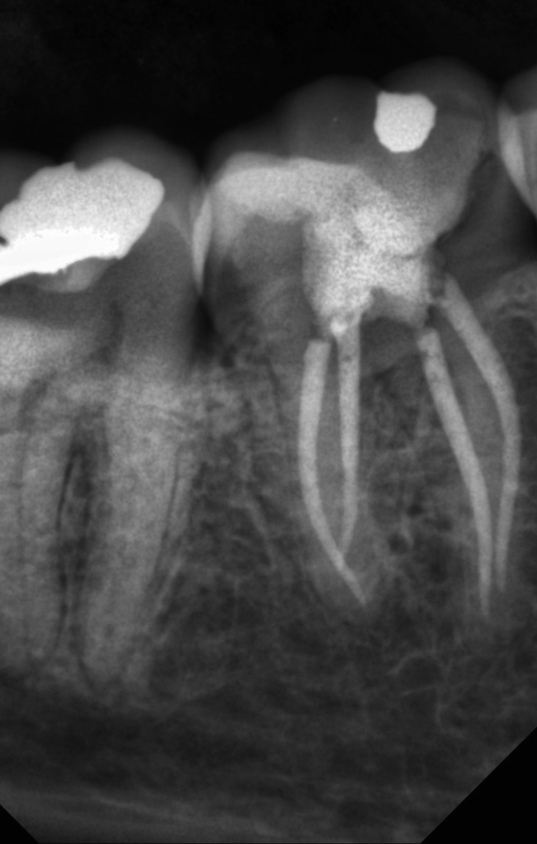
Axial CBCT slice shows radix entomolris of first mandibular permanent molar.
Distribution of prevalence of radix molaris according to the gender
Of 311 male patients included, 289 (92.9%) did not have radix molaris, 22 (7.1%) had radix entomolaris and none had radix paramolaris.
Of 389 female patients included, 362 (93.0%) did not have radix molaris, 24 (6.2%) had radix entomolaris and 3 (0.8%) had radix paramolaris.
The distribution of the prevalence of radix molaris did not differ significantly in the male and female sample population (
Distribution of prevalence of radix molaris according to age group
The distribution of incidence of radix molaris according to age group did not differ significantly in various age groups of cases studied (
Distribution of prevalence of radix molaris according to age group
Distribution of prevalence of radix molaris according to age group
Missed canals were associated with post-endodontic treatment diseases; knowledge of the prevalence and location of an extra root in the mandibular first molar is important from a clinical perspective for root canal treatment success [47]. Mandibular first molar anatomical variations have been well documented in endodontic literature which needs to be appreciated by dental clinicians [48, 49]. Finding, cleaning and shaping such anatomy is challenging in dental practice [50].
Previous studies used either extracted teeth, conventional radiography, orthopantomogram (OPG) or CBCT [51, 52, 53]. Researchers used extracted teeth to find the frequency of radix molaris which might underrate the true prevalence as the extra root could fracture easily during the extraction procedure [54, 55, 56, 57]. Periapical radiographs with different angulations could identify supernumerary roots. Endodontic diagnosis and treatment need the use of conventional radiographs taken from various angles. However, because standard radiographs are 2D images, they may not be sufficient to gather all necessary details in cases of complex anatomy [58]. Dental professionals can use CBCT to detect the structure of the root canals and then clean and shape them using the appropriate tools and methods to handle cases with complex anatomy, such as the existence of extra roots.
This study used CBCT to find the prevalence of radix molaris (RaEn and RaPa) in permanent mandibular first molars in the studied subpopulation. The presence of radix molaris (RaEn and RaPa) in mandibular first molars varied among different populations. For example, it has been documented that the prevalence of RaEn in the Malaysian population was 21.4% in mandibular first molars [59]. Radix molaris has been documented to be more common in Asian, especially Chinese and Korean with a range between 27% to 33%, populations which are considered to be normal anatomy morphology in these populations compared to other racial populations [60, 61]. A recent systematic review found that the prevalence of RE in mandibular first molars amongst the Saudi population ranged from 2% to 6.7% [21]. The wide variation of radix molaris prevalence of different ethnic populations could be because of genetic predisposition or using different research methods.
In comparison with previous studies assessing the prevalence of radix molaris, the findings of this study are higher than studies conducted using extracted teeth, conventional radiographic and CBCT evaluations with 2.3%, and 5.97% [62, 63, 64] while it is comparable to study conducted by Bahammam and Bahammam which found the prevalence of RE is 6% [65]. Thus, a correct preoperative diagnosis of radix molaris is important for proper root canal treatment to avoid missing an extra root during endodontic treatment.
Al-Nazhan [64] evaluated teeth clinically and with x-rays to determine endodontic problems associated with them. These were complicated cases that were referred to the endodontic clinic. Song et al. also arrived at comparable findings. They found that the frequency was 24.5% with computed tomography and 33.1% with periapical radiographs [60]. They came to the conclusion that the developmental theory could explain how the distolingual roots of the first and second molars connect to each other. They also stated that the presence of distolingual roots could be seen as a feature of the field in which the first permanent molar is the most important tooth.
It is unclear why mandibular molars develop additional roots. Its development during odontogenesis may be influenced by polygenic genes or environmental factors [66]. According to Kim et al. research, first permanent molars with distolingual extra-roots had crowns that were noticeably wider at the distal buccolingual end and had significantly longer distances between the points of the distobuccal and distolingual cusps [67]. In addition, Kim et al. discovered that the buccal cortical bone’s outer surface and the EM (distolingual root) apex were 12.09 mm apart [68]. They came to the conclusion that knowing the anatomy and shape of the first molar of the mandible could help plan endodontic treatment. Also, the shape of the pulp chamber is triangular in molars with 3 root canals, but it is rectangular or rhomboidal in molars with 4 canals where the extra root (RM) may be present [23]. This should be taken into account during root canal treatment so that the canal of the extra root doesn’t get missed. Before beginning a root canal treatment, a thorough clinical and radiographic examination should be carried out. Today, thanks to the use of CBCT and sophisticated technologies, software, and artificial intelligence, treatments in dentistry and all other fields of medicine are handled with greater care [69, 70, 71, 72, 73, 74, 75, 76, 77, 78, 79, 80, 81, 82]. The clinician must use caution when diagnosing and treating the lower molar teeth due to the rarity of RE. Finding an extra root might be made easier with a visual examination of the dental crown. Although, presence of an additional root is frequently linked to more cusps, more root canals, and a more pronounced occlusodistal or distolingual lobe, but not always necessary [68, 83].
Conclusion
When planning a root canal treatment for a mandibular first molar, both general dentists and endodontists should always think about the possibility of a variation in root canal morphology. Careful clinical and x-ray examinations are needed to diagnose any anatomical change in the root canal morphology. Within the limitation of this study, it can be concluded that the prevalence of RE in mandibular first molars of a Saudi subpopulation was 6.6% while the prevalence of RP was 0.4%. in addition, utilizing CBCT during endodontic diagnosis and treatment is a non-invasive, reliable and crucial tool for identifying RM in mandibular first molars.
Ethics statement
The institutional review board at the College of Dentistry of King Khalid University approved the study protocol (IRB/KKUCOD/ETH/2020-21/017).
Funding
The authors extend their appreciation to the Deanship of Scientific Research at King Khalid University for funding this work through the Small Group Research Project (grant number RGP.1/207/43).
Availability of data
Data related to this study is available from the corresponding author for academic and clinical purposes.
Footnotes
Conflict of interest
None of the authors declare any conflicts of interest.