



Abstract
BACKGROUND:
Medical imaging techniques have improved to the point where security has become a basic requirement for all applications to ensure data security and data transmission over the internet. However, clinical images hold personal and sensitive data related to the patients and their disclosure has a negative impact on their right to privacy as well as legal ramifications for hospitals.
OBJECTIVE:
In this research, a novel deep learning-based key generation network (Deep-KEDI) is designed to produce the secure key used for decrypting and encrypting medical images.
METHODS:
Initially, medical images are pre-processed by adding the speckle noise using discrete ripplet transform before encryption and are removed after decryption for more security. In the Deep-KEDI model, the zigzag generative adversarial network (ZZ-GAN) is used as the learning network to generate the secret key.
RESULTS:
The proposed ZZ-GAN is used for secure encryption by generating three different zigzag patterns (vertical, horizontal, diagonal) of encrypted images with its key. The zigzag cipher uses an XOR operation in both encryption and decryption using the proposed ZZ-GAN. Encrypting the original image requires a secret key generated during encryption. After identification, the encrypted image is decrypted using the generated key to reverse the encryption process. Finally, speckle noise is removed from the encrypted image in order to reconstruct the original image.
CONCLUSION:
According to the experiments, the Deep-KEDI model generates secret keys with an information entropy of 7.45 that is particularly suitable for securing medical images.
Keywords
Introduction
Medical images must be encrypted to confirm to the privacy and security of patient records. A medical image contains sensitive patient data such as X-rays, MRIs, and CT scans which should be protected from illegal access, alteration, and interception [1]. Security and confidentiality are guaranteed both during transmission and storage of these photos. Medical photographs often contain identifiable information [2, 3]. Transmission or storage of images must be encrypted to ensure their integrity for precise treatment and diagnosis [4]. Data breaches can be prevented by encryption, which prevents unauthorized access to medical images and ensures that patient information is only viewed by authorized healthcare personnel [5]. Healthcare providers are required by law to safeguard patient information. Medical image encryption techniques must be effective in order to prevent delays in accessing images, especially in urgent situations [6]. Research environments requiring anonymized data benefit from adding noise because it makes it harder to detect sensitive information.
Encryption methods should be compatible with different medical imaging systems and standards to ensure seamless sharing of encrypted images among healthcare providers [7, 8]. Encryption keys must be kept safe, and key management systems are required to create, store, and distribute keys in an encrypted manner. The deep learning-based techniques [9, 10] are quite efficient and secure in this medical image encryption and decryption process. Deep learning (DL) [11] and machine learning (ML) techniques [12, 13] were used for developing robust encryption techniques that enhance the privacy and security of sensitive medical data [14]. By utilizing advanced encryption approaches, deep learning models such as CNN, LSTM [15], GRU [16] and YOLO [17] contribute to safeguarding patient information and maintaining compliance with data protection regulations. Hence, in this work a deep learning-based GAN structure is introduced for key generation and encryption/decryption of medical images. The key contributions of the proposed methodology are summarized as:
The deep learning-based KEDI framework is introduced for efficient encryption/decryption of medical images with secure key generation. Initially, the medical images are gathered from different publicly available datasets (brain- CT, MRI; lung- CT, MRI, CXR; liver- CT, MRI). Afterwards, the speckle noise is added to the medical images using discrete ripplet transform to enhancing privacy and security in which it is hard to recognize sensitive information. Deep learning based zigzag GAN is proposed with the vertical, horizontal and diagonal zigzag patterns for secure encryption by generating three different patterns of encrypted images with its key. In this case, the discriminator cannot effectively differentiate whether the images are created by the generator or the original cipher image it will increase the security level of the images.
The rest of the work is organized into five sections. Section 2 presents a concise examination of existing methods. Section 3 enlightens the detailed description of proposed Deep-KEDI methodology with zigzag GAN for encryption and decryption of medical images. Section 4 shares the results and discussion and Section 5 concludes the work.
Various strategies have been developed in order to encrypt and decrypt the medical images using deep learning that are secure using computer-based methodologies. Recent developments in artificial intelligence have simplified the encryption/decryption algorithms. This section briefly discusses some of those techniques.
In 2022, Wang and Zhang [18] designed a deep neural network (DNN) for image encryption. The discrete cosine transforms (DCT) used in the weight matrix of the EDNN, a multi-layer forward network, were scrambled in order to effectively encode the raw images. The suggested DDNN decoding unit then succeeded in recovering the raw images. In the DDNN, the structure of the network was symmetric with that of the EDNN, hence the raw images were retrieved using two pursuit methods that corresponded to the activation functions of the EDNN.
In 2022, Alzamily et al. [19] encoded a large group of images with deep learning based the CNN, which was extensively used for image recognition. By using a large number of cipher images, this deep ResNet-50 model was used to progress the speed and accuracy of the appearance of outcomes. This approach allowed it to categorize and identify the encrypted images without manually decoding the raw images. ResNet-50 categorization of encoded images showed 99.75% accuracy in recognizing encrypted images.
In 2022, Huang et al. [20] designed the learnable image encryption that was used with medical images to train privacy-preserving deep neural network models in the field of medicine. The enhanced SKK image encryption method offers a parameter to regulate the trade-off between the security of the photographs. GradCAM approach was used to investigate the learnt characteristics of adaptable encryption of images for a deep neural network architecture which preserves privacy.
In 2022, Wang et al. [21] devised a DL-based V-net on 4D hyperchaotic model for encrypting the clinical image. After creating the 4D hyperchaotic pattern images, the original medical images received image segmentation, chaotic system processing, and pseudo-random order production. Chaotic patterns were trained on a V-net to get rid of their regularity. To complete the encryption processing, the chaotic sequence image was subtle to modify the pixels of the image.
In 2021, Ni et al. [22] designed a multi-image encryption system with DL and compressed sensing (CS) in the gyrator domain. Initially, many images were compressed by CS, then each measurement’s pixels were randomly jumbled to obtain many measurements. Secondly, a chaotic matrix and XOR operation were employed to disperse the scrambled measurements into a matrix. In decryption, a neural network reconstructs plaintext images from the CS metrics, achieving greater reconstruction speed and quality in comparison to the conventional technique.
In 2021, Elsharkawy and Masri [23] presented an IEDL-SCBIR model for image encryption using a secure CBIR model based on deep learning. The IEDL-SCBIR technique consists of two stages: optimum encryption of ECC and image retrieval. As a result of the proposed model, an optimal CSO algorithm is generated based on the ECC approach for the process of image encryption. For the retrieval procedure, Euclidean distance-based similarity measurements were combined with VGG-based feature extraction.
In 2019, Sirichotedumrong et al. [24] presented a pixel-based image encryption technique utilizing deep neural networks (DNNs) for privacy preservation. Their method maintains essential aspects of original photos while allowing the application of images without compromising visual information. This approach involves training a DNN model on encrypted images with separate keys, enabling the use of both plaintext and encrypted photos for testing without requiring key management. Additionally, data augmentation in the encrypted domain enhances versatility and security in image processing tasks.
In 2023, Spahić et al. [25] explored the role of artificial intelligence (AI) in medical diagnostics, focusing on its support in decision-making processes. Ultrasound, crucial for monitoring fetal development, is undergoing research to automate diagnostic procedures. To ensure consistency and minimize variability, AI-driven real-time evaluation of ultrasound recordings shows promise. The KANET test, a gold standard for assessing fetal neurodevelopmental disorders, could benefit greatly from AI automation, enhancing its diagnostic capabilities.
In 2021, Hafizović et al. [26] followed PRISMA guidelines using PubMed, ScienceDirect, and Google Scholar. They focused on recent papers meeting specific criteria. From the search, 26 out of 81 papers were included from PubMed, 205 from ScienceDirect, and 520 from Google Scholar. The observed accuracies and growing interest in the topic indicate significant potential for future medical applications.
In 2023, Akilan et al. [27] introduced an innovative architecture for secure wireless body sensor networks (S-WBSN), featuring reduced CPU usage and computational expenses. S-WBSN employs both Diffie-Hellman and OTP-Q key exchange techniques for encryption and mutual authentication. The Diffie-Hellman method facilitates mutual authentication between WBSN components, enhancing security. Meanwhile, the OTP-Q algorithm acts as a stream block cipher, utilizing sensor data for encryption. Authentication precedes encrypted data transmission, ensuring a secure communication channel. This approach demonstrates efficacy while conserving CPU resources, with significantly shorter encryption and decryption processing times compared to existing methodologies. S-WBSN stands out as a promising solution for healthcare data monitoring with robust security measures.
In 2023, Selvakumar et al. [28] introduced a novel image encryption system tailored for medical images. This hybrid system combines chaotic maps, DNA encoding, and an advanced LSB algorithm. By integrating six chaotic maps with the LSB technique, the system enhances resistance against unauthorized access. The LSB method employs arithmetic division to maintain image integrity, partitioning data into remainder and quotient components. These components are discreetly embedded within LSB regions, ensuring robust data concealment without compromising image quality. This innovative approach facilitates swift and secure data transmission via a DNA sequence generated through DNA encoding, striking a balance between data security and image fidelity.
In common conventional encryption techniques like symmetric and asymmetric encryption algorithms often require manual feature extraction and fixed algorithms, limiting their adaptability to diverse and complex image patterns for securing data in medical images. These methods lack the ability to automatically learn intricate features, making them less effective for securing detailed medical image data. Deep learning models can adapt, learn complex patterns, and enhance encryption efficiency. As a result, the current deep learning network has difficulty delivering the greatest expected performance because the computation model has to be manually adjusted for each attempt. Hence, in this work a deep learning-based GAN structure is introduced for key generation and encryption/decryption of medical images.
Proposed Deep-KEDI methodology
In this section, a Deep-KEDI method is introduced for efficient encryption and decryption of medical images for more security. The overall workflow of the proposed encryption/decryption and key generation process is displayed in Fig. 1.
Schematic portrayal of the proposed DEEP-KEDI methodology.
In this work, three different types of organ images (brain, lung, liver) are collected from publicly accessible datasets. The brain MRI images are gathered from the ADNI dataset [29] which contains 6219 MRI images with the image of
Data pre-processing
In this phase, discrete ripplet transform (DRT) is used to add and reduce the speckle noise in medical images. By introducing noise, it becomes harder to recognize sensitive information, thus enhancing privacy and security. The proposed technique is expressed in two stages for despeckling of medical image: (i) select the proper values of the two constraints for computing the ripplet coefficients from input images, (ii) chose and achieve the proper filtering process on the coefficients for attaining the improved ripplet coefficients. The following equation yields a spatially specked image,
here
Speckle noise addition process.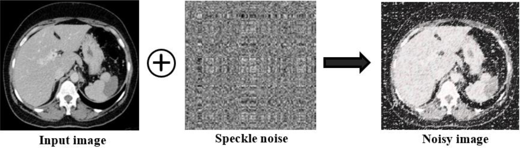
The multiplicative noise in Eq. (2) was converted to additive noise by log-transforming speckle-damaged images.
where
Where
In DEEP-KEDI, a zigzag GAN guides the learning network that generates the private key. The ZZ-GAN produces a stream cipher that is utilized with an XOR algorithm for encryption and decryption scheme. The proposed ZZ-GAN is used for secure encryption by generating three different zigzag patterns (vertical, horizontal, diagonal) of encrypted images with its key. The encryption and decryption process with the ZZ-GAN and XOR operation. The structure of the proposed ZZ-GAN is displayed in Fig. 3.
Architecture of zigzag generative adversarial network.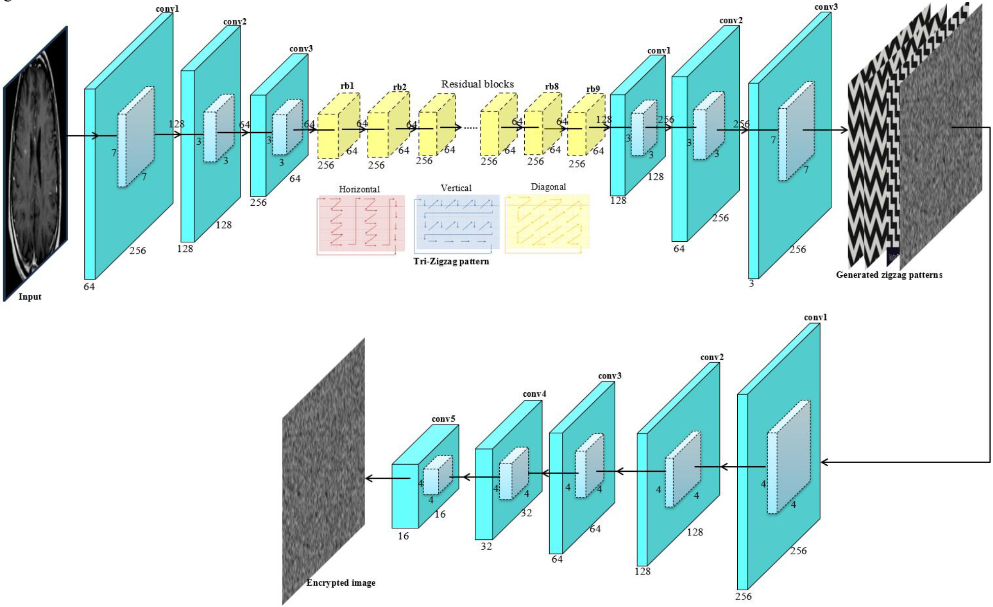
During encryption, the noisy image is encrypted through the created secret key and the XOR operation. Consequently, the encrypted image is attained and it is decrypted by the reverse process of encryption. The input medical images are encrypted using the structure of encryption network
The mapping function
where
Three different zigzag patterns-based encryption process in the generator.
In addition, the proposed ZZ-GAN attempts for ensuring that the decrypted image holds the texture details of input image even after encryption. The encoded image vector and the key are then bitwise-XOR to obtain the image. In the reconstructed loss process, the difference between
A discriminator network
In a GAN, the discriminator network is more accurate when both networks
A key generation procedure is a process of establishing a mapping between the source and transformation domains by training a network. In ZZ-GAN, each parameter of the convolutional layer is randomly initialized before training. The initialization process is derived as,
where,
In Eq. (10),
The ripplet coefficients are filtered to determine the approximate value of
Speckle noise reduction process.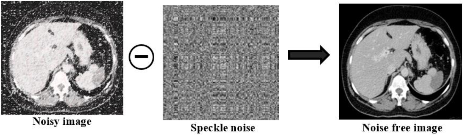
The inverse DRT is used on the filtered approximated ripplet coefficients for obtaining the estimated output as derived in Eq. (11). Finally, an enhanced despeckled image
In this section, the medical images from the various datasets such as for brain [29, 30], lungs [31, 32] and liver [33] are estimated with specific metrics. The proposed ZZ-GAN was assessed with various network metrics for the gathered datasets [37, 38]. The experimental setup of the Deep-KEDI model was executed through Spyder, an Anaconda navigator executed on a PC with Windows 10 OS on an Intel-core i7processor with 2.10 GHz processor and 8 GB RAM system. Figure 6 indicates the results of proposed model for encrypting and decrypting the medical images.
Experimental results of the Deep-KEDI system for encryption and decryption.
The experimental results of the Deep-KEDI framework for encoding and decoding the clinical images based on the generated key. In Fig. 6. (a) Plain image; (b) Speckle noise added to (a); (c) The encoded image of (b) with the generated key; (d) decoded image of (b) using the generated correct key; (e) Despeckle noise in image (d) and (f) final plain image.
The size of the key space estimates the resistance to exhaustive attacks. For the Deep-KEDI that is proposed, a private key is generated as an image. The image was
Figure 7a and b illustrates four private keys that can be distinguished visually according to their content, color, and contour with the proposed ZZ-GAN which is trained under similar experimental conditions. Afterwards, Fig. 8 shows the histograms of generated keys and the input image for the above Fig. 7c. As shown in Fig. 8, the private key histogram is fairly consistent. In this case, it means that the frequency of pixel values is small and their distribution is uniform. This suggests that the secret key that was created can withstand statistical attacks due to its high level of unpredictability.
Image sensitivity analysis
The proposed Deep-KEDI algorithm’s performance is evaluated using SSIM, PSNR, and MSE metrics. PSNR is commonly employed to assess image quality, while SSIM quantifies the similarity between the ground-truth and the output of the proposed algorithm. MSE contributes to the error ratio between decoded and encoded images, with lower values indicating greater efficiency. Table 1 illustrates the utilization of these metrics for performance analysis. The performance of ZZ-GAN in encoding and decoding medical images is evaluated. Table 1 showcases the efficiency of the proposed ZZ-GAN method, as reflected by SSIM, MSE, and PSNR values. With an average SSIM of 0.618, MSE of 2.62, and PSNR of 25.32, the ZZ-GAN system maintains high PSNR values while ensuring similarity to the source images during decryption. Results indicate accurate encryption and decryption across various datasets. Despite achieving an SSIM of 0.554 close to ground truth, the Deep-KEDI model outperforms ZZ-GAN in similarity measures for precise findings. Furthermore, Fig. 9 illustrates encrypted and decrypted data for liver, lung, and brain images alongside their respective histograms.
Scrutiny of the ZZ-GAN in terms of SSIM, MSE and PSNR
Scrutiny of the ZZ-GAN in terms of SSIM, MSE and PSNR
Portrayal of the generated keys with the same experimental conditions (a), input (b) and speckle noise addition (c) generated four private keys.
Histogram representation of the generated keys.
As observed from Fig. 9, encrypted images have rather consistent pixel value distributions, in contrast to the histogram of noisy images. These distributions closely resemble the histogram distribution of speckle noise, indicating that the statistical data in the plaintext images can be successfully protected by an encrypted image. Moreover, the produced private key may speed up and randomly encrypt the encryption of medical images. This makes extraction of usable information from encrypted images extremely difficult for attackers using statistical assaults.
Visual representation of the encryption and decryption process for (a) liver MRI image, (b) lung CXR image and (c) brain MRI image.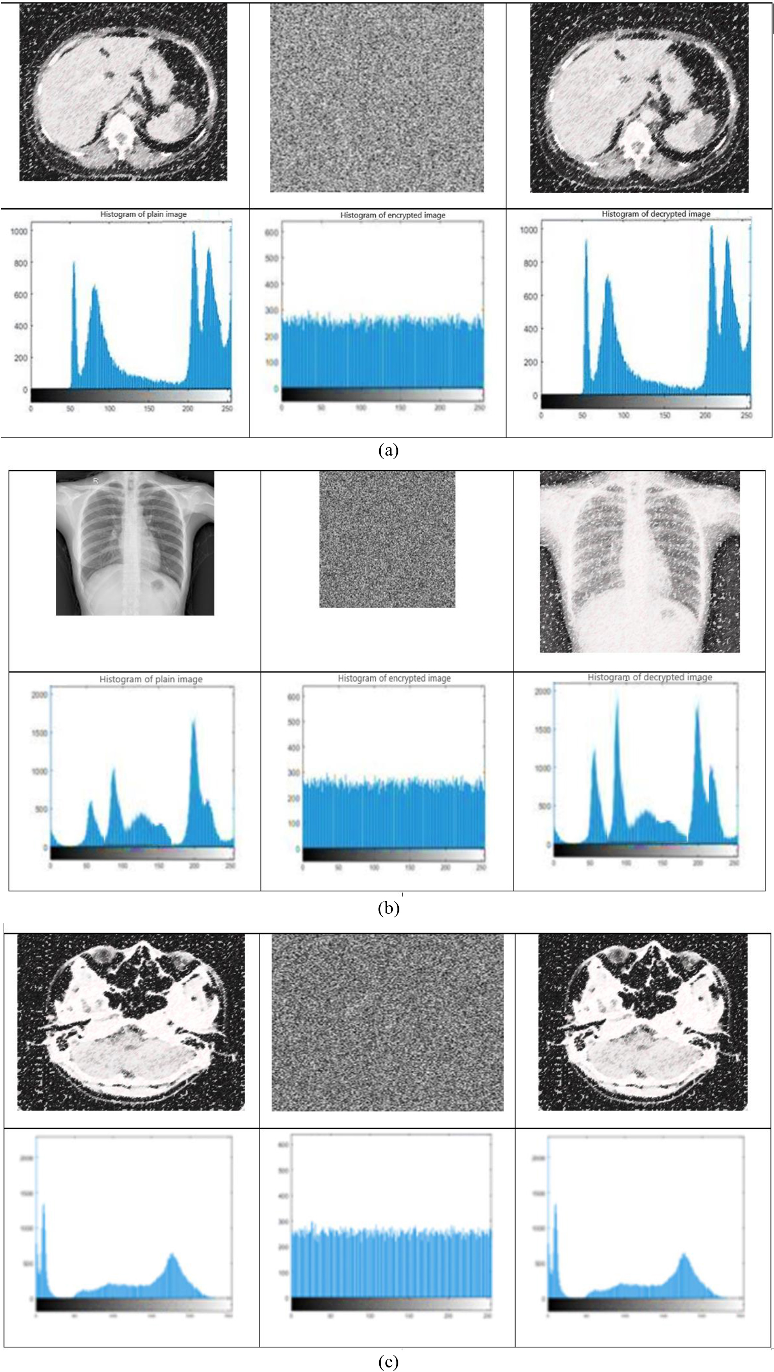
A number of pixels change rate (NPCR) and a Unified pixel change intensity (UACI) are utilized to quantify the variations among the secret keys. The NPCR indicates pixel variation rate compares the proportion of various pixel values at the identical positions in two images. The NPCR is defined as,
UACI stands for the intensity of standardized average variation that indicating the average altered density of two images. The UACI is defined as,
here,
NPCR and UACI calculation based on key id
According to Table 2, even a small change in pixel value can cause 99.5% of two generated private keys to be different. Hence, pseudo-randomness and uncertainty are achieved with the created private key generated using the proposed ZZ-GAN structure. Information entropy (IE) is used to quantify the level of uncertainty in a system for evaluating the unpredictability of secret keys,
In the equation above, the expected value of the symbol m is represented by
Information entropy evaluation of gathered medical images
Table 3 shows the entropy of private keys. Table 3(a) shows the IE value of brain MRI and CT images; 3(b) illustrates the CXR, MRI and CT images of lungs; and 3(c) shows the CT and MRI images of liver. Grayscale images have the highest possible entropy value of 8. There is a high level of randomness with the created private key having an entropy of about 7.45, which indicates the key is secure.
To restore the original noisy image, a key generated by the trained ZZ-GAN network (key A, B, C, D respectively) is used for encrypting the real noisy image. Key A is the only key that can accurately decode the initial image (Fig. 10a) encrypted by key A (Fig. 10b). The Fig. 10d–f illustrate, respectively, how the images decrypted by keys B, C, and D cannot be identified immediately. The attacker cannot produce the similar secret key and decrypt the encrypted image by creating an attack model based on knowledge of the transformation.
The proposed ZZ-GAN was evaluated against other encryption methods using NPCR, UACI, and IE measures. This comparison aimed to demonstrate the superior performance of ZZ-GAN in image processing. Table 4 presents the comparative evaluation conducted on various encryption techniques to assess the proficiency of encryption algorithms.
Comparison of traditional encryption algorithm
Comparison of traditional encryption algorithm
Analysis of attack model in encryption process. (a) Noisy medical image. (b) Encrypted cipher image with key A. (c) Decrypted noisy image with key A. (d) Decrypted noisy image with key B. (e) Decrypted noisy image with key C. (f) Decrypted noisy image with key D.
Table 4 shows the comparison traditional Encryption algorithm based on NPCR, UACI and IE. From this comparison the proposed ZZ-GAN achieves the better performance in encrypting and decrypting the medical images with high entropy value. In this work, three different types of organ images (brain, lung, liver) are gathered from publicly available datasets. The brain MRI images are gathered from the ADNI dataset [21] which contains 6219 MRI images and the brain CT images are gathered from [22] that comprises with the total of 1603 CT images. Afterwards, the lung CT and X-ray images are collected from [23] and the lung MRI is collected from [24]. Finally, the liver CT and MRI images are gathered from BioGPS and TCIA (Cancer Imaging Achieve) databases [25] are the two such databases that contain a total of 1568 CT images and 1574 MRI images taken from liver cancer suspected patients.
Performance comparison of the proposed model based on various datasets
Table 5 demonstrates the performance assessment of the proposed system with various datasets such as [29, 30, 31, 32] and real-time medical imaging datasets such as [33, 34, 35, 36]. The comparison table presents performance metrics (NPCR, UACI, IE) for various datasets. Higher NPCR and UACI values indicate better performance, while lower IE values are preferred. Datasets [29, 32] exhibit the highest NPCR, while LiTS [34] has the lowest. UACI values are relatively consistent across datasets. IE is lowest for NLST [36] and highest for [29] and [32]. Overall, [32] stands out with high NPCR and IE, while NLST shows low IE and competitive NPCR. LiTS, BRATS, NLST datasets exhibit lower NPCR values compared to the above datasets, indicating lesser pixel changes between images. UACI and IE values vary but generally show lower change intensity and entropy compared to other datasets. This analysis indicates that although the proposed network demonstrates strong performance within the gathered dataset, its performance is comparatively lower when tested on other datasets.
Efficiency comparison of state-of-the-art encryption algorithms
From Table 6, the efficiency measure was utilized for comparing various encryption techniques in terms of their IE for encryption. However, the existing model not performed well than proposed ZZ-GAN. The proposed ZZ-GAN increases the average IE range by 3.75%, 8.18% and 4.96% better than EDNN [18], ResNet-50 [19], and 4D hyperchaotic framework [21] respectively. Table 5 shows the comparison of the encryption and decryption times of various methods for [18, 19, 21] and the Proposed ZZ-GAN model. Notably, while EDNN exhibits the lowest encryption time at 7.17 ms, the Proposed ZZ-GAN method demonstrates the shortest decryption time of 45 ms. Conversely, ResNet-50 has the longest decryption time at 92 ms, while 4D hyperchaotic framework boasts the longest encryption time of 7.08 ms. Overall, these findings suggest that different encryption methods possess varying computational efficiencies, influencing their suitability for specific applications.
Clinical Integration of proposed medical image encryption model.
Figure 11 depicts a proposed system for securely sharing medical images, likely in a hospital setting. The system is labelled as “Proposed DEEP-KEDI model”. The hospital location where the patient’s medical data is stored and accessed by authorized personnel. In real-time scenario, the medical images such as X-rays, MRIs, or CT scans are gathered from the original patient. The proposed DEEP-KEDI system has two-step encryption process. Initially, the speckle noise is added to medical images using discrete ripplet transform and deep learning-based GAN structure is used in the encryption process. In this phase, Encrypted image is generated, likely using a complex mathematical formula and a secret key. Secure key represents the key needed to decrypt the confidential medical data. This key is securely stored and only accessible to authorized personnel. The medical professional who will interpret the encrypted medical images for diagnosis. Overall, the DEEP-KEDI model is an efficient method for encrypting medical images to protect patient privacy while allowing authorized doctors to access the information for diagnosis.
This paper presents a DEEP-KEDI for generating the secret key utilized for the encryption and decryption process of clinical images. Initially, the medical images are pre-processed by adding the speckle noise using discrete ripplet transform before encryption and it was removed after decryption for more security. As the learning network for DEEP-KEDI, the ZZ-GAN directs the learning network for creating the secret key. The proposed ZZ-GAN is used for secure encryption by generating three different zigzag patterns of encrypted images with its key. By combining the proposed ZZ-GAN zigzag cipher with an XOR operation, encryption and decryption can be performed. An encryption key is generated during the encryption process and used to encrypt an unencrypted image. The plain image is then reconstructed by removing the speckle noise from the decrypted image. Based on the experiments, the proposed Deep-KEDI model successfully generates secure keys for medical images. The proposed model has high level of randomness with the created private key having an entropy of 7.45, which indicates the key is secure. Moreover, the proposed ZZ-GAN increases the average IE range by 3.75%, 8.18% and 4.96% better than EDNN, ResNet-50, and 4D hyperchaotic framework respectively. In the future, further advancements will expand upon this work by integrating a hybrid methodology. This approach will merge the robustness of conventional AES encryption algorithm with the capabilities of deep learning-based GAN structures for resisting the Brute-force attack. By combining these strengths, it aims to provide the most efficient solution for encrypting and decrypting medical images. Such a hybrid approach holds promise for enhancing the security and accessibility of sensitive medical data. Its development underscores ongoing efforts to innovate within the realm of medical image encryption.
Funding
No financial support was received for this study.
Author contributions
Study conception and design: KS, SL; Data collection: KS, SL; Analysis and interpretation of results: KS, SL; Draft manuscript preparation: KS, SL. Both authors reviewed the results and approved the final version of the manuscript.
Data availability
Data sharing is not applicable to this article as no new data were created or analyzed in this research.
Footnotes
Acknowledgments
The authors would like to thank the reviewers for their careful, constructive and insightful comments in relation to this work.
Conflict of interest
The authors declare that they have no known competing financial interests or personal relationships that could have appeared to influence the work reported in this paper.