




Abstract
BACKGROUND:
Anesthesia may alter the cellular components contributing to the magnetic resonance imaging (MRI) signal intensities. Developing awake animal models to evaluate cerebral function has grown in importance.
OBJECTIVE:
To investigate a noninvasive strategy for dynamic MRI (dMRI) of awake rabbits during carbogen challenge.
METHODS:
A nonmetallic assistive device with a self-adhering wrap secure procedure was developed for the head fixation of awake rabbits. Multi-shot gradient echo echo-planar imaging sequence was applied for the dMRI on a 1.5 T clinical MRI scanner with a quadrature head coil. The carbogen challenge pattern was applied in a sequence of air - carbogen - air - carbogen - air. Twelve scans were performed for each block of carbogen challenge. T2-weighted fast-spin echo and T1-weighted gradient echo sequences were performed before and after dMRI to evaluate the head position shifts. The whole dMRI scan time was about 30 minutes.
RESULTS:
The position shift of 8 rabbits in the x-and y-direction was less than 3%. The average MRI signal intensities (SI) from the 8 rabbits during carbogen challenge was fitted well using exponential growth and decay functions. The average MRI SI increase due to carbogen inhaling was 1.51%.
CONCLUSIONS:
The proposed strategy for head dMRI on an awake rabbit during carbogen challenge is feasible.
Introduction
Magnetic Resonance Imaging (MRI) has become an important imaging modality for clinical diagnosis in the last three decades. The noninvasive characteristics makes MRI widely accepted in neuroscience studies [1–3]. Animal models are often used in MRI studies for translational researches. However, motion artifacts might be an obstacle to interpret MR images and need to be concerned [4]. For human MRI examinations, there is no need to apply anesthesia except for the extremely uncooperative patients because human subjects can usually hold still during MRI scans. On the contrary, anesthesia is commonly used in animal model MRI to exclude motion artifacts [5–7]. Anesthesia may alter the cellular components contributing to the MRI signal intensities [8, 9]. Therefore, developing awake animal models to evaluate cerebral function of conscious animals has grown in importance.
Several techniques have been proposed to study neural and hemodynamic activities on conscious rats [8–12]. In addition to rats, rabbits are commonly used as animal models for brain studies using MRI [13–15]. Nowadays, techniques published for studies on awake rabbits using MRI were limited. Furthermore, these techniques needed surgical preparation to immobilize the rabbits [16–19].
Blood oxygen level dependent (BOLD), a well-established technique for neuroscience studies, can reflect the change of the oxygenation levels of hemoglobin by assessing the signal intensity changes in cerebral regions [20]. Carbogen, a mixture of 95% O2, 5% CO2, can increase the amounts of oxy-hemoglobins and the oxygen tension (pO2) of plasma and interstitial tissue. Carbogen inhalation can be used in oncology to improve tumor oxygenation [21, 22]. Numerous studies have been performed to investigate hemodynamic response including cerebral blood flow (CBF), cerebral blood volume (CBV) changes, and cerebrovascular reactivity upon carbogen challenge [23–25].
This study aimed to develop an awake New Zealand white (NZW) rabbit MRI model without any invasive surgery. A simple strategy of using a self-designed nonmetallic assistive device and a Self-Adhering Wrap secure method was proposed to perform MRI of Carbogen Challenge on awake rabbit brain. To the best of our knowledge, this paper is the first study performed on conscious rabbit MRI without surgery. The MR images were statistically analyzed to evaluate the feasibility and fixation effectiveness of this proposed strategy.
Materials and methods
This awake rabbit MRI study was conducted with the affidavit of animal use protocol approved by the Institutional Animal Care and Use Committee of I-SHOU University (IACUC-ISU-102035).
Idea from the activity pattern of NZW rabbit’s head and limbs
Prior to design and implementation of the assistive device, a full understanding of the activity pattern of NZW rabbit head and limbs is required, so that the MRI experiments without motion artifacts can be completed. The movement of the rabbit’s head is mainly controlled by the cervical vertebra. The first cervical vertebra (the atlas and cranial skull) dominate the anteflexion and hypsokinesis activity of the rabbit’s head. The atlanto-axial joint controls the rotation activity of the head. A gear clamp described in the following section was designed to restrain the rabbit head. The activity pattern of rabbit limbs include flexion and stretching activities that can only be achieved in the front-rear manner as shown in Fig. 1. The posterior limbs provide the maximum strength of jumping. An elastic wrap (3MTM Nexcare CobanTM Self-Adhering Wrap) was used to secure the rabbit as shown in Fig. 1.

The activity pattern of NZW rabbit limbs: (a) a rabbit with forelimbs and posterior limbs marked, and (b) the secure pattern of rabbit forelimbs and posterior limbs activity. The curved arrows show the flexion and stretching activities. Self-Adhering wraps (3MTM CobanTM) were used to secure the limbs of NZL along the dotted line. (A: humerus B: radius and ulna C: metacarpals, D: femur, E: fibula and tibia, F: metatarnals).
Plastic and wood were used in the assistive device. Different sizes of gears were designed to control the direction of components movements (Fig. 2). To complete the gear linkage configuration, five large plastic gears with outer/inner diameter/thickness of 8.5/7.5/0.8 cm and one plastic pinion with outer/inner diameter/thickness of 3.0/2.0/1.2 cm were configured with wooden dowels on the centerline of a rectangle wood baseplate (40×15×1 cm3). The gears were placed sequentially from 2 cm outside the back side of the wood baseplate. Two long wooden strips (40×2×3 cm3) and one short wooden strip (11×2×3 cm3) were fixed on the boundary of the baseplate. Each long wooden strip had a 2.5 cm long groove (Fig. 2A). Two wooden racks (12.5×2×1.5 cm3) were geared by the small pinion (Fig. 2B). Each wooden rack had teeth on one side and one splat (12.5×1×1 cm3) attached on the other smooth side. The rack could pass through the corresponding groove. Two wooden plates (12.5×2.5×5.5 cm3) were fixed on the two racks, respectively. Furthermore, another two wooden plates (8×5×0.7 cm3) with C-clamps were arranged on these two wooden plates mentioned above (Fig. 2C). The angle between these two wooden plates with C-clamps was 75°. This clamp system was designed to restrain the movement of the rabbit’s head. The head gear linkage components were integrated to the gear linkage base (Fig. 2D). There were totally five arc clutches. Each clutch was made of a wood plate (15×7.5×3 cm) cut down to a semicircle 13 cm in diameter. Starting from the front end of the gear linkage base, six rectangular planks (15×3×1 cm) and the five arc clutches were arranged in the interleaved manner. The C-clamp can be adjusted to fix rabbit head by tuning the last gear and adjustable belts were used to fasten rabbit body.

Main components of the self-designed assistive device: (a) the gear linkage base, (b) the gearing scheme of gear racks and pinions (or gears), (c) two bascule C-clamps on the gearing system, and (d) the configuration of the assistive device.
A Velcro strip was added in front of the C-clamps to enhance the fixation. Furthermore, four adjustable belts were added to the assistive device to fix rabbit’s body. Finally, a cotton cushion was used to make the animal more comfortable to increase the binding capability (Figs. 3A and 3B).
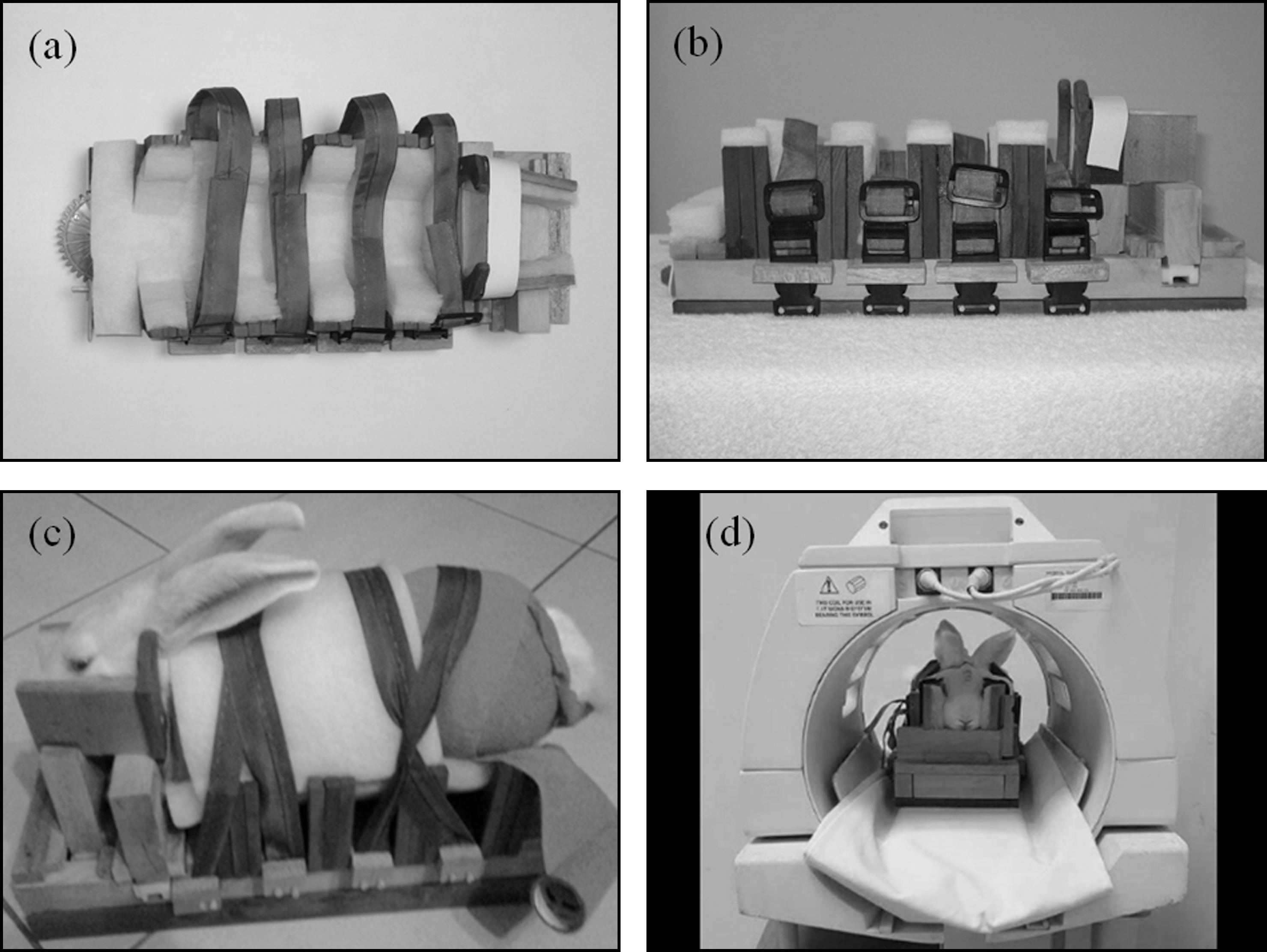
The nonmetallic assistive device with a secured rabbit: (a) the top view, and (b) the lateral view of the assistive device, (c) a NZW rabbit secured on the assistive device, and (d) the assistive device with a rabbit inside a head coil.
All scans were performed using a GE Signa HDxt 1.5T magnetic resonance imaging scanner equipped with a quadrature head coil. Eight 1.5 to 2.5 kg NZW rabbits were recruited. Each rabbit’s limbs were first carefully secured with a 4” Nexcare Coban Self-Adhering Wrap (3MTM), an elastic support bandage, to support, secure and prevent the rabbit from sprains and strains. Then, the rabbit was fixed on the exclusively designed assistive devices. The head and torso of the rabbit was carefully positioned to be parallel to the long axis of the assistive device. The assistive device with a rabbit inside was put into a head coil. The process was gently completed in order not to hurt the rabbit. Ear plugs were used during MRI scans to prevent the rabbit from hearing-impairment.
After a 3-plane localizer and calibration scan, T2-weighted (T2W) fast spin echo (FSE) and T1-weighted (T1W) gradient echo (GRE) pulse sequences were performed to obtain 17 axial images covering the whole brain. For T2W FSE, the scanning parameters were TR/TE = 2000/13.3 ms, FOV = 12×12 cm2, slice thickness = 2 mm, matrix = 256×128, Rx bandwidth = 31.25 kHz, NEX = 4, ETL = 10 and scan time = 2 minutes 6 seconds. And for T1W GRE, the scanning parameters were TR/TE = 120/3.8 ms, FOV = 12×12 cm2, slice thickness = 2 mm, matrix = 256×128, Rx bandwidth = 25 kHz, flip angle α= 90°, NEX = 4, and scan time = 3 minutes 52 seconds. Then, a multi-shot gradient echo-echo planar imaging (MSGE-EPI) sequence with time resolution of twenty seconds was used to perform dynamic (dMRI) studies. Only three consecutive axial MSGE-EPI images in the center of the brain were scanned to increase time resolution and reduce the scanning time for dynamic (dMRI) studies. The scanning parameters for MSGE-EPI were TR/TE = 260/12.3 ms, FOV = 12×12 cm2, slice thickness = 2 mm, matrix = 128×128, Rx bandwidth = 31.25 kHz, flip angle = 90°, NEX = 8, number of shots = 4, number of slices = 3, and scan time = 20 seconds.
Carbogen (95% O2 and 5% CO2) challenge was designed as the dMRI study. The pattern of the carbogen challenge: air - carbogen - air - carbogen – air is shown in Fig. 4A. Twelve scans were performed for every block of gas breath. Therefore, sixty images were obtained for five blocks per slice and totally 180 images acquired for 3 slices. The total scan time for the MRI of Carbogen challenge study was 20 minutes. Afterwards, the T2W FSE and T1W GRE pulse sequences were performed again to verify if there were motion artifacts. The total scan time for one rabbit was 34 minutes 18 seconds. Figure 5 shows the flow chart of the MRI experiments.
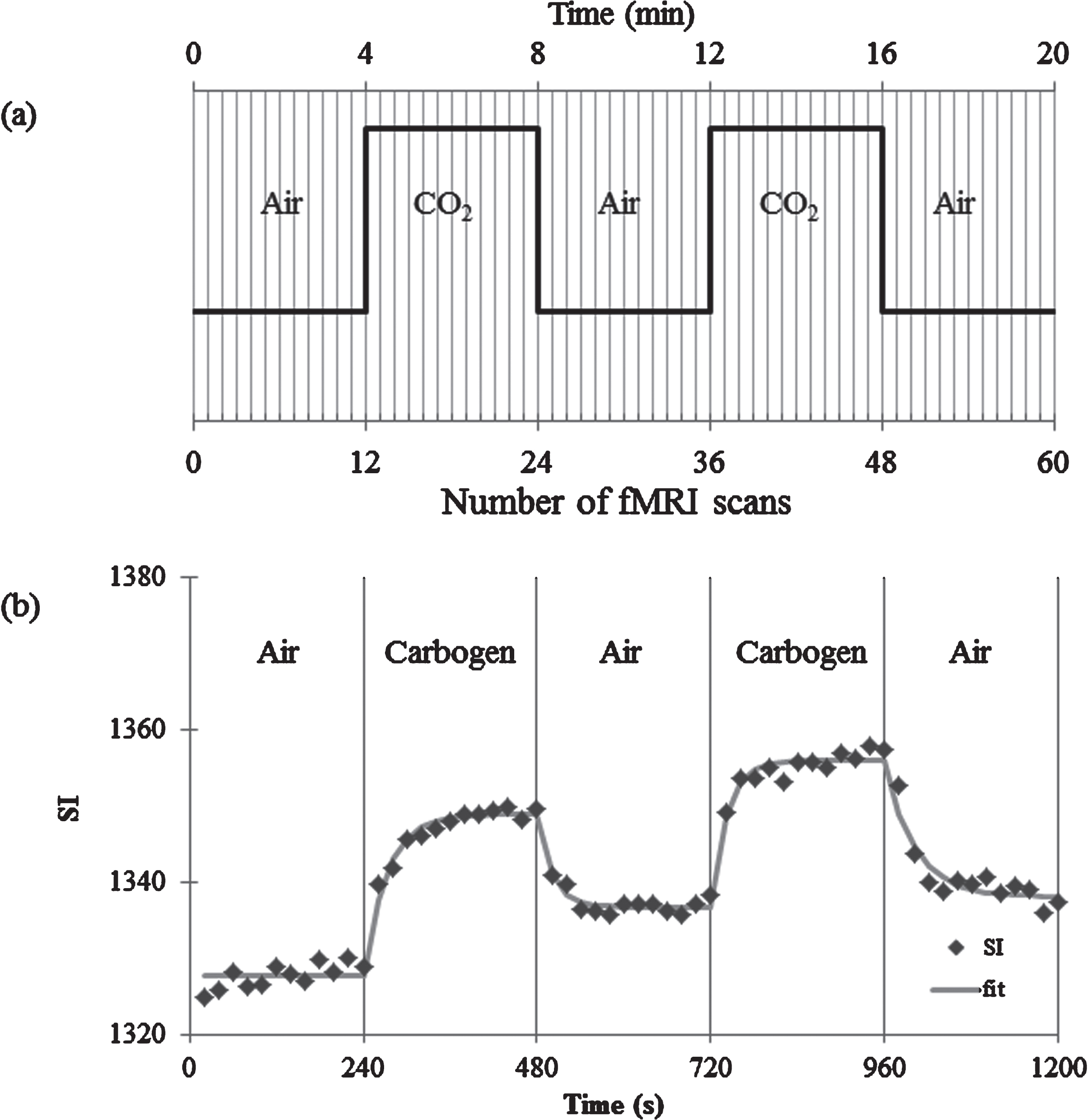
(a) The pattern of carbogen challenge, and (b) the mean SI vs. time plot for dMRI. The square dot and the solid line indicate the measured SI and curve-fitted SI for each block.
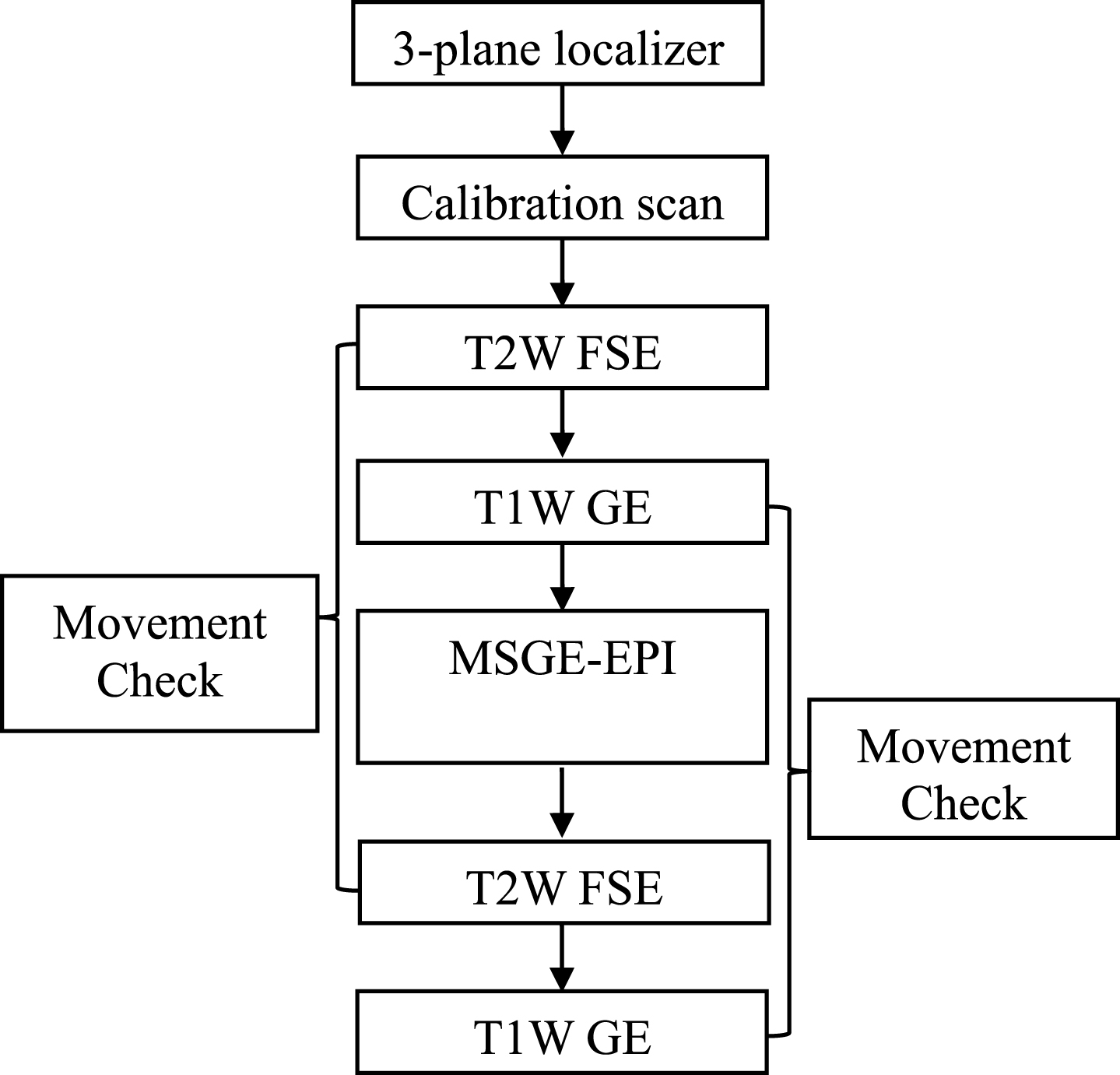
The protocol of MRI scans.
Three consecutive axial images in the center of the T2W FSE and T1W GRE axial images were analyzed to verify if there were motion artifacts within the experiment. The rabbit brains on pre-dMRI T2W FSE and T1W GRE images were segmented and fused on T2W FSE and T1W GRE images after carbogen challenge. A rectangular region of interest (ROI) was manually selected to cover the whole brain and the boundary lines of the ROI were set as the tangent lines of the boundary of the brain. The lengths in x-and y-direction (LX and LY) of the ROI before and after carbogen challenge were measured. The net position shift (PS) was defined as follows:
In the carbogen challenge study, there were totally 180 images acquired, i.e. sixty images per slice. The contours of brain obtained at the first time point for each slice were segmented and fused on all of the subsequent images for each corresponding slice. The value of the region of interest (ROI) was recorded for each time frame and each slice. The sizes and positions of ROIs were manually selected, which could be saved as a file and reopened at any time using ImageJ software. Therefore, the sizes and positions of selected ROIs could be kept consistent before and after carbogen challenge. The ROI values of all three slices from eight rabbits were averaged at the same time point, and then the scatter plot of the average ROI values versus time were obtained. The straight line of the first block was obtained by averaging the first twelve ROI values during air inhalation. Then, the average SI of each ROI as a function of time t when the inhalation gas was switched from air to carbogen and from carbogen to air could be fitted as the following Equations (3) and (4) using SigmaPlot 11 (Systat Software, San Jose, CA), respectively.
Design and fabrication of assistive device
A nonmetallic assistive device for restricting the movement of NZW Rabbits during MRI scans was designed, built and applied. Figs. 3A and 3B show the top and lateral views of the assistive device, respectively. Each rabbit was fixed in the assistive device with a Self-Adhering Wrap secure bandage (Fig. 3C). Figure 3D demonstrates the assistive device with a rabbit inside in a head coil.
MRI image processing
Figure 6 shows the segmented brain contour fused on T2W FSE images of 17 slices for pre-and post-dMRI scans. Figure 7 shows the segmented brain contour fused on the T1W GRE images of 17 slices before and after carbogen challenge. The whole MR experiment lasted about 30 minutes and there were no motion artifacts and significant movements observed throughout all the images in each experiment. The PS in x and y direction for T2W FSE and T1W GRE images are shown in Table 1.

T2 FSE images with brain ROIs before (upper) and after (lower) carbogen challenge to demonstrate a NZW rabbit stay motionless throughout the scans.

T1 GRE images with brain ROIs before (upper) and after (lower) carbogen challenge to demonstrate a NZW rabbit stay motionless throughout the scans.
The position shift in the x-and y-direction for T2W FSE and T1W GRE axial images
Figure 8 illustrates representative MSGE-EPI images with brain ROIs in each block of the carbogen challenge study which showed no significant image distortion. Figure 4B illustrates the MRI signal vs. time of the MSGE-EPI images for each block of the carbogen challenge. The signal increased when the inhalation was switched from air to carbogen, and then decreased when the gas was switched back to air. The average signal intensity in the first block and curve-fitting results in the other blocks are listed in Table 2. The relative signal increases due to the second and fourth block with carbogen inhalation were 1.59% and 1.43%, respectively. The average signal increase was 1.51%.
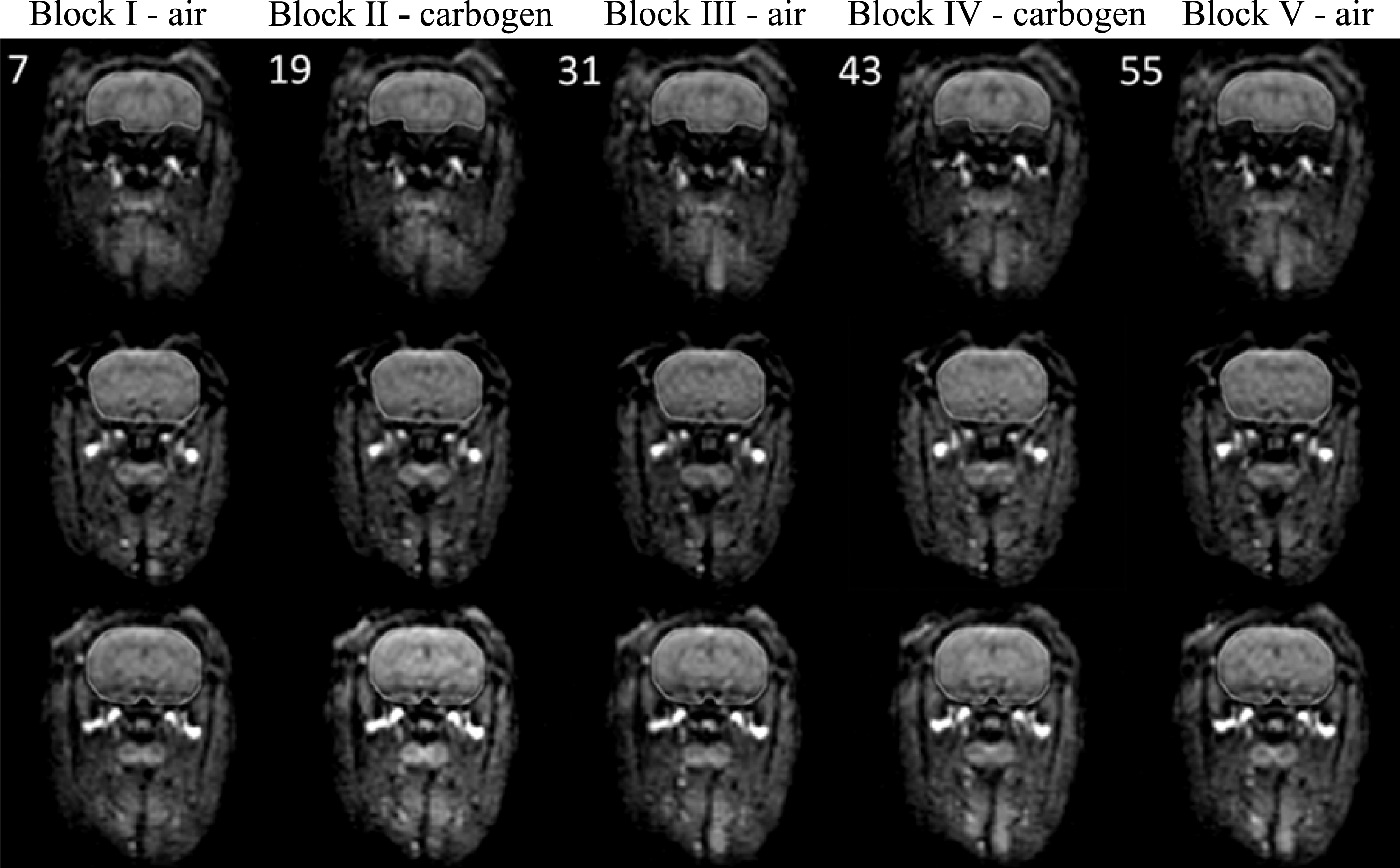
Representative MSGE-EPI images with brain ROIs from three slices of a rabbit in the dMRI study show no significant image distortion.
The curve-fitting equation of signal intensity in each inhalation block
ademonstrates air inhalation and bdemonstrates carbogen inhalation.
An assistive device together with a wrap-to-fixation method was completed to restrict the movement of NZW rabbits; therefore, no anesthesia drugs were required to perform this carbogen challenge study. From the T2W FSE and T1W GRE images before and after carbogen challenge, little motion artifacts and movements were observed. The PS(%) in the x-and y-direction for T2W FSE and T1W GRE images was less than 3%. During carbogen breathing, the MRI signal was higher than that during air breathing.
Fixation in this home-made assistive device was based on a gear mechanism that was designed according to rabbit skeletal anatomy and body movement patterns. Materials for fabricating the assisted device were carefully selected. Magnetic materials are prone to induce accidents and non-magnetic metal materials could induce a non-uniform magnetic field; hence, the image quality will be greatly degraded and artifacts might occur in the presence of metal. Consequently, the signal to noise ratio (SNR) will be affected, and metal artifacts will occur as well [26, 27]. Therefore, it is fairly important to choose the right materials to fabricate the assistive device. Nonmetallic parts were used in this assistive device including gears and racks. Most of the main parts of this assistive device were made from wood, allowing low cost and an easy-to-make process.
In order to alleviate image distortions caused by susceptibility effects using EPI, multi-shot EPI was used. Susceptibility is a scalar quantity, which stands for the extent of a substance being magnetized in a magnetic field. In other words, susceptibility can be referred as magnetizability. Susceptibility effect refers the MR signal loss caused by dephasing of magnetization due to local magnetic field inhomogeneity in magnetic resonance imaging. As a result, tissue’s signal intensity will decrease significantly as susceptibility effect exists. The MSGE-EPI has less imaging distortions than single-shot EPI [28–30]. The trade-off of using multi-shot EPI was longer scan time, which degraded the time resolution. However, time resolution 20 s was still acceptable in this study.
When the gas was switched from air to carbogen, the MR signal increased. This was because of the changes of blood oxygenation. Deoxyhemoglobin is paramagnetic and oxyhemoglobin is diamagnetic. When some deoxyhemoglobin combined with oxygen and became oxyhemoglobin, the susceptibility effect reduced and the MR signal increased. On the other hand, carbogen can cause vasodilation, which increases oxygen diffusion, and results in the increase of MR signal. Carbogen challenge can be used for the assessment of blood oxygenation or vasomotor reactivity (VR) in both normal and pathologic brains [21–25].
The fitting result was obtained according to the average SI of all 8 rabbits. The average SI increase of 1.59% was the difference between the fitted average SI at t = 480 ms in block 2 and the average SI of 12 time points in block 1 divided by the average SI of 12 time points in block 1. The average SI increase of 1.43% was the difference between the fitted average SI at t = 960 ms in block 4 and the fitted average SI at t = 720 ms in block 3 divided by the fitted average SI at t = 720 ms in block 3. The final average SI increase of 1.51% was simply the average of 1.59% and 1.43%. The other method to obtain the final average SI increase after carbogen challenge of all 8 rabbits was to obtain the fitted results first and then calculate the signal increase as mentioned above from each rabbit. Afterwards, the final average SI increase was obtained from the average results of all 8 rabbit (1.58% with standard deviation 0.25%). Two methods have the similar results. However, better fitting result could be obtained from the average 8 rabbits, i.e. with high R square values, because the signal to noise was increased.
The limitations of the assistive device were as follows: first, the device was better fit for small size NZW rabbits, i.e. less than 2.5 kg. Constraining bigger rabbits imposed additional challenges. Second, this device was designed for the quadrature head coil. It might need some modification for fitting various multi-channel volume coils. The device was made of wood which could be replaced, reshaped and resized with acrylic material based on the similar design idea. Even so, the use of the assistive device with Self-Adhering Wrap to restrain the rabbit’s activity was feasible. The rabbit could remain still for about 30 minutes, throughout carbogen challenge.
Conclusion
A nonmetallic assistive device with a Self-Adhering Wrap secure procedure for fixing NZW Rabbits during MRI scans with carbogen challenge has been demonstrated to be feasible. In the future, more MRI studies using this assistive device with Self-Adhering Wrap on awake rabbit brains could be performed to obtain useful information about neuroscience. This device can also be modified to fit various coils used in various MR scanner.
Disclosure
The authors declare that they have no conflict of interest. All applicable international, national, and/or institutional guidelines for the care and use of animals were followed.
Footnotes
Acknowledgments
The authors would like to thank the financial support of the Ministry of Science and Technology in Taiwan, R.O.C. (Grant No. NSC101-2221-E-214-010)