
Abstract
BACKGROUND:
Traditionally, X-ray systems for capturing moving objects consist of a continuous X-ray source and a detector that operates at a predetermined frame rate.
OBJECTIVE:
This study investigates the possibility of using pulsed X-ray source with an inductive energy storage device and a semiconductor opening switch for shooting moving objects.
METHODS:
The study uses a high-voltage pulse generator that has the following parameters namely, the pulse voltage amplitude up to 320 kV, the pulse current up to 240 A, the current pulse duration of about 50 ns, and the pulse repetition rate up to 2 kHz. The duration and intensity of glow for standard CsI:Tl and Gd2O2S:Tb X-ray phosphors after their irradiation with X-ray flashes of about 50 ns duration are investigated. After X-ray radiation is converted into light, the signal is recorded using semiconductor detectors. We acquired several images of an object moving at a speed of about 20 m/s. A semiconductor detector with phosphor, which operates in the mode of continuous signal accumulation, is used.
RESULTS:
When using the pulsed X-ray source and phosphors with a short afterglow, the individual frames can be obtained at the pulse repetition rate of several kilohertz, and the detector does not contain the residual luminescence from the previous frame by the arrival of the next frame.
CONCLUSIONS:
The X-ray source shows good pulse-to-pulse reproducibility of X-rays, and can be used to capture objects in motion at a frame rate of several kHz.
Keywords
Introduction
Typical X-ray systems for scanning moving objects consist of a continuous X-ray source and a linear detector, the detector having a relatively large pixel size so that the detector receives sufficient X-ray intensity and the signal-to-noise ratio is high. The detector pixel size is chosen based on several important conditions: the X-ray image resolution, the speed of the object, and the power of the X-ray source. In practice, linear detectors with the pixel size of about 0.2–0.8 mm are widely used, for example, XV-LDA 1.3 / 400 W HE by SPS Inspection Systems. Linear detectors made by this company provide line exposure times from 2 ms to 4000 ms, from which it follows that the maximum speed of the object is not higher than 0.1–0.4 m/s. Such systems are set at airports [1], railway stations and subways to control baggage. Computed tomography uses high-frequency X-ray detectors and continuous X-ray sources.
With the development of pulsed X-ray technology, much interest is taken in diagnostics of fast processes using X-ray methods [2]. To obtain X-ray images of moving objects, pulsed X-ray sources could be used instead of constant radiation sources, and flat-panel high-resolution detectors with dimensions from 200×200 mm and the pixel size of 0.1–0.2 mm instead of linear detectors, which would significantly increase the maximum speed of the object. These sources generate a high power X-ray flash, the pulse current can reach several kiloamperes, whereas for continuous-radiation sources this parameter is tens of milliamperes. They are smaller, since high voltage is formed only for a period of a few nanoseconds, and there is not enough time for the breakdown to occur, the dielectric strength of the gaps increases significantly. Pulsed X-ray tubes are based on the explosive electron emission effect [3]. In these generators the output pulse is formed by a gas-filled gap therefore the pulse repetition rate is very low, about 10 Hz, for example, the devices of the Arina series, by Spectroflah [4]. When shooting fast processes, such devices allow you to get one frame with a good resolution, because they have a low pulse repetition rate. As the result it will not be possible to significantly increase the shooting speed of moving objects. For the time between frames to be a few milliseconds or less, the operation of several sources must be synchronized. Also, gas-filled spark gaps are triggered at different voltages (the error is more than 15%), which leads to generating X-ray flashes of different power. To obtain images in one X-ray pulse, it is important that the radiation dose is constant from pulse to pulse.
Recently, X-ray sources have appeared which have the pulse repetition rate up to 5 kHz and the flash duration of about 10–70 ns. It should be mentioned that pulse X-ray devices with the pulse repetition rate up to several kilohertz became possible thanks to discovering the semiconductor opening switch (SOS) [5–7]. High-voltage pulse sources with solid-state switching based on a SOS make it possible to generate pulses with the frequency up to 20 kHz [8]. X-ray devices with the described generator type proved themselves as worthy portable devices [9, 10]. In the world, only we develop pulsed X-ray sources based on SOS. However, the sources that we made before were of low power, and in order to obtain good quality images with high resolution, it was necessary to generate a series of pulses. These types of portable devices are used in hospitals in Russia [11]. In this article, we demonstrate a powerful pulsed X-ray source with the pulse duration of 50 ns, the peak power of 70 MW, and the pulse repetition rate of up to 2 kHz, which allows you to obtain images of objects in one pulse, presumably these devices will allow you to shoot moving objects with the speed of tens of meters per second.
We carried out a number of experiments to study the interaction of the developed source with X-ray detectors. Modern X-ray flat panel detectors use phosphors to amplify the signal. The afterglow of X-ray phosphors is important for shooting high-speed processes using the described pulsed sources. For a quick frame change, it is necessary that the glow of the phosphor ends before a new frame is shot. In this work, the brightness and afterglow duration for 2 types of CsI:Tl and Gd2O2S:Tb phosphors were investigated after irradiation with a series of X-ray flashes.
Experiment
Pulsed X-ray source
The pulsed X-ray source has a high-voltage pulse generator with an inductive energy storage device and a SOS, the circuit is shown in Fig. 1. This source was developed in our laboratory, it has a higher power compared to previous developments. To obtain an X-ray image of object equivalent in thickness to acrylic glass 150 millimeters thick, one pulse is sufficient when the distance from the source to the detector is 600 mm.
The circuit of the high-voltage generator with an X-ray tube.
The power source for this generator is a three-phase alternating current supply system with the voltage of 380 V and a frequency of 50 Hz. The capacitor C0 charges through the diodes D1-D6 from alternating current. Then through the resonant charging choke L1 the capacitor C1 is charged to 900 V. After the control signal has been sent to the thyristor VS1, the capacitor C1 is completely discharged. We can send trigger signals to the thyristor with the pulse repetition rate up to 2 kHz. The time during which a flash is generated after the pulse has arrived at the thyristor is about 14μs, which is caused by the inductance of the subsequent circuits. The principle of operation is that in each subsequent circuit, the pulse is transformed: the pulse duration decreases, and the amplitude increases. This process takes several microseconds, while the current amplitude reaches 2 kA. The element TV1 is both a transformer and a magnetic switch (the inductance drops when the core is magnetized), i.e. the voltage amplitude increases and the pulse duration decreases. The circuit is set in such a way that at the moment the voltage on the capacitors in series reaches 87 kV, the core TV1 is not magnetized, and the magnetic switch MS1 is magnetized (inductance is minimal). The capacitors are discharged through the element TV2, which is also a transformer and a magnetic switch. In the last part of the circuit we use a SOS, which increases the voltage on the load up to 320 kV. The duration of the voltage pulse front is about 30 ns. Also in the last circuit, an additional inductor L4 is set, so that the amplitude of the voltage pulse on the magnetic switch TV2 decreases when the current is broken by a SOS. This simplifies the design of the magnetic switch and increases the reliability of the entire device.
We use a coaxial pulsed X-ray tube as a load. This tube is experimental, it is not sealed. Dynamic vacuum is maintained by a turbomolecular pump. The tube body is combined, it consists of alternating metal and dielectric rings, metal rings are introduced to equalize the potential and prevent breakdown through the tube body. The production of sealed tubes is planned, we have a positive experience in the production of sealed tubes in a glass case, the power is 7 times lower than in this experiment. In this experiment, the tube has a pointed anode and a metal dielectric explosive emission cathode. The cathode is a tantalum disk, which has the shape of a comb. The petals of the comb are in contact with a ceramic disc (see Fig. 2), it ensures the creation of a triple point (ceramic, metal, vacuum) and the stability of electron emission [12, 13]. The pulse voltage is 320 kV, the tube current reaches 240 A, as shown in Fig. 3. The dose per flash is about 1μSv while the distance between the source and the detector is 1 m. The radiation dose of one flash is equivalent to the one received during the operation of a constant radiation source at a power of 10 kW for 10–20 ms.
The electrode system of the experimental pulsed X-ray tube, developed in our laboratory, the pulse voltage is up to 320 kV, the current is up to 250 A. The case consists of metal and dielectric rings. In the experiments a variant of an unsealed tube is used and high vacuum is maintained by a turbomolecular pump.
The oscillograms of voltage and current pulses for the X-ray tube.
We calculated the X-ray bremsstrahlung spectrum for this pulsed source. The comparison of our source with a continuous one, which has a supply voltage equal to the maximum voltage of the described pulsed X-ray source, is shown in Fig. 4. It is seen that the spectrum of the pulsed source has a large fraction of quanta from the long-wavelength region. This can be explained by the fact that voltage and current are not constant, but have zones of rise and fall. In certain cases, radiation with such a spectrum will allow obtaining more informative images.
Bremsstrahlung X-ray spectrum for the pulsed source and a continuous source with the same maximum voltage.
Thanks to the use of a SOS, the pulse repetition rate for this unit reaches 2 kHz. The closest analogues are pulsed X-ray sources with gas dischargers, which have a pulse repetition rate of only 10–20 Hz.
We measured the intensity and duration of the glow for two different X-ray phosphors after irradiating them with X-ray flashes, which were generated by the described X-ray source. The X-ray flash duration is about 50 ns, however, X-ray to light converters (scintillators), which use a phosphor, have a long afterglow. For the study, we chose the CsI:Tl and Gd2O2S:Tb phosphors, which have different levels of brightness and different duration of the glow after irradiation with X-ray pulses, it depends on the substance from which the phosphor is made. In addition, these types of phosphors are most often found as scintillators in modern flat-panel X-ray detectors. The distance between the source and the phosphor under investigation is 1 meter; apart from light radiation, the detector is also hit by X-ray radiation. The signal from a direct X-ray flash does not affect the reliability of the experiment, although the amplitude reaches 10% of the useful signal. It can be explained by the fact that the attenuation time is about 1μs (Fig. 5), whereas for the investigated phosphors, it is hundreds of microseconds [14]. The solid photomultiplier SensL’s C-Series low-light sensor is used as a detector [15].
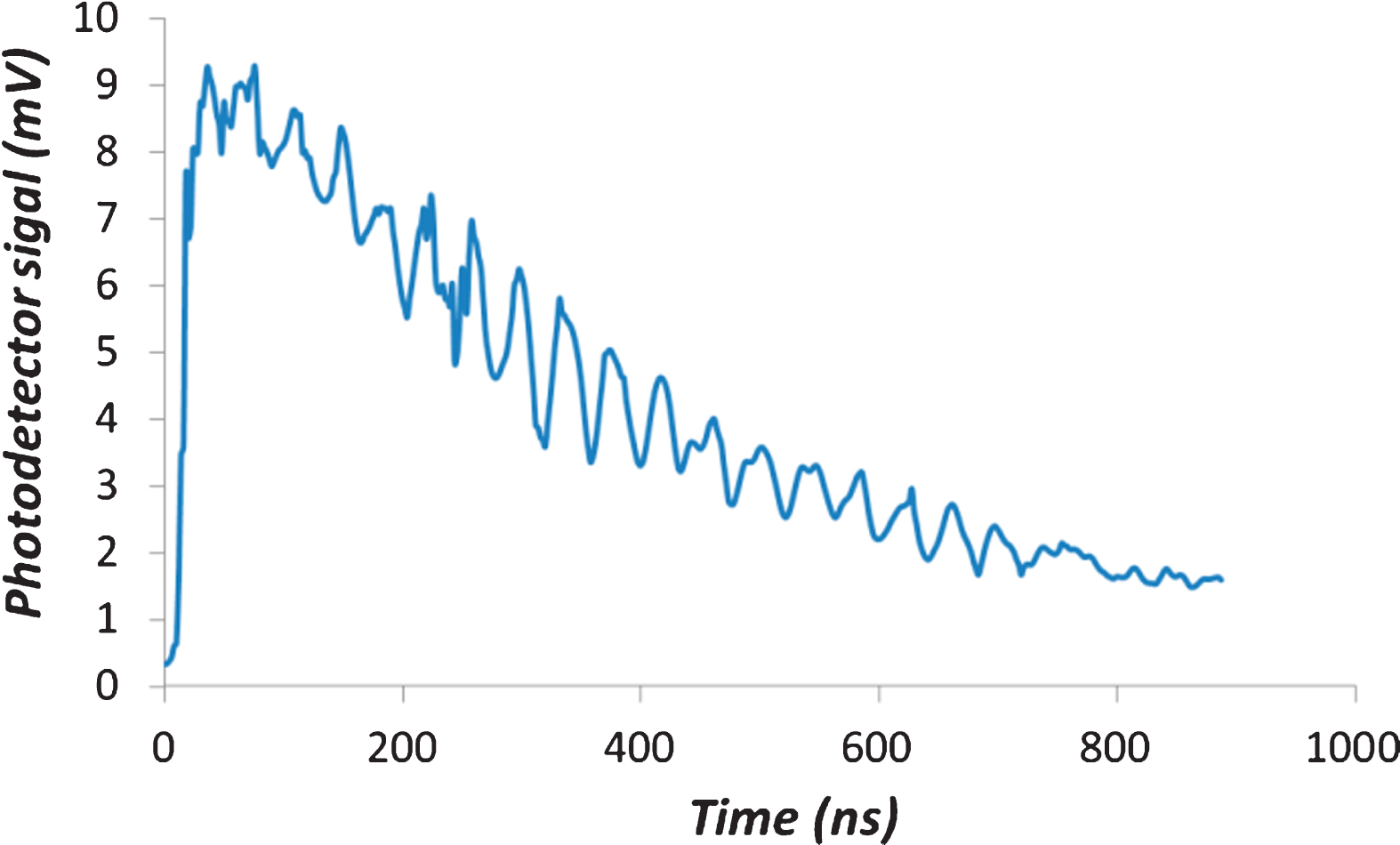
Afterglow of the detector when it is hit with a direct X-ray flash.
The device consists of numerous photosensitive in-parallel microcells with typical dimensions of 35μm. We use the photodetector with the surface area up to 36 mm2. Each microcell is an avalanche photodiode with its own quench resistor and a capacitively coupled fast output. These microcells are arranged in a close-packed array with all of the like terminals summed together. In our case the array of microcells can thus be considered as a single photodiode sensor, as shown in Fig. 6. We did not use the fast output, since the afterglow duration of the phosphors is quite long.
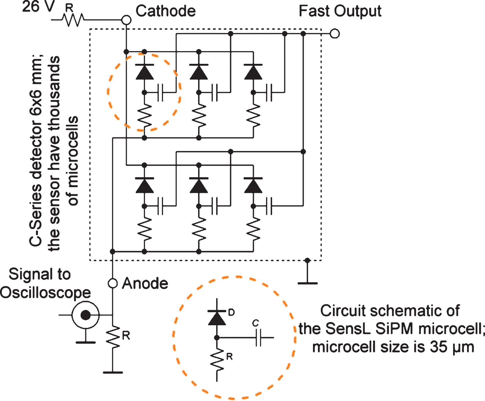
Registration scheme for studying the glow of phosphors based on SensL’s C-Series sensor.
The time dependences of the afterglow for 2 types of phosphors after their excitation by X-ray flashes at the pulse repetition rate of 700 Hz are shown in Fig. 7. The detector amplifies the signal linearly up to 120 mV, then the real signal is much stronger than the signal shown in the graph. Actual luminescence values for CsI:Tl reach 3 V. The total glow intensity after one X-ray pulse is similar for each phosphor, but the duration is different. As for the CsI:Tl phosphor the glow 500μs after the X-ray flash is less than 10% of the total glow intensity per 1 pulse, and for the Gd2O2S:Tb phosphor it is about 25%.
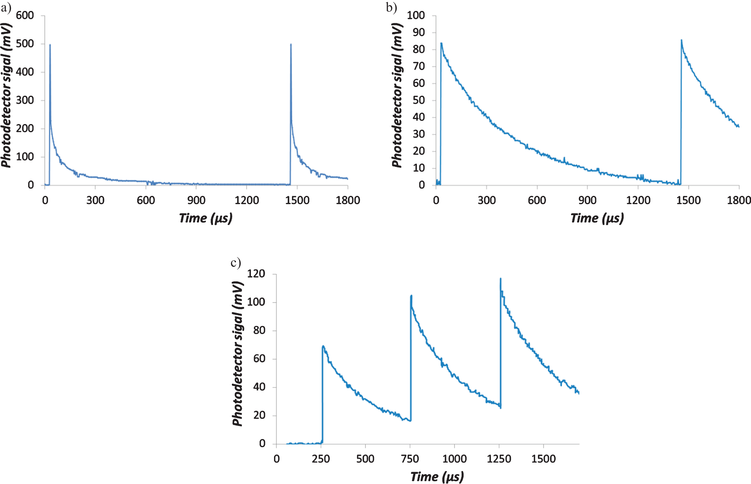
Typical afterglow for a) CsI:Tl and b) Gd2O2S:Tb phospors, c) the process of imposing afterglow from pulse to pulse for Gd2O2S:Tb phosphor at the pulse repetition rate of 2 kHz.
To choose the best X-ray fluorescent converter for the fluoroscopic system, which is used for high-speed shooting it is important to find suitable phosphor. As the result of using Gd2O2S:Tb at a pulse repetition rate above 700 Hz the residual glow of the previous pulse is imposed on the glow of the next one as shown in Fig. 7.c). Therefore, there is additional noise, because the detector is registering the frame with the new position of the object, while the residual glow from the previous pulse is still significant.
We curried out an experiment to obtain individual frames of a moving object. To register a shadow X-ray image, we used the CareStream DRX-1 detector (X-ray scintillator type is Gd2O2S:Tb) operating in the continuous mode. The object of study is a steel washer attached to a fan blade, which moves in a circle at a speed of about 11 m/s.
The pulsed source generates X-ray flashes with frequencies of 40 Hz and 1000 Hz and the X-ray flash duration is 50 ns Individual frames with the image of the washer are clearly visible in the picture made by the detector as shown in Fig. 8. Shooting at such a frame rate using a detector that operates in the continuous mode in order to obtain individual frames is only possible at a pulsed X-ray source with a SOS. During the X-ray flash, the object practically does not move. Using an X-ray continuous source, it is impossible to obtain such an image of a moving object, it will be significantly blurred.
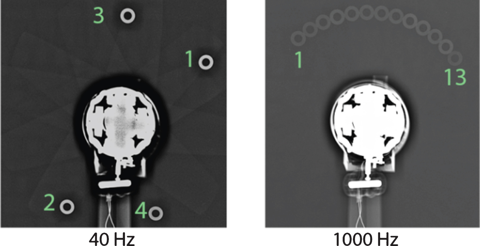
Frames of the washer moving while being attached to one of the fan blades. The frames were obtained by a semiconductor detector of the CareStream DRX-1 system, with the X-ray pulse repetition rate of 40 and 1000 Hz. The detector in this experiment accumulates the signal for one second, during which X-ray pulses are being generated, therefore, the overexposure effect is observed.
Figure 9 shows an X-ray image of a fan that rotates at 3000 rpm during the experiment. The extreme points of the blades move at the speed of about 20 m/s. We used the VIVIX-V 2323D flat panel detector, the X-ray scintillator type is CsI:Tl (Thallium doped Caesuim Iodide), which was investigated above. In this case the detector operates in the recording mode of 60 frames per second. This experiment demonstrates the stability of the radiation dose from pulse to pulse, the images of the blades have clear boundaries and there is no blur.
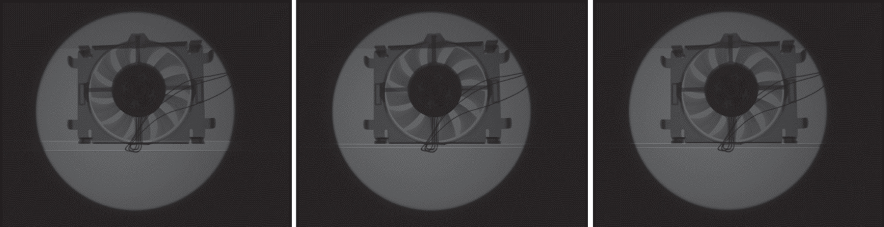
Frames of a fan that rotates at 3000 rpm. The detector operating frequency is 60 Hz, the X-ray pulse repetition rate is synchronized with the detector operation. In each image, the blades are in a different position, there is no blur, the brightness is constant at each flash.
Based on the obtained data on the afterglow of X-ray converters, we can conclude that it is possible to obtain frames of with X-ray images at frequencies up to several kilohertz. If the pulse repetition rate of the source is synchronized with the frame recording frequency of the detector, then an X-ray flash of about 50 ns in duration can illuminate a moving object, and the detector, taking into account the afterglow of the phosphor, will record a signal with the duration of up to 500μs. In the image we will practically have a stationary object. Then we can generate the next flash, because there is almost no residual illumination from the previous pulse, and register the signal again thus obtaining an image of the object at another point. We believe that this mode is very beneficial for computed tomography (CT) [16, 17]. In spiral CT scanners continuous X-ray sources are used. In this case, the source with the detector rotates around the object with the speed of more than 10 m/s, and the signal is recorded at the frequency of several kHz. The image of each frame contains information about the movement of the source and the detector relative to the object which is several millimeters. This causes significant image blurring. There is also increased noise due to the overlap of the afterglow from the previous frame on the next one. As the result the resolution decreases while the radiation dose increases. In the case of using a pulsed x-ray source, the movement of the source and the detector relative to the object is up to 1μm.
An experiment was performed to obtain tomographic slices. The object of study is made of acrylic glass and has the diameter of 110 mm. The location of the holes in the test object and their diameters are shown in Fig. 10.a). This object imitates a child’s head. In this experiment, a VIVIX-V 2323D flat panel detector was used. To simplify the experiment, it was the object of study which was rotated. The detector and generator were synchronized and operated at the frequency of 30 Hz, this limitation is due to the detector. At the distance of 60 cm between the source and the detector, there was enough X-ray power to obtain a projection image in one flash. Thus 100 images were obtained, i.e. 100 X-ray pulses were generated. Images of the test object at different rotation angles from 0 to 180 degrees with the discrete step of 1.8 degrees were obtained. Next, we created a program that selected a strip one pixel thick from each projection image, then we compiled a sinogram from those 100 obtained projection images and using the inverse Radon transformation we obtained a slice, which was done for each layer. To reconstruct the slices we used the Python programming language, and open source libraries. A 3D image of this test object is shown in Fig. 10.b), it contains 30 slices. The resolution of the system is 0.1 mm. The thickness of one slice is equal to the detector pixel size of 0.18 mm. The extreme points of the research object rotated at a speed of about 0.1 m/s, which was limited by the detector speed. Previous experiments have shown that when the object is moving at tens of meters per second, the images have practically the same resolution, since the X-ray flash is very short - 50 ns. If we use modern detectors for tomographs that operate at the frequency of 2 kHz, the scanning speed can be increased by hundreds of times compared to this laboratory experiment. And unlike using a constant radiation source, the resolution of projection images obtained at different angles with the help of a pulsed X-ray source will be higher, and as the result, the resolution of each slice will be higher.
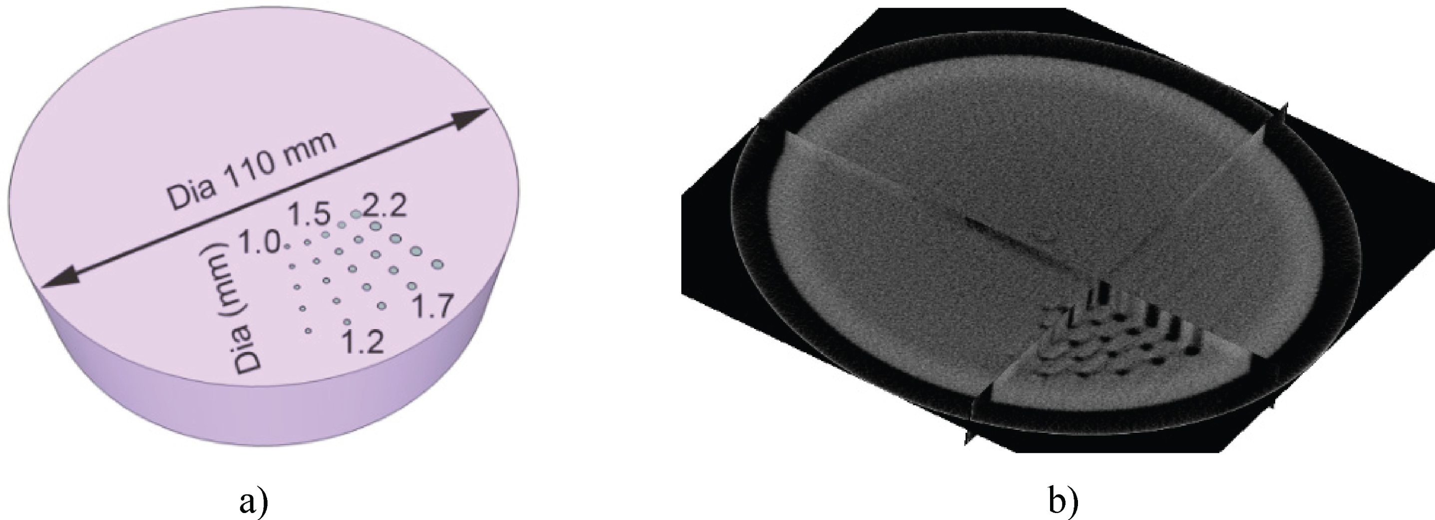
CT scan: a) study object made of acrylic glass, b) 3D image.
These experiments show great potential of using the described pulsed X-ray source for a CT scanner. The problem for pulsed X-ray sources is the instability of the radiation from pulse to pulse, up to the point of skipping of X-ray flashes. Thanks to the use of a semiconductor opening switches, instead of gas-filled spark gaps and the developed X-ray tube, the power of X-ray flashes from pulse to pulse is practically constant.
This article presents possible uses of pulsed X-ray sources with inductive energy storage devices and a semiconductor opening switch for X-ray shooting of dynamic processes, including computed tomography. In the experiments we use the high-voltage pulse generator with the following parameters: the pulse voltage amplitude up to 320 kV, the pulse current up to 240 A, the current pulse duration at 50% of the peak height of about 50 ns, the pulse repetition rate up to 2 kHz. We use a SOS as a high voltage driver. This pulsed X-ray source, in contrast to our previous designs, allows generating powerful X-ray flashes, the radiation dose is 1μSv per X-ray flash at the distance of 1 m between the source and the detector. The sources developed earlier provided the radiation dose up to 100 nSv. The radiation energy in one flash corresponds to the energy released during the operation of a 10 kW continuous radiation source in 10 –20 ms. World analogues provide such flash power, however, the pulse repetition rate is only 10–20 [4], this limitation arises from using gas-filled spark gaps as the final pulse shaper. In addition, using a SOS, as well as the X-ray tube we developed, makes it possible to provide a stable radiation dose from pulse to pulse.
It is important to understand the operation principle of modern detectors, specifically how a signal is formed after exposure to powerful X-ray flashes generated by our pulsed X-ray source. We carried out experiments to determine how the signal intensity and afterglow duration of the scintillators used in the detectors differ: VIVIX-V 2323D flat panel detector, the X-ray scintillator type in this detector is CsI:Tl; CareStream DRX-1 detector, the X-ray scintillator type is Gd2O2S:Tb. For 2 types of X-ray phosphors, the intensity and duration of the glow after irradiation with X-ray flashes were measured. The CsI:Tl phosphor has a short afterglow duration and can be irradiated with flashes at the repetition rate of X-ray up to 2 kHz, there will be no residual glow from the previous frame. When the Gd2O2S:Tb phosphor is irradiated with X-ray flashes at the repetition rate of more than 700 Hz, residual glow is observed. Experimentally, we managed to obtain a superposition of the signals from 2 x-ray pulses, when the signal of the previous pulse did not decay and the next pulse was generated. Such a result cannot be obtained using other types of pulsed X-ray sources, since they do not provide a high pulse repetition rate.
We obtained several clear frames of an object moving at the speed of 20 m/s to the detector, which operates in the continuous mode. This was achieved by a pulsed X-ray source, which operates at the pulse repetition rate of about 2 kHz with the pulse duration of about 50 ns. Based on the images of a moving object which we obtained on the detector that operates at 60 frames per second, we can conclude that the pulse-to-pulse dose stability is high. These pulsed sources can be used for security inspection systems and demonstrate a higher performance as compared to continuous radiation sources.
In modern computed tomographic scanners, a continuous X-ray source rotates relative to the test object at the speed of about 12 m/s. In tomographs they use continuously working X-ray tubes and detector systems which operate at the frequency of several kHz. During one exposure the detector moves several millimeters relative to the test object, so the signal is not obtained at one angle, but is smeared. If a pulsed radiation source is used, then over the course of 50 ns this shift will be 0.0006 mm, thus there will be no dynamic blurriness. Also, detectors have a time interval when they transmit a signal to a computer. During this time the constant radiation source is still generating X-ray radiation, so the patient is under unnecessary exposure, although the signal to the detector is not registered.
Using a pulsed X-ray source can increase the resolution and reduce the radiation exposure of the patient when the spiral CT method is used. Synchronization of the source with the detector will allow us to eliminate the dead time of the detectors, which occurs when the detectors are used in combination with sources of continuous X-ray radiation. In the article we obtained a tomographic slice of an object that simulates a child’s head. For each fixed angle, an exposition was obtained for one X-ray flash.
In the future, we will increase the power of our pulsed X-ray sources by modernizing the electrical circuit so that the pulse repetition rate is 5 kHz while the energy stays the same. We will also increase the permissible heat power that the X-ray tube can dissipate, and possibly will introduce a system with a rotating anode.
Footnotes
Acknowledgments
The investigation is made with financial support from the Russian Science Foundation (RSF), grant No 20-79-00036.