



Abstract
BACKGROUND:
Medical image processing has gained much attention in developing computer-aided diagnosis (CAD) of diseases. CAD systems require deep understanding of X-rays, MRIs, CT scans and other medical images. The segmentation of the region of interest (ROI) from those images is one of the most crucial tasks.
OBJECTIVE:
Although active contour model (ACM) is a popular method to segment ROIs in medical images, the final segmentation results highly depend on the initial placement of the contour. In order to overcome this challenge, the objective of this study is to investigate feasibility of developing a fully automated initialization process that can be optimally used in ACM to more effectively segment ROIs.
METHODS:
In this study, a fully automated initialization algorithm namely, an adaptive Otsu-based initialization (AOI) method is proposed. Using this proposed method, an initial contour is produced and further refined by the ACM to produce accurate segmentation. For evaluation of the proposed algorithm, the ISIC-2017 Skin Lesion dataset is used due to its challenging complexities.
RESULTS:
Four different supervised performance evaluation metrics are employed to measure the accuracy and robustness of the proposed algorithm. Using this AOI algorithm, the ACM significantly (p≤0.05) outperforms Otsu thresholding method with 0.88 Dice Score Coefficients (DSC) and 0.79 Jaccard Index (JI) and computational complexity of 0(mn).
CONCLUSIONS:
After comparing proposed method with other state-of-the-art methods, our study demonstrates that the proposed methods is superior to other skin lesion segmentation methods, and it requires no training time, which also makes the new method more efficient than other deep learning and machine learning methods.
Introduction
Image analysis is a technique to examine images using computer technology while dividing them into segments or objects [1]. Not every image can be studied with the same algorithm as the images are made up of different particles and patterns [2]. Image segmentation is used in various areas including biomedical where it is used to identify lung diseases [3], classify brain tumor and skin lesion etc., identify white blood cells [4] and detect cancer-causing cells [5]. Numerous skin diseases can be identified by image segmentation [6–9]. On the daily basis, human skin is exposed to various DNA damaging bacteria which may result in numerous changes. Some of these changes are visible to human eye while others can cause internal changes [10].
Skin lesion is one of the commonly found diseases which refers to skin area being different to its surrounding skin in terms of color, size, or texture. The disease may start with a local skin exposure to damaging sources such as sunburns or contact dermatitis [11]. Not only this, but lesion may also be a result of underlying disorders such as infection, diabetes, or genetic disorders. In most cases, lesion can be harmless or for a short period of time but some of them may result in life-taking skin cancer [11]. Cancer is one of the deadliest diseases to be discovered by the science. American Cancer Society (2020) calculated over 100,000 new cases out of which 7,000 people are expected to die in coming years. Dermatologists currently are trying to identify the root cause of this disease and the possible measures that can be taken to avoid further deaths.
There are different techniques to identify image segmentation, these techniques can be further categorized into different classes such as characteristic featuring thresholding or cluster, region extraction, edge detection, Otsu thresholding and active contour.
Characteristic featuring thresholding or cluster: For threshold technique, there are some pre-processing and post-processing steps [12]. Few of the frequently used techniques are Mean Method, P-tile method, Histogram Dependent Technique (HDT), Edge Maximization Technique (EMT) and visual technique. However, HDT and EMT are considered as the most reliable techniques for thresholding. [13]. For efficiency, threshold-based segmentation Particle Swarm Optimization (PSO) and 2-d Otsu algorithm (TOPSO) are also very popular.
Region extraction: As the name states, it divides the image into small regions, based on different criteria such as color, intensity, or object. Regional extraction is comparatively simple than other techniques. Region technique is sub-divided into categories like region growing, region splitting and region merging [14]. Unsupervised image segmentation methods are also in practice which employs level set methods and texture statistics [15].
Edge detection: Detection of edge is an initial step in image segmentation [16, 17]. This process divides the image into objects and background. The image is further divided into pixels by observing the change of intensity. The key steps are histogram and gradient computation [18]. The most popular techniques of edge detection include classical edge detectors, zero crossing, Laplacian of Gaussian [16] and color edge detection [19].
Active contour: Active control model (ACM), also referred to as the snake model, was introduced in 1998 by Demetri Terzopoulos, Michael Kass and, Andrew Witkin [20]. It is widely used in applications like object-tracking, shape recognition, image segmentation, edge detection and object detection. It enables the model to come out of a local minimum and move towards the end goal. The model works under the effect of image forces and external constraint forces. The image forces basically push the active contour model towards several features of images, like contours, lines, edges, outlines. The total image energy is expressed as the sum of three different energy functions as given in Equation 1.
OTSU thresholding: OTSU thresholding performs automatic thresholding on the image and divides the image into foreground and background. Using pixel’s spatial information, including mean and median, 3D Otsu improves the segmentation results and has better immunity as compared to 1D and 2D Otsu.
The current research focuses on skin lesions segmentation from digital images. A new algorithm Adaptive Otsu-based Initialization (AOI) is proposed for fully automated initialization scheme for skin lesion segmentation.
In summary, the major research contributions of the current research are as follows:
A new algorithm, “Adaptive Otsu-based Initialization (AOI)” is proposed for computer aided initialization of the active contour models of skin lesion segmentation. A fully automated process is defined to segment skin lesions from skin images. An efficient and unsupervised method is proposed that does not require any training data for the segmentation. Asymptotic analysis of AOI is computed to measure the efficiency of the proposed algorithm. The proposed method is validated on a benchmark dataset [21] to prove its robustness as a method which can handle challenging artifacts present in skin images which makes lesion segmentation a challenging task
The remaining paper is organized as follows: In Section 2, Related work is presented in detail for skin lesion segmentation. A fully automated method for skin lesion segmentation is explained in Section 3. Section 4 described Experiments and Results which are used to validate the proposed method and empirically compare results with other methods. Discussion related to this research is presented in the Section 5. Lastly, Section 6 describes conclusion of this research.
A lot of image segmentation methods are currently being used in the medical field such as the ones specified in [22]. It is usually considered a pre-processing step for classification techniques. Classification is a supervised machine learning technique that attempts to assign a class to an unknown image. Image classification is widely used in the medical field to classify, for example, an image of a brain tumor as benign or malignant. This requires taking a large number of labeled samples for benign and malignant tumor and then training a classifier on them. For classifiers to perform well, the tumor part of the brain needs to be extracted using segmentation techniques. Several image segmentation methods have been proposed.
Neeraj Sharma et al., have discussed the medical image segmentation techniques in depth [22]. The authors have classified the medical image segmentation techniques into two classes: gray level-based techniques and textural feature-based techniques. The gray level techniques are used for edge detection in images or region-based segmentation. The objective of region-based segmentation is to extract a homogeneous region from the image, depending on the region’s properties. Region-based segmentation is classified into three types: region merging, region splitting and split and merge. Textural feature-based segmentation relies on the division of images based on the regions having a certain textural property. The authors conclude that every type of segmentation technique or model is implemented based on its suitability, considering several conditions. Although the gray level technique is a bit limited, textural level techniques require domain knowledge experts for manual input.
Alireza Norouzi et al have reviewed several medical image segmentation methods and their applications in their research [23]. The authors raised a very crucial concern of global thresholding. In case of an image with uneven background, the global thresholding method does not produce good results, since the threshold value differs for every part of the image. To solve this problem, local thresholding method is proposed. Local thresholding method works under the principle of dividing one image into several different images with different backgrounds, takes both, background, and foreground. It selects the threshold value for each sub-image accordingly and apply thresholding. Finally, it combines the sub-images into one image with appropriate result. However, time complexity is not as efficient as global thresholding, but it produces fairly good results for images with diverse backgrounds. The selection of threshold value is also an issue which causes poor results. Otsu’s thresholding value is proposed to automate the process of selecting threshold value for images. After discussing the commonly used medical image segmentation methods, researchers have said hybrid image segmentation method to be performing better than other methods. It utilizes the boundary as well as region of interest to perform image segmentation. Graph cut is a hybrid image segmentation method. The user predefines the region of interest and then the model separates the object from the background. This approach is also called as region and boundary detection. Snake model also works under the similar principle.
Withey et al. have explained the three generations of medical image segmentation [24]. Each generation has its own limitations and complexities. The first generation consists of low-level techniques with pre-defined information. This generation includes techniques like thresholding and region extraction. The second generation includes models such as the snake model [20], deformable models or graph cut models. The third generation consists of hybrid models like Atlas and rule-based image segmentation models. Another comprehensive study on medical image segmentation was done by [25] that compares the proposed model for medical image segmentation with the state-of-the-art medical image segmentation models. They discussed the conventional image segmentation models like active contour models, active contour models without edges and Chan Vese active contour model [26].
Convolutional neural networks (CNN) have gained a lot of popularity for image segmentation because of their remarkable results. Several architectures for CNN have been proposed such as VGG-16, VGG-19, Dense-Net, etc. U-Net is also a CNN-based architecture for image segmentation however, it works under the principle of an image-based approach instead of a pixel-based approach. U-Net takes a single image as input and returns the segmented image. U-Net has been shown to produce better results as compared to the pixel-based CNN approaches.
In [27] U-Net architecture has also been used in combination with the ACM for image segmentation. Their proposed model’s loss function is taken from ACM and the image segmentation model is inspired by U-Net. The idea of proposed active contour loss is to efficiently find an active contour which is a global minimization of active contour energy for automated image segmentation. The proposed base model for image segmentation is U-Net. The proposed model was tested on cardiac magnetic resonance dataset (CMR). It is a very popular medical imagery dataset. The comparison of results with the existing state-of-the-art image segmentation models and the proposed model made it evident that the proposed model produced comparatively better results.
M. Khelif et al have discussed two types of active contour models: parametric models and geometric models [28]. Original snake models or active contour models [20] introduced by Kass is an example of a parametric active contour model. It takes an initial contour as an input, and then there is an objective function which is the energy of the active contour model. James A. Sethian introduced the concept of geometric model [29] which defines the front evolving which depends upon speed function or the image gradient. However, both, parametric and geometric models have one thing in common, both are driven by the forces extracted from the image itself.
While concluding their research, the author also highlights the initialization problem of active contour models. Considering the initialization problem of active contour models, the model Chan Vese model is capable of handling images with weak boundaries however, it fails with images having intensity inhomogeneity.
Tingting Liu et al worked specifically to overcome the problem of wrong segmentation of images with intensity inhomogeneity [30]. The model developed by the authors is a hybrid, region-based model. To avoid the curve, movement around the noise or unwanted objects in the image, a regularization function is added takes the arc length of the curve during the evolution. The proposed model is tested against the images having intensity inhomogeneity, with other models; Local region based active contour model and CV model. The results after testing the proposed model and then comparing it with other models showed that the proposed model produced fairly good results and the computation cost was reduced. However, the proposed model still suffers from the initialization problem. Skin Lesions datasets have been quite popular in medical image processing applications because of their challenging complexities. The skin lesion datasets images contain noise in most of the images, and the region of interest differs in various aspects, such as locality, size, shape, and even intensity in-homogeneity.
In [31], a convolutional-deconvolutional neural network (CDNN) model is proposed to perform automatic skin lesion detection. The model is trained on ISIC 2017 skin lesion dataset [9]. Loss function is calculated using the Jaccard distance. The proposed model can perform well with images having noise. Initialization is not an issue since the model is trained to take an image and output the region of interest (ROI). CDNN produced a JI value of 0.784.
The only issue with ACM is its initialization, or in other words, the placement of initial contour. Inaccurate segmentation occurs if the initial contour is placed in the wrong position. Manually placing the initial contour significantly improves the results but this is time consuming and requires an expert annotator. There is a need to automate the initialization process.
In [32], genetic algorithm (GA) is used for contour initialization. With the help of GA, a circular area is found that is the initial contour and it is optimized using GA. The population size was 10 and was chosen after trial and error. JI was used as a fitness function. Each chromosome contains the random initial contours that are changed during the optimization process until a higher value of JI is achieved. Data preprocessing was also done and included hair removal using Dullrazor.
To improve initialization robustness, [33] proposed a simple method that is based on local region fitting energy. It extracts local information within an image and segments the regions with non-homogeneous intensity. This study does not propose an initialization technique but proposes how to make the best use of the initialization once it is done. It uses a curve evolution technique using local image fitting (LIF) that calculates the mean intensity of the homogeneous regions and then builds on it. This technique can also work with images with brighter backgrounds and darker objects. The method can also be applied to other models. As per the LIF model, the fitting functions that are donated as m1 and m2 can be mapped onto f1 and f2 as per the RSF model.
Querios et al managed to introduce an automatic initialization algorithm. This was introduced for mid-ventricular slices, and this was highly based on the LV localization method as well as the matching algorithm of the elliptical annular template [34]. LV centroid can be detected through this method. It must be followed by class decomposition. Moreover, it is also followed by a search procedure. The most circular object is known as LV. Moreover, it is close to the center of the image. It is important to obtain less than 2 elliptical initial contours. This is done to be used in segmentation process. Furthermore, with the help of template method to look for elliptical ring of the darker side. The following strategy can be used to look for the template. In the case of every kernel, a search related to block matching is done with the help of cross-correlation that is normalized. It also defines optimal parameters. The optimal parameters help in drawing two ellipses that may be used as initial contours. Evaluation of the models is something which contains high significance. It shows the quality of the model. Choosing the right evaluation metrics while conducting research is something that should be taken in account. The accuracy of the model is proved by the results of evaluation metrics. However, it is important to verify if the evaluation metric can evaluate the model, as there are different evaluation metrics for different kinds of models.
In addition, Zhaobin Wang et al. have conducted a detailed study on evaluation metrics for image segmentation [35]. The authors have classified image segmentation evaluation metrics into two major classes: supervised evaluation methods and unsupervised evaluation methods. In supervised evaluation metrics, the final output is compared with a corresponding ground truth image. The ground truth images are mostly prepared manually by domain experts. Some of the supervised evaluation metrics are dice score, Jaccard index, F-1 score, precision, recall, sensitivity, and specificity. Whereas unsupervised evaluation metrics calculate the indicators or measures of the final output image to evaluate the model. In this case, ground truth images are not required. Some of the unsupervised evaluation metrics are peak signal-to-noise ratio (PSNR), root mean squared error (RMSE), mean absolute error (MAE), etc.
Supervised evaluation metrics are costly, time-taking and limited by the availability of the ground truths. Since ground truth images are prepared by domain experts, and performing manual segmentation also requires domain experts, the process can be time taking and costly. In some cases, it is very difficult to prepare ground truth images, for example, ground truth images of natural images. On the contrary, un-supervised evaluation metrics do not require ground truth images at all, hence it makes the unsupervised evaluation process faster and cheaper. However, the reliability in terms of accuracy of unsupervised evaluation is low as compared to supervised evaluation models.
Thus, considering our research nature and the dataset being used, we chose to implement supervised evaluation metrics. The ISIC-2017 dataset [9] used in this study has manually segmented ground truth images to each corresponding training and testing image. To assure maximum accuracy of our results, we decided to implement supervised evaluation models. V. Mezaris et al proposed an evaluation metric for still image objective segmentation [36]. The proposed methodology is based on the measure of spatial accuracy using the manually segmented ground truth image. The proposed metric was quite successful, as it was able to highlight issues like over-fitting, under-fitting, and inaccurate boundary localization, etc.
Methodology
Recently many methods have been published for skin lesion segmentation [37, 38]. The proposed methodology aims to automate the initialization of active contour models and to produce accurate and appropriate image segmentation results. The proposed methodology is divided into three major stages:
Dataset selection Proposing a new model Evaluation of proposed model
Dataset selection
The detection of several kinds of diseases makes use of medical imagery datasets frequently. After a thorough research, ISIC 2017 Skin lesion dataset is used [9] for the experimentation of proposed technique. ISIC 2017 Skin lesion is a medical imagery dataset having diverse types of skin lesions with various artifacts. These artifacts make the segmentation task very challenging. Training and testing images are the actual images captured of skin lesion disease, and the ground truth images are the actual binary segmentation of the relevant images. These ground truth images are used for the evaluation of the final segmentation by the proposed technique. The segmentations are marked by experts, and it is a very time-consuming process to determine the lesion boundaries manually. The dataset consists of a wide range of skin lesion images, in terms of the size, shape, and locality of the skin lesion/region of interest as well as noise or unwanted objects in the images. ISIC 2017 is a publicly available dataset with 2000 skin lesion training images along with their corresponding ground truth images.
Proposed method
The proposed method (AOI) is developed to accurately segment skin lesions from different images in the presence of different artifacts. The working of the AOI model can be divided into three main phases or steps:
Preprocessing Automated initialization Final Segmentation using ACM
During preprocessing, the input image is rescaled to the size 125×125 to make it computationally efficient. The contrast of the input image is enhanced to identify outliers and noise in the image.
After preprocessing, the proposed algorithm takes an input of an image and creates an initial boundary mask that is used to perform the final image segmentation. So, the first step is to accurately produce the initial contour. To get an optimal thresholding value, the Otsu’s thresholding method is implemented, as it produces a single intensity threshold image to divide the input image into two distinct regions (background and foreground). Using the prior information of the skin lesion images, the region of interest is chosen based on the area and locality of all binarized components which are obtained from the Otsu’s thresholding method. The output of the Otsu is used as input to the ACM to obtain a more refined final segmentation. The high-level diagram of the proposed AOI model is given in Fig. 1.

The high-level diagram of the proposed AOI model.
Figure 2 highlights algorithm which contains all the steps of the proposed method for automated initialization and final segmentation. In this algorithm initially, a 2D image I and ground truth image M are taken as inputs. To apply thresholding on the test image I histogram H is computed along with the probabilities of each intensity level. Next, both ω
i
(0) and μ
i
(0) are initialized. After initialization, all possible thresholds t from 1 to maximum intensity are computed by updating ω
i
, μ
i
and computing

An Adaptive Initialization Algorithm for Active Contour Models of Skin Segmentation: Adaptive Otsu-based Initialization (AOI).
The quality of the proposed method is estimated using standard evaluation metrics. The images from ISIC 2017 dataset are used for the estimation of the proposed technique. These images were different, in terms of skin lesion size, shape, locality, artifacts and noise in the image. In supervised evaluation metrics, the final output is compared with a corresponding ground truth image, with actual results. The ground truth images are prepared manually by domain experts. The standard measures used for the evaluation are the dice score (Equation 2), Jaccard index (Equation 3) and recall or sensitivity (Equation 4). These three different supervised performance evaluation metrics are calculated using the expressions given below.
The above-mentioned evaluation metrics return quantitative values, so it becomes easier to compare and analyze our proposed model. To compare it with other possible methods, the initialization methods are changed, results are stored, and compared them with our proposed method to ensure the novelty of our research work.
The experimentation is performed on Skin images from ISIC, 2017 dataset using Python on 2.5 GHz Intel Core i7 processor with 8 GB of RAM. The results obtained from the proposed method are compared with the state-of-the-art active contour model. Table 1 shows the results using DSC, JI and sensitivity for different methods.
Comparisons of different segmentation methods. The best value is shown in bold
Comparisons of different segmentation methods. The best value is shown in bold
From Table 1, it can be analyzed that the average dice score obtained from Otsu thresholding is 0.768, that is quite poor as compared to the average dice score of the proposed model, that is 0.8840. These results allow us to conclude that our proposed model has made significant improvements. The two-sample t-test was used to test the significance of the segmentation results obtained at a confidence interval of 95% (p≤0.05). The initialization process, after being automated is producing accurate and appropriate image segmentation results. All results show significantly better results than Otsu (p≤0.01) and snake model (p≤0.05).
Figure 3 shows some test images along with their ground truths taken from the ISIC 2017 Skin Lesion dataset and compares the actual ground truths with the segmentation results qualitatively produced by our proposed model and Otsu threshold method. We have carefully selected different types of images with different backgrounds, sizes, colors, and artifacts.
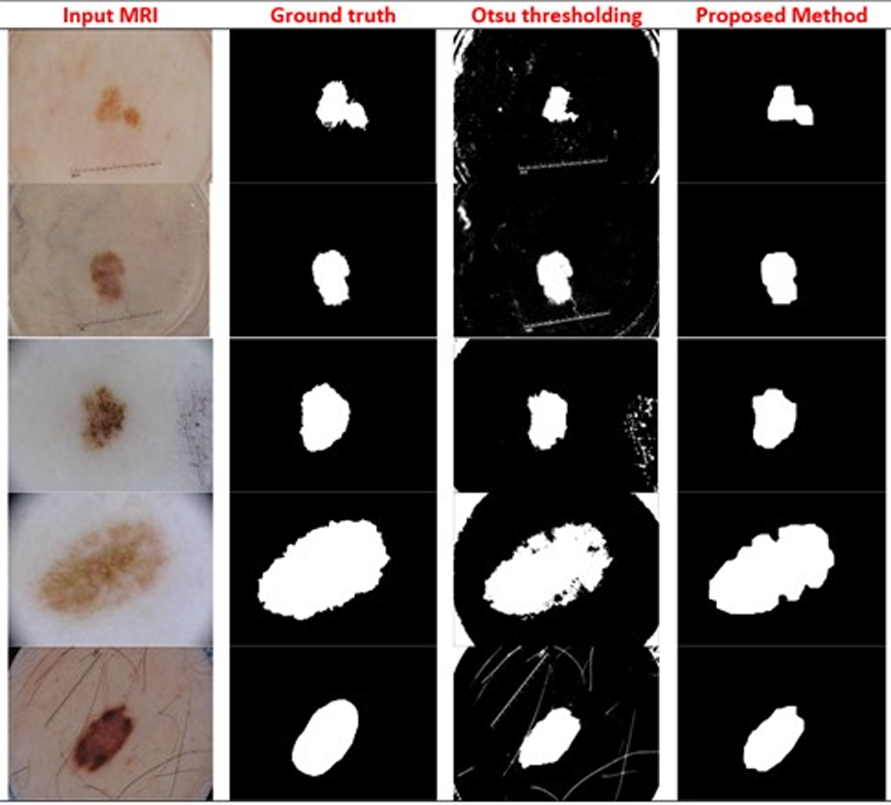
Segmentation result from ground truth, Otsu thresholding method and proposed algorithm.
In this subsection, we have shown the impact of a good initialization vs. bad initialization on the segmentation results. From Fig. 4, it can be observed that a bad initialization produces undesirable segmentation while a good initial contour is more likely to produce a more accurate extraction of the region of interest (ROI).

Comparison of segmentation results produced from a bad initialization and from the proposed initialization method.
In this subsection, we compare the performance of the proposed model with the other available models and algorithms using the same ISIC 2017 Skin lesion dataset [9]. The results are shown in Table 2. In this table, the proposed method (AOI) is compared with some recently presented CNN models [40–44]. Different CNN models with variation in the architectures are used, it can be observed that this proposed non deep learning approach producing better result than many existing deep learning models [13, 45]. The evaluation metric used is JI. The studies were short-listed because they used the same dataset, thus allowing a fair comparison of the results produced by these previous studies with our proposed model.
Performance of the proposed segmentation model with the other available models using the ISIC 2017 dataset
Performance of the proposed segmentation model with the other available models using the ISIC 2017 dataset
In the following section, the comparison of the proposed technique and other state-of-the-art techniques is done. Figure 5 shows mostly deep learning models are producing good segmentation results.
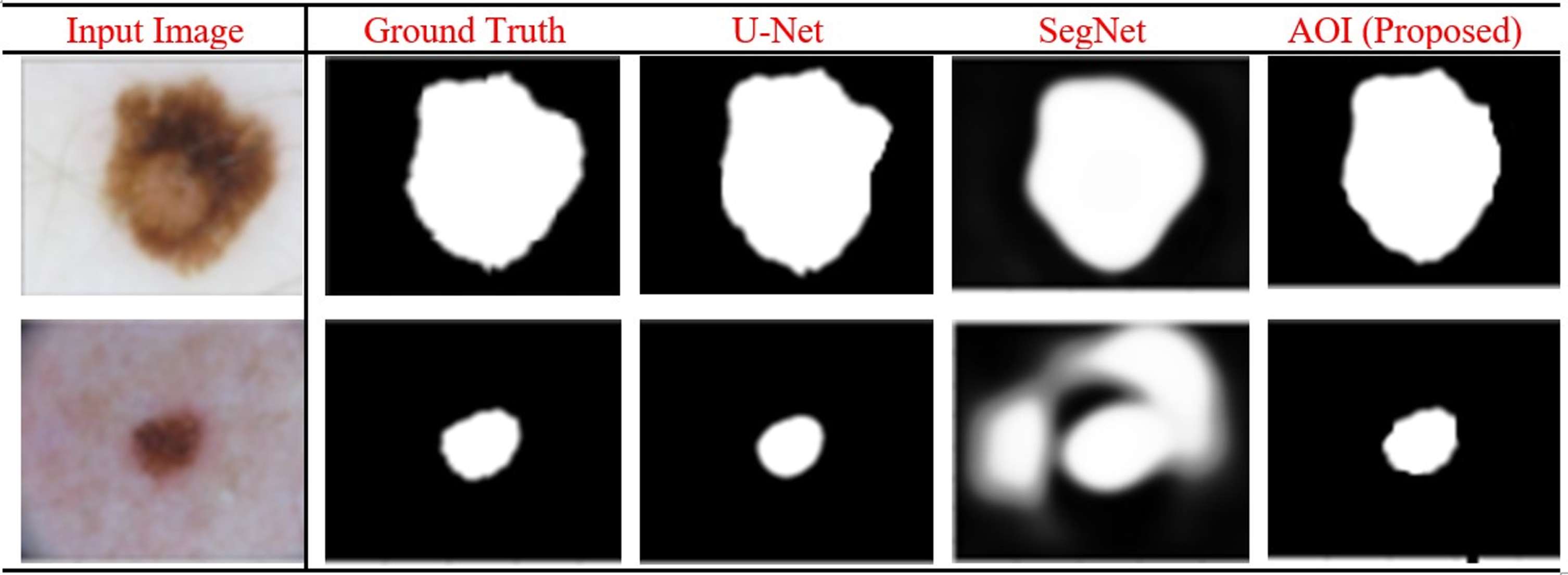
Comparison of segmentation results produced from the proposed method (AOI) and other state-of-the-art methods.
The time complexity for the worst case is also analyzed and it is observed that this is computationally efficient method for the segmentation. The total time taken by the proposed method is O (mn + n + k) = O (mn), where m and n are image dimensions and, k is the number of regions.
Discussion
Active contour models have been quite popular in the field of computer vision and image processing and has many applications such as shape recognition, object tracking, object detection and image segmentation [20, 52]. These applications have made it possible to detect several diseases using x-rays, MRIs, CT-Scans, and other imagery data. Every imaging modality has its own challenges, some are common in all medical images like intensity inhomogeneity [53–56], but some are specific to the type of input images. For instance, skin images have some common challenges that are specifically related to them such as the presence of hair, markers, and scales [6, 57]. ACM has also been used for image segmentation in the medical field. However, the final segmentation results are highly dependent on the initial placement of the evolving contours [51, 58–60]. Although manual initialization by user input produces a more accurate segmentation of images, still it is a very time-consuming process and requires domain experts [61, 62]. It has been observed that the active contour model fails to produce accurate segmentation results if the initialization contour is not close to the region of interest (ROI) [58, 60]. The objective of this research is to automate the initialization process of ACM for improving final segmentation results. To achieve that, an adaptive thresholding-based algorithm (AOI) is proposed which works on region-based principle [51].
In this study, the ISIC-2017 Skin Lesion dataset is used. This dataset is quite complex and has a variety of images which could be challenging for image segmentation because of its inconsistency in terms of object locality, shape, size, and patterns [63].
The proposed method gives promising results for all the three measures (dice score, Jaccard index and sensitivity) when compared to state-of-the-art techniques in Table 1. Apart from these techniques the proposed method shows comparable results with thirteen techniques proposed in recent years by different researchers in Table 2.
Over the last many years, deep learning is giving promising results in medical image segmentation [40] however, these models are computationally very expensive and require a large amount of annotated data. Parameter optimization is also another challenge in designing a new deep learning architecture or fine-tune the existing ones for some new dataset. From Table 3, it can be observed that each CNN model requires training time (in hours) even for simple 2D skin images however, the proposed model (AOI), does not require any labeled data to train the model. This method is used to segment any skin lesion in the presence of several challenging artifacts without using any training time and examples.
Comparison of Training time required for different state-of-the-art deep learning models using the ISIC 2017 dataset
Comparison of Training time required for different state-of-the-art deep learning models using the ISIC 2017 dataset
Since the research objective is to solve the initialization problem of active contour models [51, 58]. However, there is still a lot of room of improvement in the proposed model as our model is sensitive to extreme similarity of color intensity of background and foreground. In addition to that, the Otsu thresholding is also sensitive to artifacts present in skin images like ill-defined boundaries, low contrast, and intensity inhomogeneity. In the future, some image processing pipeline can be used to handle those artifacts and then apply Otsu thresholding.
In future, the proposed fully automated method can be applied for other imaging modalities like for lung tumor classification, brain tumor classification and Left Ventricle (LV) segmentation for the early diagnosis of several diseases [56].
Accurate skin lesion segmentation plays a significant role in diagnosing several skin diseases like Melanoma, Basal cell carcinoma and many more. Early detection of diseases helps to remove cancer at the early stages. Automated segmentation models are useful to reduce the time-consuming effort of manual segmentation of skin lesions which is done by experts from the medical field. Skin lesion images have many intrinsic artifacts which make the segmentation process very challenging.
In order to help medical professionals, the present study proposes a fully automated method called AOI to perform accurate skin lesion segmentation. The proposed model uses an automated thresholding method that binarizes input skin images. This thresholded image is further refined using the ACM. AOI gives promising results on a publicly available benchmark skin dataset. The performance is verified using different evaluating metrics and the proposed method shows significantly improved segmentation results than state-of-the-art methods.
In the future, the proposed fully automated method can be applied to other imaging modalities like lung tumor, brain tumor and left ventricle (LV) segmentation for the early diagnosis of several diseases [56]. Finally, the research concludes that:
The new algorithm, “Adaptive Otsu-based Initialization (AOI)” can successfully initialize the active contour models of skin lesion segmentation. The proposed method is useful in fully automating the process of skin lesions segmentation. The unsupervised method AOI does not require any training data for segmenting skin lesions from skin images. With the help of a benchmark dataset, the proposed algorithm is validated and found that it is robust in handling challenging artifacts which are present in skin images. The computation complexity of AOI also reveals that it can work efficiently and can produce good results. The research achieves up to 0.79 JI and 0.88 DSC scores which are better than the state-of-the-art techniques.