
Abstract
BACKGROUND:
Recently, deep learning reconstruction (DLR) technology aiming to improve image quality with minimal radiation dose has been applied not only to pediatric scans, but also to computed tomography angiography (CTA).
OBJECTIVE:
To evaluate image quality characteristics of filtered back projection (FBP), hybrid iterative reconstruction [Adaptive Iterative Dose Reduction 3D (AIDR 3D)], and DLR (AiCE) using different iodine concentrations and scan parameters.
METHODS:
Phantoms with eight iodine concentrations (ranging from 1.2 to 25.9 mg/mL) located at the edge of a cylindrical water phantom with a diameter of 19 cm were scanned. Data were reconstructed with FBP, AIDR 3D, and AiCE using various scan parameters of tube current and voltage using a 320 row-detector CT scanner. Data obtained using different reconstruction techniques were quantitatively compared by analyzing Hounsfield units (HU), noise, and contrast-to-noise ratios (CNRs).
RESULTS:
HU values of FBP and AIDR 3D were constant even when the iodine concentration was changed, whereas AiCE showed the highest HU value when the iodine concentration was low, but the HU value reversed when the iodine concentration exceeded a certain value. In the AIDR 3D and AiCE, the noise decreased as the tube current increased, and the change in noise when the iodine concentration was inconsistent. AIDR 3D and AiCE yielded better noise reduction rates than with FBP at a low tube current. The noise reduction rate of AIDR 3D and AiCE compared to that of FBP showed characteristics ranging from 7% to 35%, and the noise reduction rate of AiCE compared to that of AIDR 3D ranged from 2.0% to 13.3%.
CONCLUSIONS:
The evaluated reconstruction techniques showed different image quality characteristics (HU value, noise, and CNR) according to dose and scan parameters, and users must consider these results and characteristics before performing patient scans.
Keywords
Introduction
Computed tomography angiography (CTA) has been developed, along with various reconstruction techniques such as filtered back projection (FBP) and iterative reconstruction (IR), as the first-line imaging study for neurovascular morphology and disease diagnosis. Its unmatched availability, minimal invasiveness, high resolution, and, when necessary, its combination with cerebral perfusion CT make it a versatile imaging tool for rapid evaluation of vessels. The major drawbacks of CTA are the increased cumulative dose as its use increases and the side effects of contrast agents used on patients, ranging from mild discomfort such as itching to life-threatening emergencies [1–3]. Therefore, to obtain optimized CTA image quality, users must understand the characteristics of image quality according to the reconstruction technique, radiation dose, and contrast agent concentration.
The FBP algorithm has been used for decades owing to its low computational power because it assumes that the acquired projection data are noise-free [4]. However, when examining obese patients with meager or excessive radiation doses, FBP produces noisy images that are prone to artifacts [5]. Along with advances in computational power, CT reconstruction techniques have made impressive progress over the past 20 years, especially IR techniques, which are now becoming the new standard [6–8].
A new and more sophisticated version of this hybrid IR (Adaptive Iterative Dose Reduction 3D [AIDR 3D] (Canon Medical, Ottawa, Japan) is now available in our hospital. Because the image noise is lower and artifacts are fewer on IR images than on FBP images, IR is widely used to reduce radiation exposure and improve diagnostic ability [9, 10]. Although it significantly improves image quality, especially in low-dose CT studies, the image texture, spatial resolution, and object detection potential are still unsatisfactory. Conversely, the properties of this IR algorithm change the noise texture and characteristics and render the spatial resolution dependent on dose and contrast [11, 12]. Many studies have shown that the image quality obtained with these IR algorithms was reported by radiologists to be impaired, modified, and smoothed [13, 14].
Recently, artificial intelligence has generated significant interest in several imaging applications, ranging from detection, recognition, and segmentation to a new type of reconstruction technique based on deep learning reconstruction (DLR) [15, 16]. The Advanced Intelligent Clear-IQ Engine (AiCE) (Canon Medical, Otawara, Japan) was the first commercialized DLR tool and uses a deep convolutional neural network to distinguish real signals from noise within an image. AiCE uses high-quality IR patient datasets, high-quality FBP phantoms, and patient datasets. Many studies on phantoms and patients have demonstrated the image-quality capabilities and dose-reduction opportunities of these algorithms [17–21]. Although AiCE has higher CT values and contrast-to-noise ratios (CNRs) than those of AIDR 3D in CTA, detailed characteristics of AiCE and AIDR 3D are required for accurate evaluation and clinically valid data acquisition [22]. To our knowledge, there is a lack of quantitative data for accurate image quality for various tube currents and voltages based on different iodine concentrations. In this phantom study, we performed an in-depth quantitative evaluation of image quality characteristics, such as Hounsfield units (HU), noise, and CNR in FBP, AIDR 3D, and AiCE at different iodine concentrations and scan parameters.
Materials and methods
Phantom setup and image acquisition
To evaluate the image quality according to iodine concentration, nine 3 cc syringes were attached to the edge of a cylindrical water phantom with a diameter of 19 cm. A 3 cc syringe with a diameter of 1 cm was filled with 8 various iodine concentrations (1.2, 2.9, 4.9, 6.9, 10.4, 14.3, 18.4, and 25.9 mg/mL). The contrast medium (Omnipaque 350 mg I/mL; GE Healthcare, Oslo, Norway) used in this study was adjusted to the concentration of iodine by adding distilled water.
Figure 1 shows the phantom setup for scanning and a cross-sectional image of the phantom. This study was performed using a 320-row-detector CT scanner (Aquilion One PRISM edition; Canon Medical Systems, Otawara, Japan). The detector collimation was 0.5×80 mm, the field of view was 280 mm, the gantry rotation speed was 0.35 s, and the wide-volume scanning pitch factor was 0.813. The X-ray tube voltage was 120 kVp; the X-ray tube current was 80, 100, 150, 200, 250, and 350 mA; and the CTDIvol for each tube current was 4.2, 5.2, 7.8, 10.4, 13, and 18.2 mGy. The tube voltages were 80, 100, 120, and 135 kVp; the X-ray tube current was 280, 140, 80, and 60 mA; and the CTDIvol for each tube voltage was the same as 4.2 mGy. The tube current was manually selected without using an automatic exposure control.
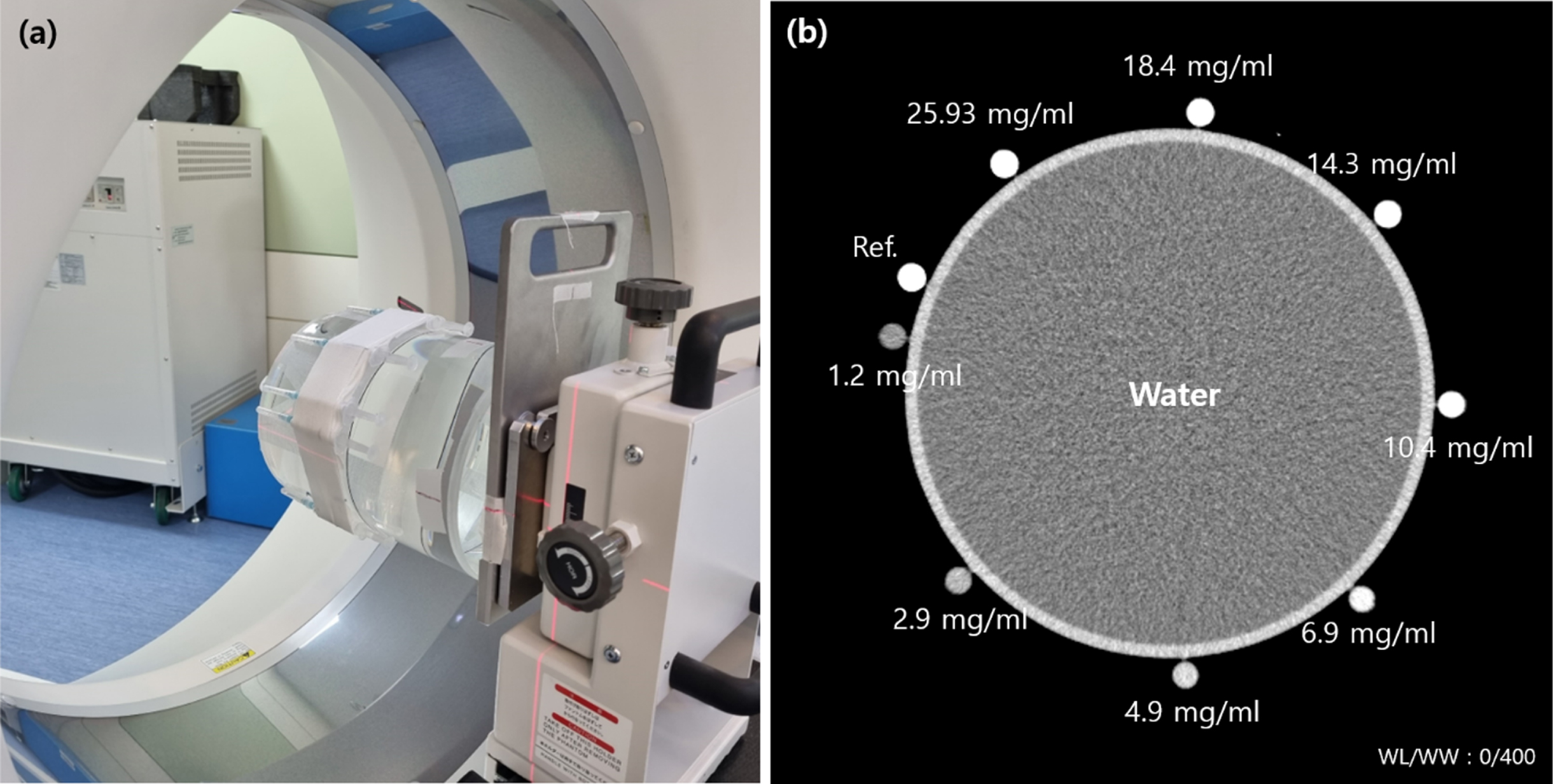
(a) The setup and (b) cross-sectional CT images of a water phantom (diameter: 19 cm) containing nine vials of 1 cm diameter filled with different iodine concentrations (1.2, 2.9, 4.9, 6.9, 10.4, 14.3, 18.4, and 25.9 mg/mL).
Raw data were reconstructed using the FBP with kernel FC13, hybrid IR with medium-sharp kernel FC14 (AIDR 3D), and DLR with body-sharp option (AiCE). AIDR 3D and AiCE have three strengths: Mild, Standard, and Strong. The detailed acquisition parameters and reconstruction settings of the CT scanner are listed in Table 1.
Scan acquisition and reconstruction settings for FBP, AIDR 3D and AiCE
AIDR, Adaptive Iterative Dose Reduction; AiCE, Advanced Intelligent Clear-IQ Engine; FBP, filtered back projection.
We measured image noise, defined as the standard deviation (SD) of the mean CT number within the region of interest (ROI), in the water and iodine phantoms (Fig. 2b and 2c). Noise measurements were obtained manually by drawing a circular ROI of 15.5 mm2 in the center of each iodine phantom using ImageJ (version 1.45, NIH, Bethesda, MD, USA) [23]. The noise of the water phantom was measured in the central ROI, with a phantom diameter of 50% in the water-filled area. CNRs were calculated using the formula CNR = (HUcontrast – HUwater)/SDwater, where HUcontrast represents the mean pixel value in the iodinated contrast-filled vials of a certain diameter, and HUwater and SDwater represent the mean pixel value and standard deviation in the water phantom filled vials of the same diameter and in the same position in the phantom.
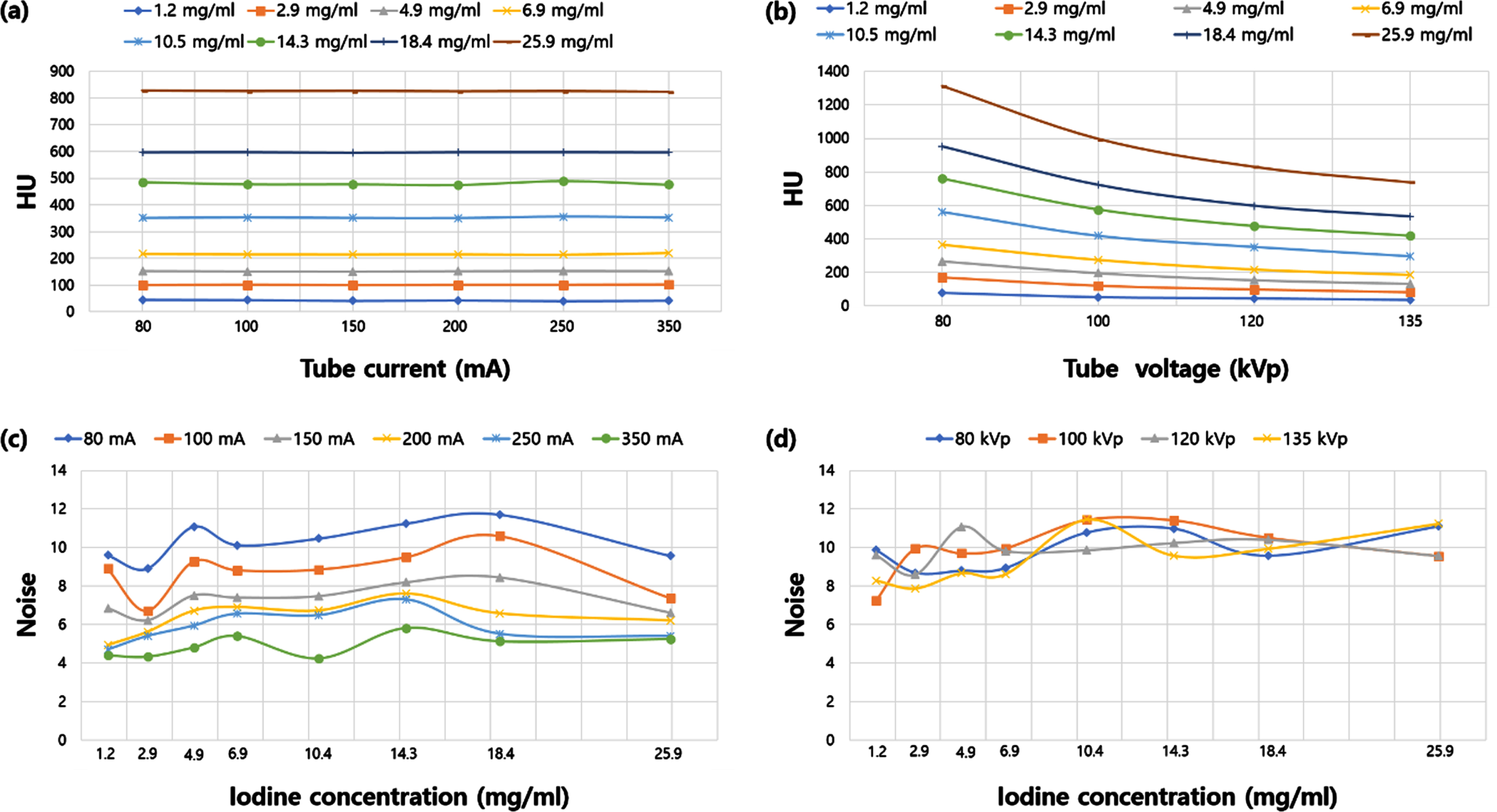
HU values (a and b), noise (c and d), and CNR (e and f) for various tube currents (80, 100, 150, 200, 250, and 350 mA) and tube voltages (80, 100, 120, and 135 kVp) at different iodine concentrations with FBP.
Figure 2 shows the HU values (a and b), noise (c and d), and CNR (e and f) for various tube currents (80, 100, 150, 200, 250, and 350 mA) and voltages (80, 100, 120, and 135 kVp) at different iodine concentrations with FBP. The HU value increased as the iodine concentration increased, but the HU value remained constant as the tube current increased (Fig. 2a). However, as the tube voltage increased, the HU value decreased nonlinearly (Fig. 2b). Noise decreased as the tube current increased, but there was no consistency in the noise changes according to various tube voltages and iodine concentrations (Fig. 2c and d).
Figure 3 shows the measured noise in three straightforward settings (mild: a and d, standard: b and e, and strong: c and f) of the AIDR 3D and AiCE at various iodine concentrations and tube currents (80, 100, 150, 200, 250, and 350 mA). In AIDR 3D and AiCE, the noise decreased as the tube current increased; however, there was no consistency in the change in noise according to the iodine concentration. Figures 4 and 5 show the characteristics of the HU values and noise according to various iodine concentrations and tube currents (80, 100, 150, 200, 250, and 350 mA) using FBP, AIDR 3D, and AiCE. The tube voltage was fixed at 120 kVp without any change. Thus, the higher the iodine concentration, the higher the HU values. The HU values of FBP and AIDR 3D increased almost equally, while AiCE was lower than FBP and AIDR 3D values when iodine concentration was low and higher than FBP and AIDR 3DA values when iodine concentration increased (Fig. 4). Overall, the noise decreased in the order of FBP, AIDR 3D, and AiCE, but there was no noise characteristic of iodine concentration (Fig. 5). In Fig. 6, CNRs according to various iodine concentrations and tube currents (80, 100, 150, 200, 250, and 350 mA) in FBP, AIDR 3D, and AiCE were calculated using Figs. 4 and 5.
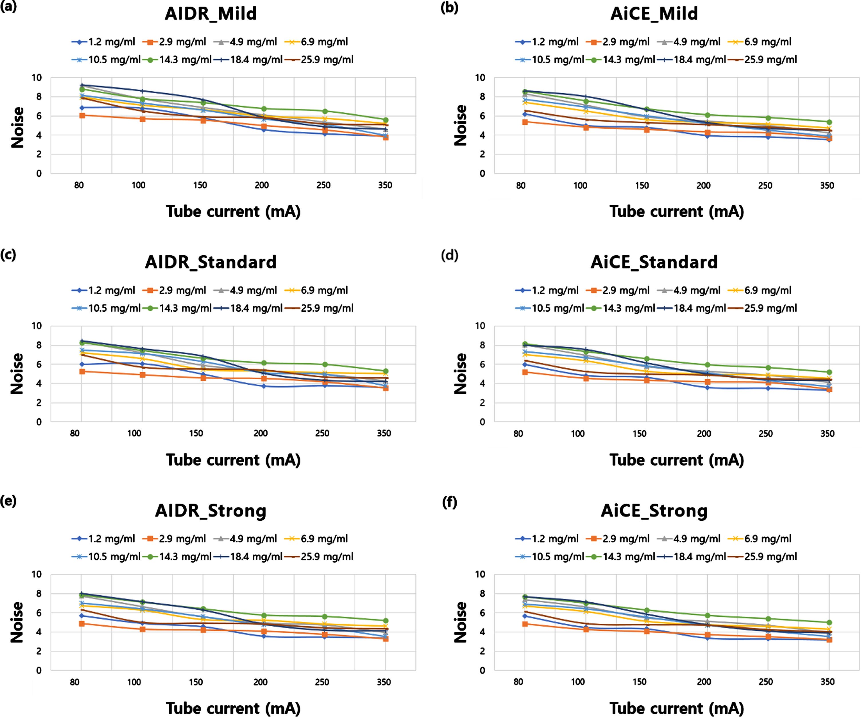
Measured noise in three straightforward level settings of AIDR 3D and AiCE under various iodine concentrations and tube currents (80, 100, 150, 200, 250, and 350 mA). The three straightforward level settings are mild (a and b), standard (c and d), and strong (e and f). The tube voltage is fixed at 120 kVp.
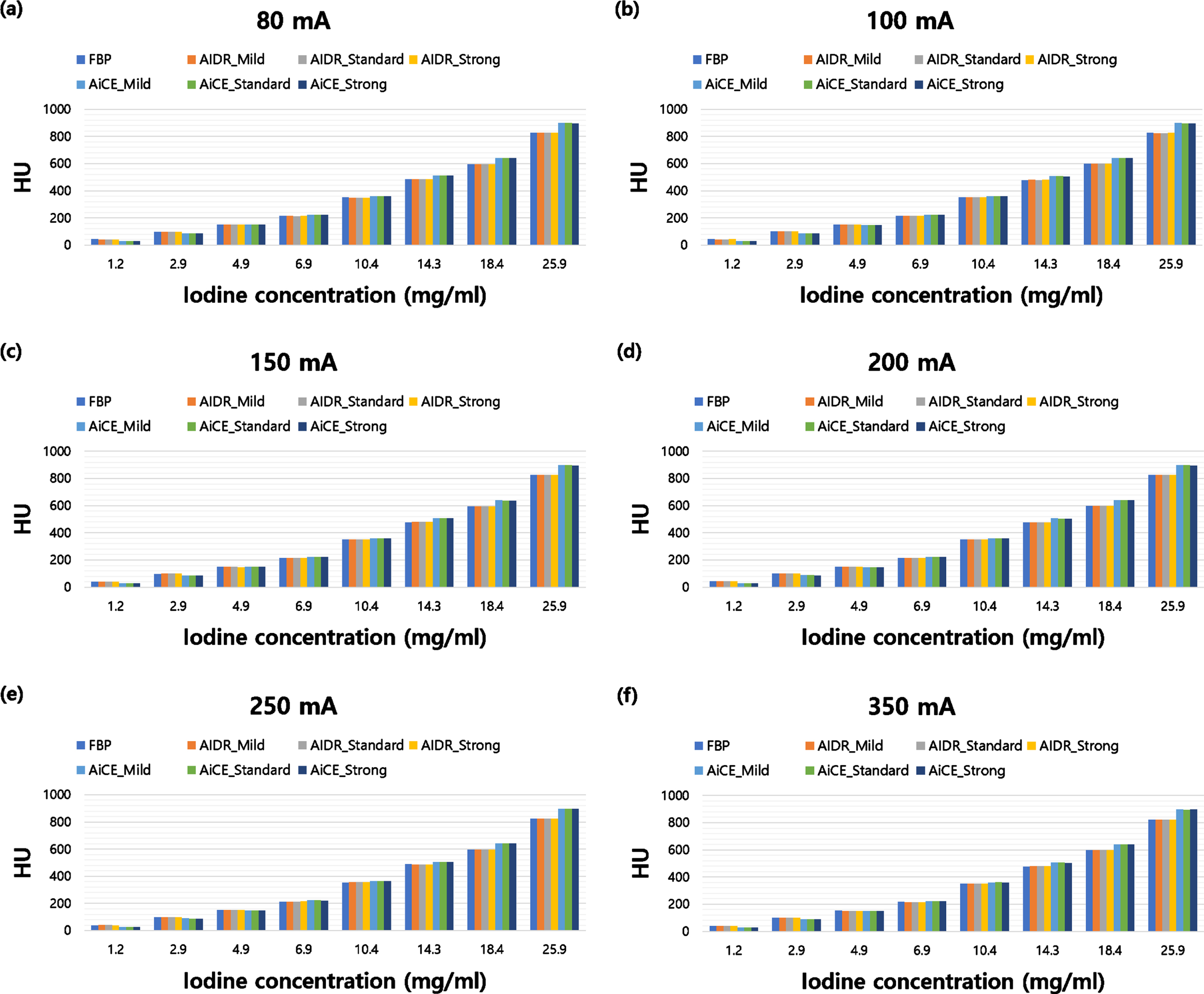
HU values according to various iodine concentrations and tube currents (80, 100, 150, 200, 250, and 350 mA) with FBP, AIDR 3D, and AiCE. The tube voltage is fixed at 120 kVp.
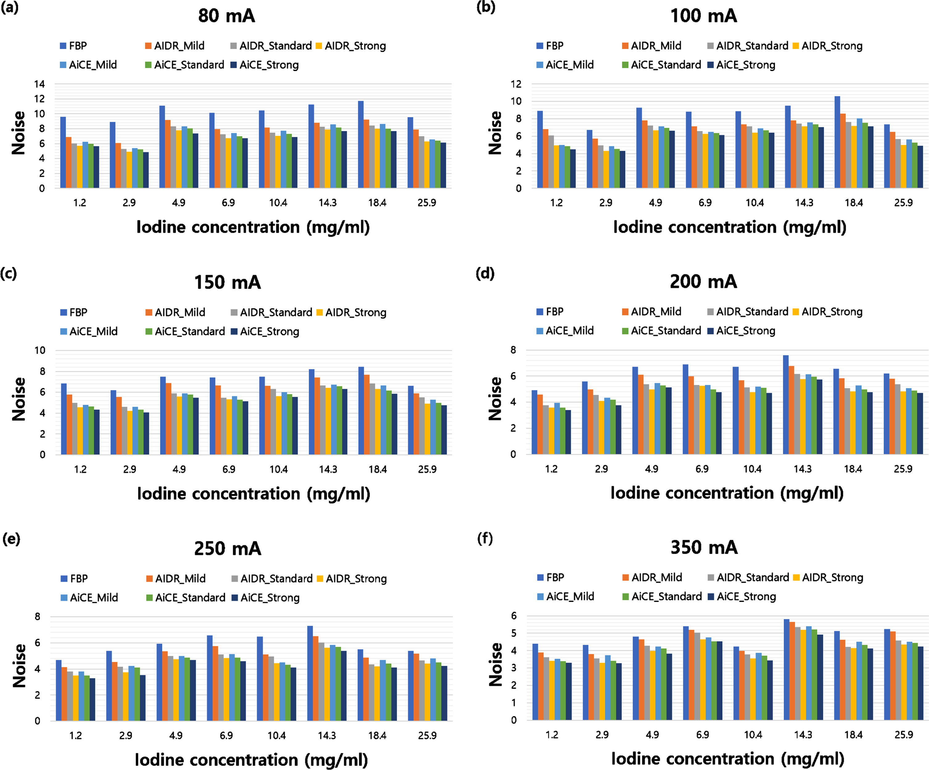
Measured noise according to various iodine concentrations and tube currents (80, 100, 150, 200, 250, and 350 mA) with FBP, AIDR 3D, and AiCE. The tube voltage is fixed at 120 kVp.
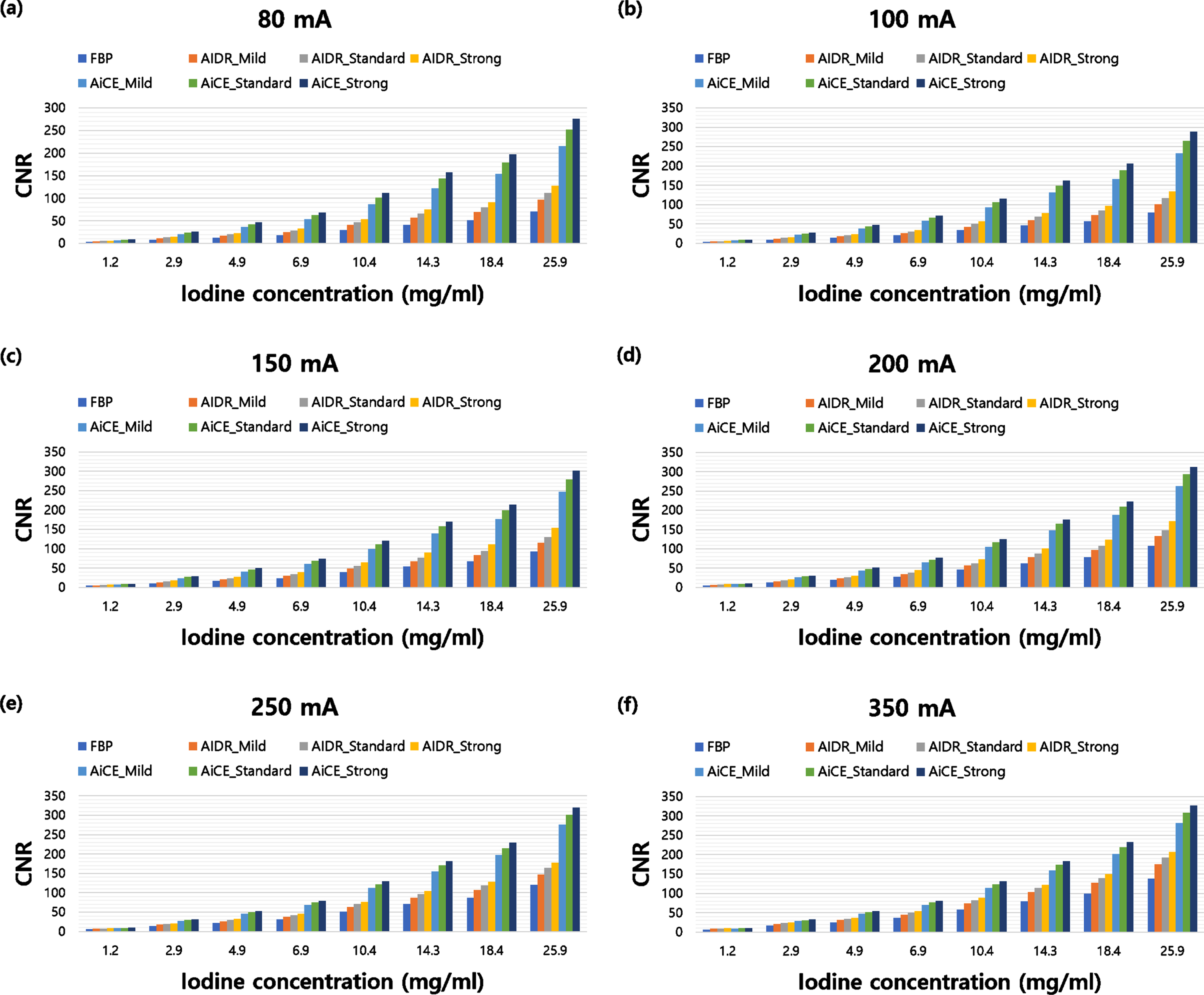
CNR according to various iodine concentrations and tube currents (80, 100, 150, 200, 250, and 350 mA) with FBP, AIDR 3D, and AiCE. CNR was calculated using figures 4 and 5, and the tube voltage is fixed at 120 kVp.
Figures 7 and 8 show the HU values and noise according to various iodine concentrations and tube voltages (80, 100, 120, and 135 kVp) in FBP, AIDR 3D, and AiCE. As the iodine concentration increased, the HU values also increased. At low iodine concentrations, the HU values of FBP and AIDR 3D were higher than those of AiCE; however, as the iodine concentration increased, the AiCE values increased (Fig. 7). Noise decreased in the order of FBP, AIDR 3D, and AiCE, but there was no noise characteristic of iodine concentration (Fig. 8). In Fig. 9, CNRs according to various iodine concentrations and tube voltages (80, 100, 120, and 135 kVp) in FBP, AIDR 3D, and AiCE were calculated using Figs. 7 and 8.
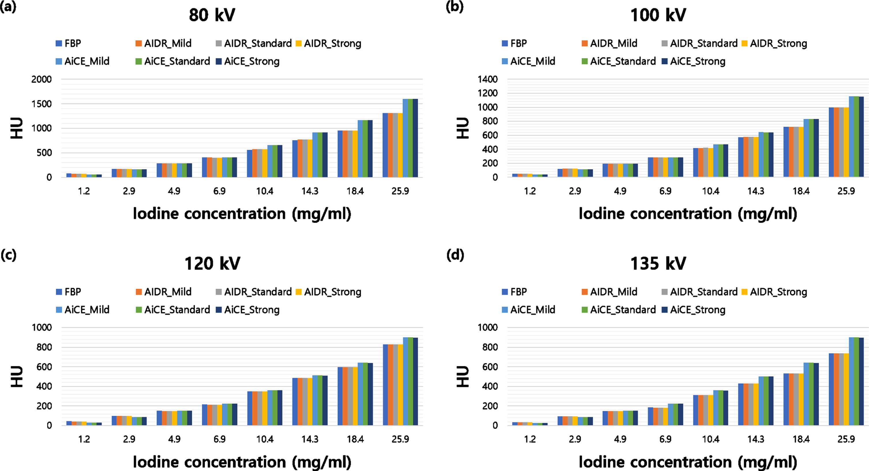
HU values according to various iodine concentrations and tube voltages (80, 100, 120, and 135 kVp) with FBP, AIDR 3D, and AiCE. The dose (CTDIvol: 4.2 mGy) is the same for all.
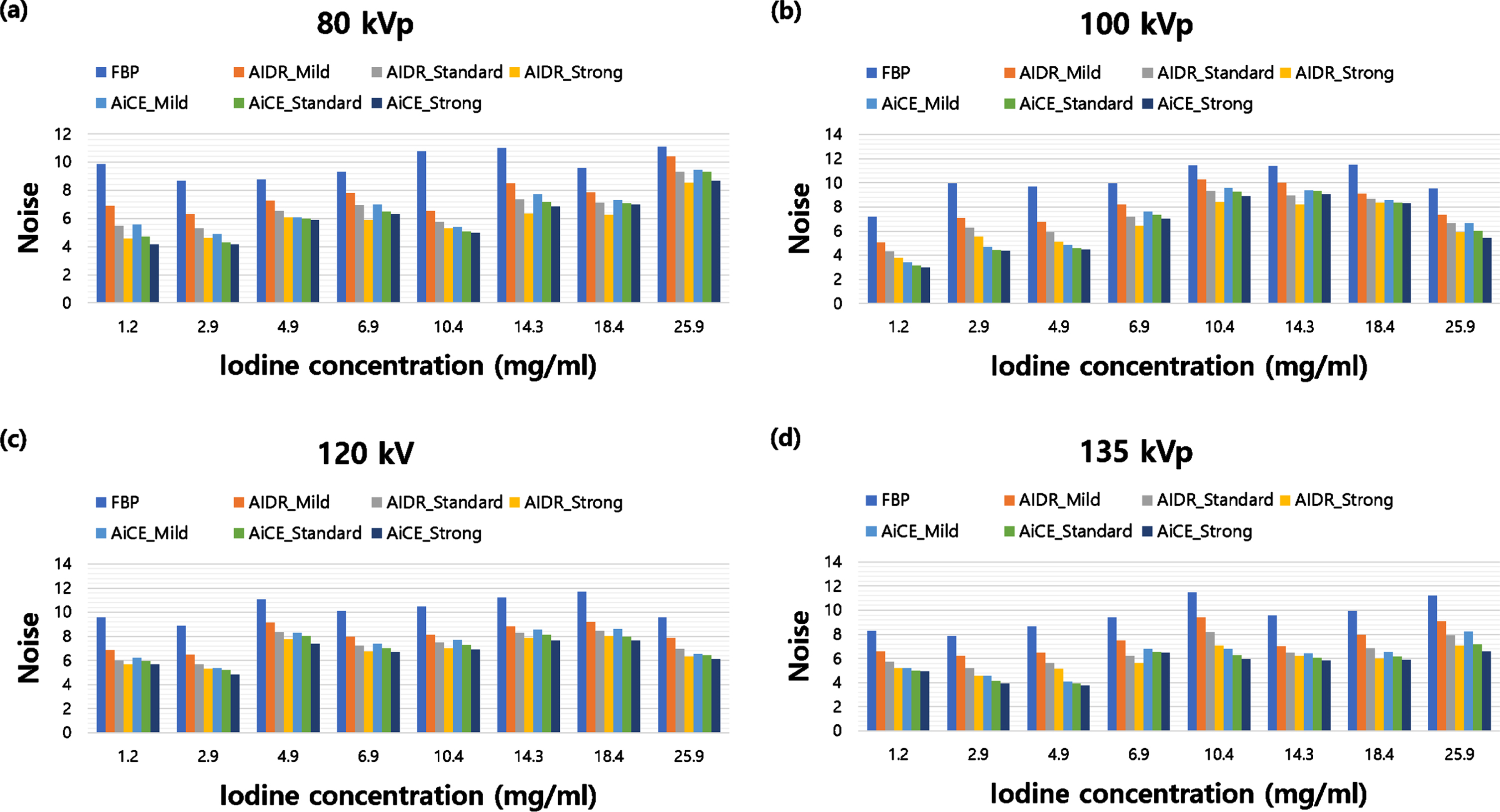
Measured noise according to various iodine concentrations and tube voltages (80, 100, 120, and 135 kVp) with FBP, AIDR 3D, and AiCE.

Figure 10 shows representative phantom images scanned at 2.5 mGy; (a) FBP, (b) AIDR 3D (standard), and (c) AiCE (standard). The image noise is lowest on the AiCE (standard) image, and the object boundary is sharper than that on the other images. (a′), (b′), and (c′) are magnified images of (a), (b), and (c) focusing on some iodine-enhanced areas.
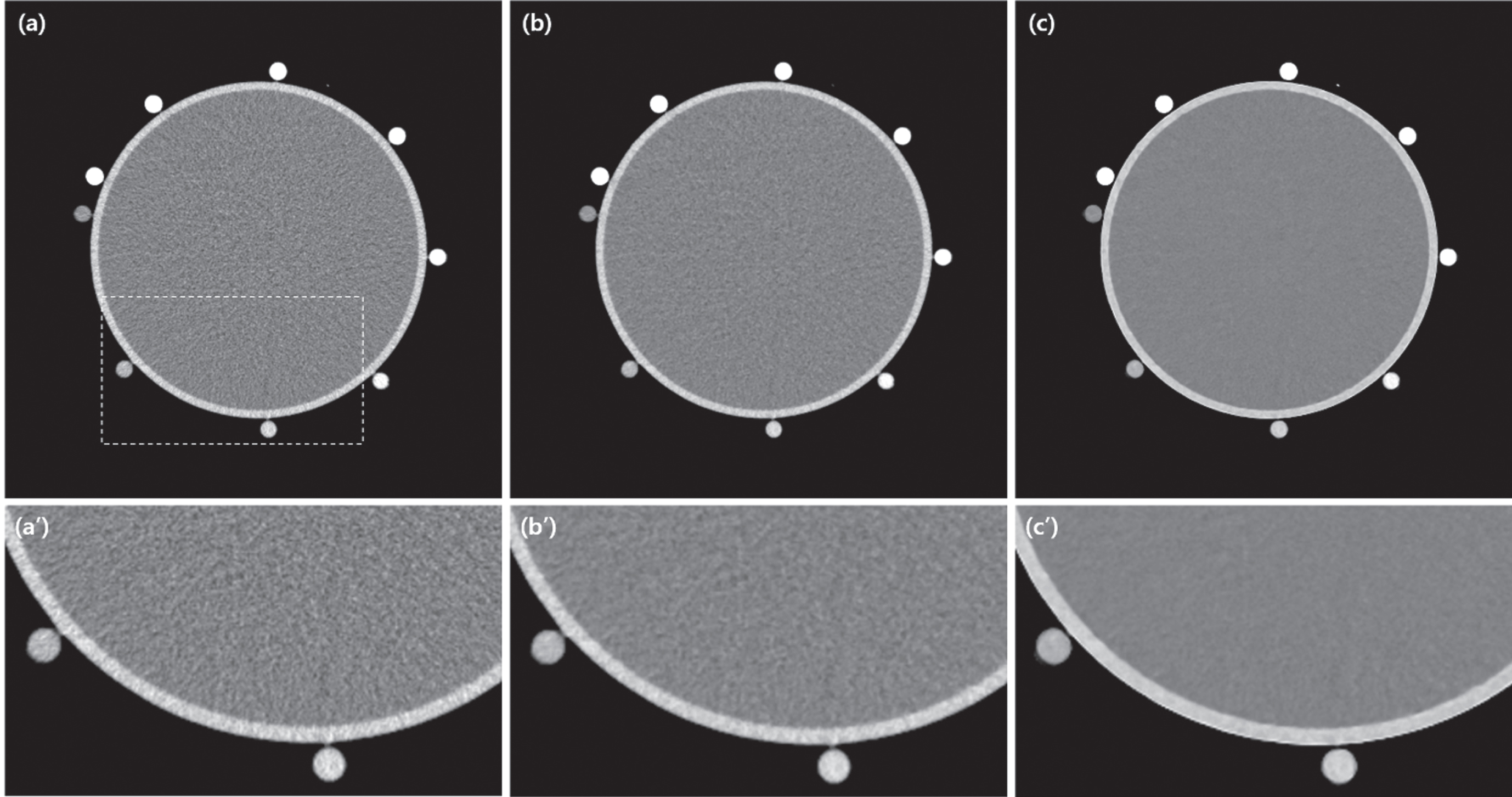
Representative images acquired at CTDIvol: 2.5 mGy; (a) FBP, (b) AIDR 3D (standard), and (c) AiCE (standard). (a'), (b') and (c') are magnified images of (a), (b) and (c) focusing on some iodine-enhanced areas; 120 kVp, 80 mA, slice thickness: 0.5 mm, display window: WL/WW: 0/400.
Figure 11 shows the noise reduction ratio for various tube currents, reconstruction techniques, and levels. Figure 11 (a) shows the noise reduction rate of AIDR 3D and AiCE compared with the noise of FBP, and Fig. 11(b) shows the noise reduction rate of AiCE compared with the noise of AIDR 3D. The noise reduction rate compared to FBP according to the tube current change showed the highest noise reduction rate at 80 mA among AiCE and was mild (22.6%), standard (30.1%), and strong (34.6%). In contrast, the highest noise reduction rates at 350 mA were mild (6.4%), standard (12.9%), and strong (17.6%) (Fig. 11a). The noise reduction rate of AiCE compared to AIDR 3D was as follows: mild was lowest at 350 mA (6.3%) and highest at 150 mA (13.3%). In the standard, 80 mA (3.3%) was the lowest and 100 mA (6.2%) was the highest, and in the strong condition, 100 mA (2.0%) was the lowest and 250 mA (3.7%) was the highest (Fig. 11b).
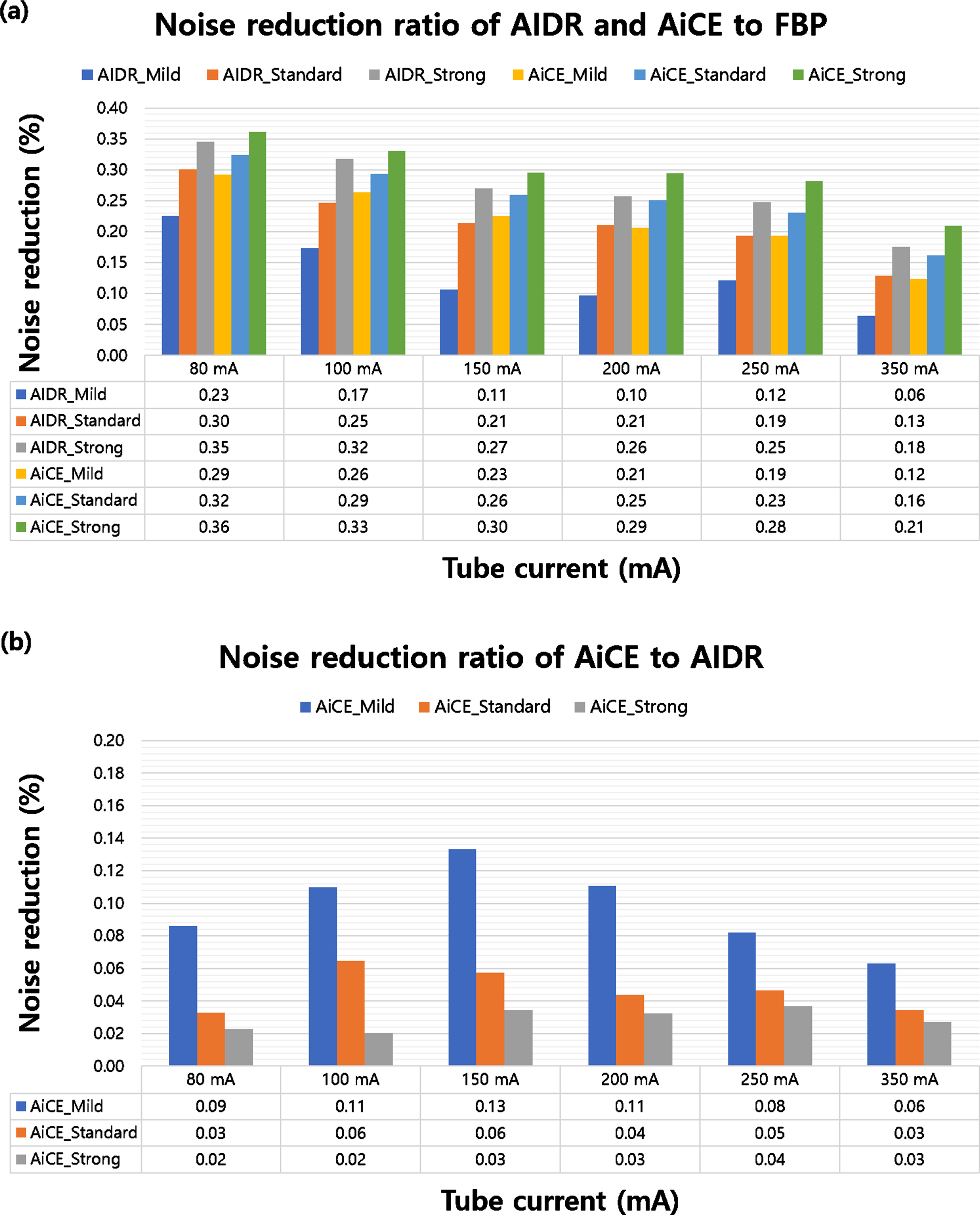
Noise reduction rate for various tube currents and iodine concentrations. (a) The noise reduction ratio of AIDR 3D and AiCE to FBP and (b) noise reduction ratio of AiCE to AIDR 3D.
Understanding the noise characteristics and improving the spatial resolution are required to accurately diagnose CT images. Furthermore, users should consider the characteristics based on different CT scan parameters and contrast medium concentrations to achieve optimal image quality while minimizing the patient dose.
A comparison of the noise reduction rates of hybrid IR (AIDR 3D) and DLR (AiCE) versus FBP showed a better noise reduction rate at a low tube current. Low-contrast detectability, which is particularly important in abdominal CT studies, is highly affected by image noise [24]. Therefore, AiCE may outperform other reconstruction methods in abdominal CT studies in terms of diagnostic performance. In addition, since high-dose radiation exposure must be avoided in pediatric scans, good noise characteristics at reduced radiation doses are clinically important. As noise is reduced and resolution is improved through various reconstruction techniques, the possibility of clinical application of low-concentration iodine contrast agents is being studied [25].
We first analyzed the characteristics of HU values and noise at various tube currents and voltages based on various iodine concentrations in FBP reconstruction technology. The HU value was constant according to various tube currents but decreased according to the tube voltage, and the HU value was the highest at 80 kVp. Lowering the tube voltage reduces the penetration of X-rays and increases attenuation after reaching the organ, allowing lower X-ray energies to be brought closer to the K-edge of iodine (33 keV). Therefore, as shown in Fig. 2, the detection efficiency of the iodine signal can be increased, and HU values of the iodine contrast agent can be increased. In addition, as the tube current increased, the noise decreased (Fig. 2c); however, even if the kVp increases, the noise does not change significantly when the final radiation dose is the same (Fig. 2d). In our study, the iodine concentration in the FBP did not have a significant effect on the noise, but the tube current (i.e., radiation dose) and tube voltage had a significant effect on the CNR (Figs. 6 and 9). Similar to the characteristics of FBP, the noise at the three levels of AIDR 3D and AiCE decreased as the tube current increased; however, it was not related to the iodine concentration (Fig. 3).
In Figs. 4 and 7, the HU values of FBP and AIDR 3D, according to changes in tube current and voltage, were constant even with changes in iodine concentration. However, AiCE showed a higher HU value than FBP and AIDR 3D when the iodine concentration was low; however, the HU value reversed when the iodine concentration exceeded a certain value (reversing iodine concentration: 4.9 and 6.9 mg/mL). The average HU value of the contrast agent according to the body and body parts is generally determined, and the diagnosis is sometimes made by referring to the HU value when diagnosing patients [26].
In Figs. 5 and 8, FBP, AIDR 3D, and AiCE have in common that the noise decreases as the tube current increases, but there is no clear characteristic of noise change at various iodine concentrations. Similarly, when the radiation dose was the same for the tube voltage change, there was no clear change in noise and no effect due to the change in iodine concentration. However, the characteristics of the CNR were different. As the iodine concentration increased, the CNR increased rapidly; as the tube current increased, the average CNR also increased (Fig. 6). As the iodine concentration increased, the CNR increased; however, as the tube voltage increased, the average CNR decreased. The reason for the decrease in the CNR is that the HU value decreased as the tube voltage increased, even at the same dose (Fig. 9). These results may be applied to the CTA. Peripheral blood vessels with small HU values in CTA can obtain better peripheral vascular resolution images when AiCE reconstruction technology is applied than FBP or AIDR. Furthermore, FBP or AIDR are more efficient than AiCE for imaging vessels with high HU values. It was found that the resolution of peripheral blood vessels depended on iodine concentration rather than noise. And since the CNR is proportional to the HU value, the higher the HU value, the more advantageous it is for peripheral vascular diagnosis. Therefore, in imaging diagnosis considering the HU value, the HU characteristics of iodine concentration according to the reconstruction technique should be considered.
We not only analyzed the characteristics of the three reconstruction technologies (FBP, AIDR 3D, and AiCE) but also compared the noise reduction rates with FBP. Consequently, AIDR 3D and AiCE showed better noise characteristics and improved CNR at a low tube current. The noise reduction rate of AIDR 3D and AiCE compared to that of FBP showed characteristics ranging from 6.4% to 34.6%, and the noise reduction rate of AiCE compared to that of AIDR 3D ranged from 2.0% to 13.3%.
There are limitations in evaluating various reconstitution techniques and iodine concentration characteristics with the iodine phantom used in this study. First, since one phantom size was used, it may be limited for small children or large patients. Second, there was no direct comparison between supply sources, and only the tube current and tube voltage results according to iodine application could be used. Therefore, it can be used as a guideline for CTAs at the supplier level used in this study. Third, it was a simple phantom study, not a human study with various attenuation factors, and it was not a qualitative evaluation based on the images.
According to previous studies, there is a close relationship between image quality, the concentration and amount of injected iodine, and the radiation dose in CT imaging [27]. Lowering the tube current and voltage can greatly reduce the patient’s radiation dose [28, 29], but it is necessary to select an appropriate voltage according to the patient’s characteristics (age, sex, body mass index) without lowering the tube voltage as much as possible. However, the main factors affecting CT image quality, patient radiation dose, and image evaluation results are not only the manufacturer’s CT system characteristics and model but also the scan protocol and parameter settings selected by the user [30]. Therefore, analyzing the characteristics of various tube currents, tube voltages, and iodine concentrations according to the reconstruction technology analyzed in this study will be useful in obtaining optimal image quality with minimal radiation dose.
Conclusions
This study quantitatively evaluated and compared the properties of FBP, hybrid IR (AIDR 3D), and DLR (AiCE) at various doses and iodine concentrations. In the AIDR 3D and AiCE, the noise decreased as the tube current increased, and the change in noise according to the iodine concentration was inconsistent. A comparison of the noise reduction rates of hybrid IR (AIDR 3D) and DLR (AiCE) with FBP showed better noise reduction rates at a low tube current. Therefore, the user must carefully scan the patient considering the image quality characteristics of these reconstruction techniques.
Declaration of interests
The authors declare no conflicts of interest.
Footnotes
Acknowledgments
We would like to express our sincere gratitude to Wonju Severance Christian Hospital for their generous support of this experiment.