
Abstract
To isolate specific genomic regions that retain their molecular interactions, allowing direct identification of chromatin-bound molecules, we developed two locus-specific chromatin immunoprecipitation (locus-specific ChIP) technologies, insertional ChIP (iChIP) and engineered DNA-binding molecule-mediated ChIP (enChIP) using the clustered regularly interspaced short palindromic repeats (CRISPR) system or transcription activator-like (TAL) proteins. Essentially, a locus-specific ChIP consists of locus-tagging and affinity purification and can be combined with downstream analyses to identify molecules associated with the target genomic regions. In this review, we discuss the applications of locus-specific ChIP to analyze the genome functions, including transcription and epigenetic regulation.
Keywords
Introduction
Biological activities involving the functions of genomic DNA play vital roles in all aspects of living organisms. These functions include transcription, epigenetic regulation, genomic imprinting, dosage compensation such as X-chromosome inactivation, and others.1–8 To understand the molecular mechanisms underlying the genome functions, it is essential to identify the molecules associated with genomic regions of interest. Recently developed techniques have enabled us to detect molecular interactions on genomic regions. For example, chromosome conformation capture (3C) and its derivatives9,10 have been used to detect interactions between genomic regions. In addition, proteomics of isolated chromatin (PICh) has been used to identify proteins associated with multicopy loci such as telomeres and centromeres. 11
In this review, we discuss two locus-specific chromatin immunoprecipitation (locus-specific ChIP) technologies, insertional ChIP (iChIP) and engineered DNA-binding molecule-mediated ChIP (enChIP), which we developed for locus-specific biochemical analysis of genome functions such as transcription and epigenetic regulation.
iChIP
Scheme of iChIP
Tagging of loci with the insertion of recognition elements of an exogenous DNA-binding protein has been widely used in live imaging. 12 Such locus-tagging was utilized for biochemical purification of specific genomic regions in yeast by Kornberg's group. 13 His group used the Cre-loxP system to excise and circularize a target genomic region for affinity purification. In this regard, although Cremediated excision and circularization can generate fragments of genomic regions with defined ends, such manipulation may disrupt interactions between the target locus and its interacting molecules in the native conformation, making it difficult to use this approach to detect interactions between genomic regions. In addition, it is not clear whether the Cre recombinase is compatible with cross-linking aimed at preserving molecular interactions. Furthermore, the insertion of loxP sites takes time and effort.
Using the locus-tagging strategy, we developed iChIP technology,14–19 which overcomes many of the drawbacks of previous methods. The original scheme of iChIP is as follows (Fig. 1):
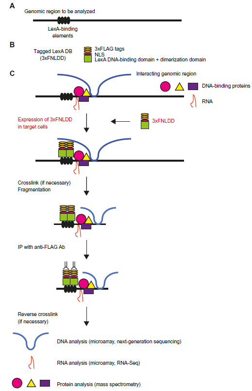
iChIP. (
A repeat of the recognition sequence of an exogenous DNA-binding protein is inserted into the target genomic region of the cell to be analyzed. Examples of these exogenous DNA-binding proteins include LexA, LacI, and Tet repressor.
The DNA-binding domain of the exogenous DNA-binding protein can be fused with a tag(s) and a nuclear localization signal (NLS) and is expressed in the cell to be analyzed.
If necessary, the cells are cross-linked with a compound such as formaldehyde.
Chromatin fraction is prepared, and chromatin DNA is fragmented by sonication or digestion with a endonuclease(s).
Chromatin complexes containing the DNA-binding domain are subjected to affinity purification using an antibody (Ab) or an affinity reagent against the tag(s) or the DNA-binding domain.
Isolated chromatin complexes are reverse cross-linked if a cross-linker was used in step 3.
Proteins contained in the isolated complexes are identified by mass spectrometry (MS), whereas RNAs and DNA are identified by next-generation sequencing (NGS) or microarray analysis.
In our initial attempts, using the first-generation tagged LexA, the yields of the target loci were about 2.5% of input. 14 After optimization of tags and LexA domains, we could achieve yields greater than 10% of input using 3xFNLDD, our second-generation tagged LexA, which consists of a 3xFLAG-tag, an NLS, and the DNA-binding domain and dimerization domain of LexA. 16 The yields of iChIP are important to decide how many cells are necessary for downstream analysis. In our experience, we were able to identify proteins associated with a single-copy locus from 5 × 10 7 cells when the yield (% input) was around 10%. Therefore, we suggest iChIP users to change the cell numbers to be used according to the yield. In this regard, if the yield is significantly lower than 1%, it might be difficult, if not impossible, to harvest sufficient number of cells. In that case, we would suggest choosing other genomic regions for locus-tagging. In this regard, enChIP is much more flexible in testing multiple target genomic regions (see below). In contrast, for the identification of nucleic acids associated with the target locus, smaller number of cells can be used.
in vitro iChIP
In addition to the original iChIP procedure, in which the tagged exogenous DNA-binding protein is expressed in the cells to be analyzed, we developed in vitro iChIP (Fig. 2). 18 In this method, cells, in which a repeat of the recognition sequence of an exogenous DNA-binding protein is inserted into the target genomic region, are subjected to cross-linking, if necessary. Fragmented chromatin from the cells is incubated with a recombinant exogenous DNA-binding protein fused with a tag(s). Subsequently, the chromatin complexes associated with the tagged recombinant exogenous DNA-binding protein are subjected to affinity purification. Using the in vitro iChIP technology, we were able to achieve as much as 30% of the input. 18 Thus, the yields of in vitro iChIP were comparable or even higher than those of the original iChIP method. In addition, in our analysis of the chicken Pax5 gene promoter, we detected nascent Pax5 transcripts in the isolated Pax5 genomic region, 18 suggesting that in vitro iChIP can also isolate target genomic regions while retaining molecular interactions. Because in vitro iChIP does not require the expression of the tagged exogenous DNA-binding protein in cells, the iChIP procedure can be markedly simplified. This is advantageous especially when a specific genomic region is isolated from primary cells obtained from organisms such as mice because transgenic expression or viral transduction of the tagged exogenous DNA-binding protein is not necessary.
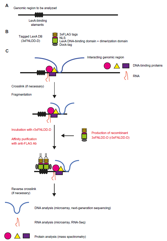
in vitro iChIP. (
Applications of iChIP
iChIP can be performed using cells in which recognition sequences of an exogenous DNA-binding molecule have been knocked in at the endogenous locus as well as using cells harboring transgenes that retain such recognition sequences (Fig. 3). If a transgene contains functional sequences such as gene regulatory elements, random integration of transgenes could be beneficial because the cells might acquire multiple copies of the transgene, allowing more target genomic regions to be obtained by iChIP. On the other hand, tagging the endogenous locus would be physiologically more relevant. It has not been easy to perform knock-ins in cell lines, but the advent of genome engineering technologies makes the knock-in approach much more straightforward.
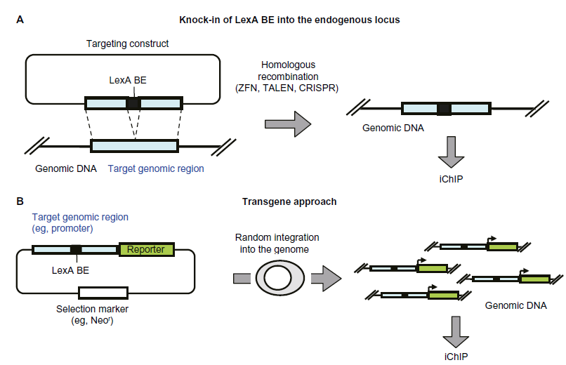
Knock-in and transgene approaches of iChIP. (
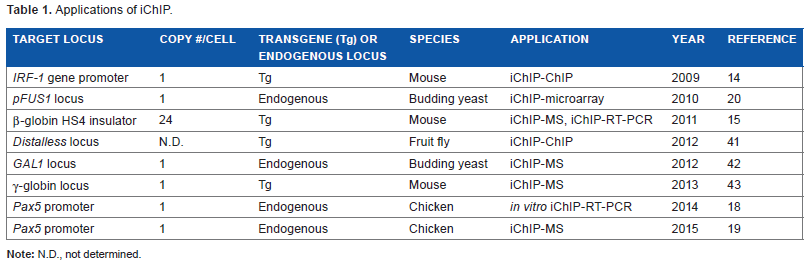
We hold patents on iChIP (“Method for isolating specific genomic regions,” US patent 8,415,098; Japan patent 5,413,924). Since we developed iChIP in 2009, 14 it has been used to identify molecules associated with specific genomic regions (Table 1). For example, iChIP combined with MS (iChIP-MS) enabled us to identify proteins associated with specific genomic regions, 15 including a single-copy locus in cells of multicellular eukaryotes. 19 In our analysis of chicken β-globin HS4 insulator (cHS4), we detected p68 as a known cHS4-interacting protein. 15 In addition, we found that the cHS4 complex binds to a nuclear matrix protein, Matrin-3, mediated by CCCTC-binding factor (CTCF), 15 suggesting that the tethering of cHS4 insulator complexes to the nuclear matrix by Matrin-3 might be important for the function of the cHS4 insulator. In our analysis of chicken Pax5 gene promoter, we identified the Thy28 protein as a binding protein to the Pax5 gene promoter and showed its importance in the expression regulation of the Pax5 gene. 19 In addition, RNAs associated with a given locus have been detected using iChIP combined with reverse transcription-polymerase chain reaction (iChIP-RT-PCR).15,18 In our analysis of cHS4, we detected SRA1 RNA, which has been shown to interact with cHS4, in the cHS4 insulator complex. 15 We are undertaking nonbiased identification of RNAs associated with a single-copy genomic region of interest using iChIP combined with RNA sequencing (iChIP-RNA-Seq). Furthermore, iChIP combined with microarray (iChIP-microarray) has successfully identified genomic regions interacting with pFUS1, a target locus of a transcription factor, Ste12, involved in pheromone-response pathway, in budding yeast. 20 Their analysis suggested that the regulation of gene expression through long-range gene interactions might come at the cost of increased noise. 20 We are currently undertaking iChIP combined with NGS (iChIP-Seq) to identify genomic regions interacting with a given genomic locus. Thus, iChIP is a powerful tool for the identification of multiple types of molecules associated with specific genomic regions as it can be flexibly combined with appropriate downstream analysis methods.
Applications of iChIP.
enChIP
Scheme of enChIP
Although iChIP is a powerful technique, it requires the insertion of recognition sequences of an exogenous DNA-binding protein such as LexA into target loci. The advent of engineered DNA-binding molecules such as zinc-finger proteins, 21 transcription activator-like (TAL) proteins, 22 and the clustered regularly interspaced short palindromic repeat (CRISPR) system23,24 prompted us to utilize such engineered DNA-binding molecules for the isolation of specific genomic regions, leading to the development of enChIP.25–31 The scheme of enChIP is as follows (Fig. 4):
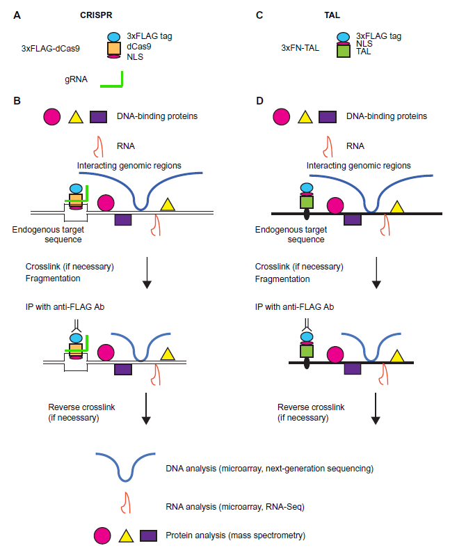
enChIP. A locus to be analyzed is tagged with engineered DNA-binding molecules such as the CRISPR complex, consisting of a catalytically inactive form of Cas9 (dCas9) and gRNA (
An engineered DNA-binding molecule recognizing the target genomic sequence is generated.
The engineered DNA-binding molecule can be fused with a tag(s) and an NLS, and the fusion protein is expressed in the cell to be analyzed.
If necessary, the cells are cross-linked with a compound such as formaldehyde.
The chromatin fraction is prepared, and chromatin DNA is fragmented by sonication or digestion with a endonuclease(s).
Chromatin complexes containing the engineered DNA-binding molecule are subjected to affinity purification using an Ab or an affinity reagent against the tag(s) or the engineered DNA-binding molecule.
Isolated chromatin complexes are reverse cross-linked, if a cross-linker was used in step 3.
Proteins contained in the isolated complexes are identified by MS, whereas RNAs and DNA are identified by NGS or microarray analysis.
Because enChIP does not require the insertion of recognition sequences of an exogenous DNA-binding molecule, which is necessary for iChIP, the time and cost of the analysis can be significantly reduced. This is one of the greatest advantages of enChIP over iChIP.
enChIP using CRISPR
In our first report of the use of an engineered DNA-binding molecule for the purification of specific genomic regions, we utilized the CRISPR system, which consists of a catalytically inactive form of Cas9 (dCas9) and guide RNA (gRNA). 25 dCas9 contains two mutations that abolish endonuclease activities but preserves the DNA-binding activity. 32 Because the recognition of target genomic sequences is solely determined by gRNA, we can easily perform enChIP in a short period of time. The yields of enChIP using CRISPR were comparable with those of iChIP.25,27
enChIP using TAL
enChIP can be performed using other engineered DNA-binding molecules. For example, when we performed enChIP using a tagged TAL protein that recognized the telomeric repeat to isolate telomere regions, we were able to successfully isolate telomere regions. 26 The yields of enChIP using TAL proteins were about 1%, 26 lower than those of enChIP using CRISPR. At this stage, it is not clear whether the difference in yields is intrinsic to the system employed or because of the difference in the target loci. in vitro enChIP using recombinant engineered DNA-binding molecules is also feasible, although yields of this system using TAL proteins were very low, and future technical improvement may be required. 18
Identification of proteins associated with a specific genomic region using enChIP
In our initial attempt, using transient expression of dCas9 and gRNA targeting the promoter region of interferon (IFN)-inducible IFN regulatory factor-1 (IRF-1) gene in 293T cells, we could achieve yields of the target locus as high as 8% of input. 25 Subsequent enChIP analysis using retroviral expression vectors had comparable yields. 33 We then performed enChIP combined with MS (enChIP-MS) to identify proteins associated with this region using 5 × 10 7 cells.25,33 When we performed enChIP combined with stable isotope labeling by amino acids in cell culture (SILAC), a quantitative form of MS (enChIP-SILAC), to identify proteins specifically associated with the IRF-1 promoter in an IFNγ-dependent manner, we detected proteins involved in transcriptional regulation as well as acetyl-transferases, DNA topoisomerases, histone variants, and histone deacetylation/corepressor components. 33 The histone deacetylation/corepressor components are of interest because they have been implicated in IFNγ-induced gene expression.34,35 In our analysis of the mammalian telomere regions, we could successfully detect known telomere-binding proteins by enChIP using a TAL protein. 26 In addition, we detected novel telomere-binding proteins, whose localization in telomeres was confirmed by fluorescent microscopy. 26 Thus, it is feasible to identify physiological interactions between a given genomic region and chromatin proteins with enChIP-MS. In addition, these results show that enChIP can be used to elucidate molecular mechanisms of genome functions such as transcription and epigenetic regulation.
Identification of RNAS associated with a specific genomic region using enChIP
In addition to proteins associated with specific genomic regions, we could detect RNAs associated with specific loci using enChIP combined with RT-PCR (enChIP-RT-PCR). 26 For example, we detected the RNA component of telomerase (Terc) using enChIP-RT-PCR. 26 Furthermore, the combination of enChIP with RNA sequencing (enChIP-RNA-Seq) allowed us to identify RNAs associated with specific genomic regions in a nonbiased manner. 28 The detected RNAs included known telomere-associated RNAs such as Terc, the RNA component of mitochondrial RNA processing endoribonuclease (Rmrp), telomeric repeat-containing RNAs (TERRAs), and small Cajal body-specific RNAs (scaRNAs). 26 We could also detect novel telomere-associated RNAs, whose telomeric localization was confirmed by RNA fluorescent in situ hybridization. 26 Given that accumulating evidence suggests that the RNAs (especially, noncoding RNAs) play important roles in the regulation of genome functions, enChIP-RNA-Seq is a powerful tool for elucidating the RNA-mediated mechanisms underlying genome functions.
Comparison of iChIP and enChIP
As described earlier, both iChIP and enChIP enable the isolation of specific genomic regions that retain molecular interactions, allowing the identification of associated molecules. One of the advantages of enChIP over iChIP is that it does not require knock-in of the recognition sequence of an exogenous DNA-binding molecule, such as LexA, which is necessary for iChIP. This is important because the insertion of such sequences might abrogate normal functions of the targeted genomic regions; if this was the case, it would be necessary to insert the sequences into different genomic regions. In this regard, even if enChIP is used, the binding of an engineered DNA-binding molecule might affect the function of the genomic region. However, in this case, engineered DNA-binding molecules that recognize different DNA sequences could be tested more easily. In this regard, the CRISPR system is most flexible because many different gRNAs can be generated and tested easily and economically. By contrast, generating multiple TAL proteins recognizing different target sequences can be expensive and time consuming.
On the other hand, iChIP has some advantages over enChIP. First, because only one allele can be tagged with an exogenous DNA-binding molecule and its recognition sequences, allele-specific analysis can be performed by iChIP. To perform allele-specific analysis in enChIP, sequence differences such as single nucleotide polymorphisms must exist between alleles of the target genomic region, and therefore, careful evaluation is necessary to confirm allele-specific isolation. Second, the in vitro iChIP system is already available, making the expression of tag molecules unnecessary. This is an important issue, especially when locus-specific ChIP might be performed using primary cells from individual organisms, such as mice, because it avoids the tedious mating procedure required to generate mice for analysis. In addition, the sophistication of genome editing technologies has made it much easier to generate knock-in cells or knock-in animals. Therefore, the need to insert the recognition sequences of an exogenous DNA-binding molecule might not be a fatal flaw of iChIP.
Management of Detection of Nonspecific and off-Target Interactions in Performing Locus-Specific ChIP
One of the major problems of detecting specific interactions of genomic regions with proteins, RNAs, and other genomic regions is nonspecific binding. In addition, it has been shown that dCas9 exhibits significant binding to off-target sites.36–39 Therefore, it should be necessary to cancel out the effects of nonspecific and/or off-target interactions. We suggest putting the following comparison sets in performing locus-specific ChIP:
Comparison of different conditions such as different stimulations (eg, the absence or presence of IFNγ stimulation in reference 33) or different cell types (eg, B cells and macrophages in reference 19).
Comparison of different engineered DNA-binding molecules for a target region in enChIP. Molecules detected commonly using different engineered DNA-binding molecules would represent true positives. In this regard, enChIP using CRISPR would be advantageous over that using zinc finger and TAL proteins because different gRNAs can be easily tested for each genomic region. For enChIP using CRISPR, it would be better to include cells expressing only the tagged dCas9 but not gRNA as a negative control to exclude molecules associated with dCas9.
Conclusions
Locus-specific ChIP technologies, including the complementary approaches iChIP and enChIP, are powerful tools for the isolation of specific genomic regions to identify their interacting molecules. Detailed protocols of iChIP and enChIP are attached as Supplementary Files and found in our published methods papers.27,29,40 Updated versions of these protocols in the future can be downloaded from our homepage (http://www.biken.osaka-u.ac.jp/lab/microimm/fujii/iChIP_protocols/english.html).
Author Contributions
Wrote the first draft of the manuscript: TF, HF. Contributed to the writing of the manuscript: TF, HF. Jointly developed the structure and arguments for the paper: TF, HF. Made critical revisions and approved final version: TF, HF. Both authors reviewed and approved of the final manuscript.
Supplementary Material
Supplementary File 1
Procedure to purify genomic DNA with iChIP/enChIP.
Supplementary File 2
iChIP/enChIP SILAC procedures.
Supplementary File 3
iChIP/enChIP MS procedures.
Supplementary File 4
iChIP/enChIP RNA-Seq procedures.
Supplementary File 5
Preparation of retroviral vector plasmids.
Supplementary File 6
Design of gRNA and construction of gRNA expression vectors.