Editorial
Select search scope: search across all journals or within the current journal


An important event in the pathogenesis of Alzheimer's disease (AD) is the deposition of the amyloid β (Aβ)1–40 and 1–42 peptides in a fibrillar form, with Aβ42 typically having a greater propensity to undergo this conformational change. A major risk factor for late-onset AD is the inheritance of the apolipoprotein E (apoE) 4 allele [3,14,31]. We previously proposed that apoE may function as a “pathological chaperone” in the pathogenesis of AD (i.e. modulate the structure of Aβ, promoting or stabilizing a β-sheet conformation), prior to the discovery of this linkage [7,40,41,42]. Data from apoE knockout / AβPPV717F mice, has shown that the presence of apoE is necessary for cerebral amyloid formation [1,2], consistent with our hypothesis. However, in AβPPV717F mice expressing human apoE3 or E4 early Aβ deposition at 9 months is suppressed, but by 15 months both human apoE expressing mice had significant fibrillar Aβ deposits with the apoE4 expressing mice having a 10 fold greater amyloid burden [8,9]. This and other data has suggested that apoE, in addition to having a facilitating role in fibril formation, may also influence clearance of Aβ peptides. In order to address if apoE affects the clearance of Aβ peptides across the blood-brain barrier (BBB) and whether there are differences in the clearance of Aβ40 versus Aβ42, we performed stereotactic, intra-ventricular micro-injections of Aβ40, Aβ42 or control peptides in wild-type, apoE knock-out (KO) or human apoE3 or apoE4 expressing transgenic mice. We found that consistent with other studies [5], Aβ40 is rapidly cleared from the brain across the BBB; however, Aβ42 is cleared much less effectively. This clearance of exogenous Aβ peptides across the BBB does not appear to be affected by apoE expression. This data suggests that Aβ42 production may favor amyloid deposition due to a reduced clearance across the BBB, compared to Aβ40. In addition, our experiments support a role of apoE as a pathological chaperone, and do not suggest an isotype specific role of apoE in exogenous Aβ peptide clearance from the CSF across the BBB.
Progressive cell loss in specific neuronal populations is a pathological hallmark of neurodegenerative diseases, but its mechanisms remain unresolved. Apoptosis or alternative pathways of neuronal death have been discussed in Alzheimer disease (AD) and other disorders. However, DNA fragmentation in human brain as a sign of neuronal injury is too frequent to account for the continuous loss in these slowly progressive diseases. In autopsy cases of AD, Parkinson's disease (PD), related disorders, and age-matched controls, DNA fragmentation using the TUNEL method and an array of apoptosis-related proteins (ARP), proto-oncogenes, and activated caspase 3, the key enzyme of late-stage apoptosis, were examined. In AD, a considerable number of hippocampal neurons and glial cells showed DNA fragmentation with a 3- to 6-fold increase related to amyloid deposits and neurofibrillary tangles, but only one in 2.600 to 5.650 neurons displayed apoptotic morphology and cytoplasmic immunoreactivity for activated caspase~3, whereas no neurons were labeled in age-matched controls. Caspase~3 immunoreactivity was seen in granules of cells with granulovacuolar degeneration, in around 25% In progressive supranuclear palsy, only single neurons but oligodendrocytes in brainstem, around 25% TUNEL-positive and expressed both ARPs and activated caspase 3. In PD, dementia with Lewy bodies, and multisystem atrophy (MSA), TUNEL-positivity and expression of ARPs or activated caspase~3 were only seen in microglia and oligodendrocytes with cytoplasmic inclusions in MSA, but not in neurons. These data provide evidence for extremely rare apoptotic neuronal death in AD and PSP compatible with the progression of neuronal degeneration in these chronic diseases. Apoptosis mainly involves reactive microglia and oligodendroglia, the latter occasionally involved by deposits of insoluble fibrillary proteins, while alternative mechanisms of neuronal death may occur. Susceptible cell populations in a proapoptotic environment, particularly in AD, show increased vulnerability towards metabolic or other noxious factors, with autophagy as a possible protective mechanism in early stages of programmed cell death. The intracellular cascade leading to cell death still awaits elucidation.
The principal protein component of paired helical filaments (PHFs) in Alzheimer disease is abnormally hyperphosphorylated tau (PHF-tau). The stress activated protein kinases JNK and p38 have been shown to phosphorylate tau at some sites only seen in PHF-tau. If JNK and p38 are involved in the abnormal hyperphosphorylation of tau, they should be activated in neurons undergoing neurofibrillary degeneration. In the present study, we determined the intracellular and regional distribution of the active forms of JNK and p38 kinase in entorhinal, hippocampal, and temporal cortices of brains staged for neurofibrillary changes according to Braak and Braak. Neurons with tangle-like inclusions positive for active forms of JNK and p38 kinase were found to appear first in the Pre-α layer of the entorhinal cortex, and then extend into other brain regions co-incident with the progressive sequence of neurofibrillary changes. The intraneuronal accumulation of active forms of JNK and p38 kinase apeared to precede the deposition of amyloid in the extracellular space. These data indicate that increased activation of the stress related kinases JNK and p38 occurs very early in the disease and might be involved in the intraneuronal protein phosphorylation/dephosphorylation imbalance that leads to neurofibrillary degeneration in Alzheimer disease.
A morphometric study of amyloid-ß-positive plaques in the neocortex of eight non-demented people from 68 to 82 years of age and 17 subjects with late-stage Alzheimer disease (GDS stage 7/FAST stages 7a–f) from 73 to 93 years of age shows a shift from prevalence of fibrillar plaques to prevalence of nonfibrillar plaques. In the aged, non-demented subjects, about 4/mm2 plaques are detectable in the neocortex, and the majority are fibrillar plaques. Specifically, 64% found to be classical fibrillar and Thioflavin-S-positive bright primitive plaques. A lower percentage of pale primitive plaques (35%) relatively small proportion of plaques that are poor in thioflavin S-positive fibrils. The numerical density of plaques in the severe stage of AD increases to about 41/mm2. Severely demented subjects appear to maintain an active process of fibrillar plaque formation. This is reflected in the presence of 3% bright primitive plaques. Severely demented subjects also manifest plaque degradation, reflected in the presence of 22% and 48% percentages of classical fibrillar plaques in non-demented subjects and in the end stage of disease suggest that once activated, the process of fibrillar plaque formation persists at a somewhat stable rate during the whole course of brain amyloidosis.
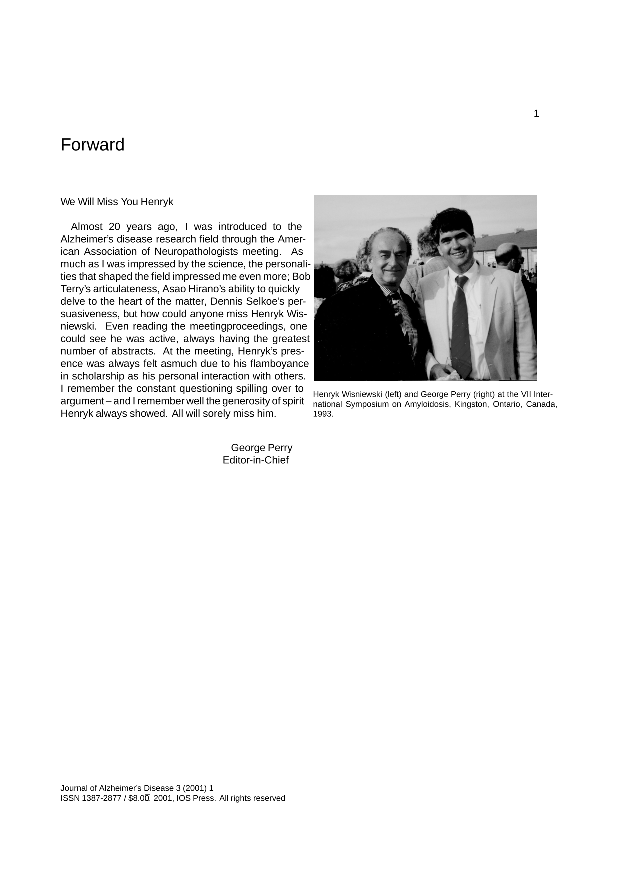
This article is a review of scientific work on Alzheimer neurofibrillary degeneration and Aß-amyloidosis that was done in collaboration with Dr. Henryk Wisniewski, in part at the Institute for Basic Research in Developmental Disabilities. Our work on paired helical filaments and the tau protein spans from basic immunocytochemical analyses of brain tissue to clinical application as a biological marker used in diagnostic tests. Even though only a small part of Dr. Wisniewski's scientific oeuvre, these data illustrate how a great scientist opens the field to his student, collaborator and friend, how basic science can evolve, and how results can be applied in clinical practice to the benefit of our patients.
Cerebral amyloid angiopathy (CAA) is the common term used to define the deposition of amyloid in the walls of medium- and small-size leptomeningeal and cortical arteries, arterioles and, less frequently, capillaries and veins. CAA is an important cause of cerebral hemorrhages although it may also lead to ischemic infarction and dementia. It is a feature commonly associated with normal aging, Alzheimer disease (AD), Down syndrome (DS), and Sporadic Cerebral Amyloid Angiopathy. Familial conditions in which amyloid is chiefly deposited as CAA include hereditary cerebral hemorrhage with amyloidosis of Icelandic type (HCHWA-I), familial CAA related to Aß variants, including hereditary cerebral hemorrhage with amyloidosis of Dutch origin (HCHWA-D), the transthyretin-related meningocerebrovascular amyloidosis of Hungarian and Ohio kindreds, the gelsolin-related spinal and cerebral amyloid angiopathy, familial PrP-CAA, and the recently described chromosome 13 familial dementia in British and Danish kindreds. This review focuses on the various molecules and genetic variants that target the cerebral vessel walls producing clinical features related to stroke and/or dementia, and discusses the potential role of amyloid in the mechanism of neurodegeneration.


Robust activation of the neuronal lysosomal system and cellular pathways converging on the lysosome, such as the endocytic and autophagic pathways, are prominent neuropathological features of Alzheimer's disease. Disturbances of the neuronal endocytic pathway, which are one of the earliest known intracellular changes occurring in Alzheimer's disease and Down syndrome, provide insight into how ß-amyloidogenesis might be promoted in sporadic Alzheimer's disease, the most prevalent and least well understood form of the disease. Primary lysosomal system dysfunction in inherited disorders is commonly associated with prominent neurological phenotypes and neurodegeneration. New studies now directly implicate lysosomal cathepsins as proteases capable of initiating, as well as executing, cell death programs. These and other studies support the view that the progressive alterations of lysosomal system function in Alzheimer's disease have broad relevance to the neurodegenerative processes occurring during the disease.
With the application of molecular genetics, we are now beginning to understand the etiology and the early stages of pathogenesis of the major neurodegenerative diseases, including Alzheimer's disease, Parkinson's disease, Pick's disease and Progressive Supranuclear Palsy. Surprisingly, these studies are showing that these diseases share pathogenic mechanisms which involve tau or synuclein aggregation. In this article, I review the progress in the molecular genetic analysis of these major neurodegenerative diseases and discuss how they are related to each other.

Synaptic damage is an early pathological event common to many neurodegenerative disorders such as Alzheimer's disease (AD) and is the best correlate to the cognitive impairment. Several molecules involved in AD and in other neurodegenerative disorders play an important role in synaptic function and when misfolded aggregate and form amyloid fibrils. Synaptic proteins with an amyloid domain include amyloid ß-protein precursor, prion protein, huntingtin, ataxin-1 and α-synuclein. Two of the possible mechanisms by which alterations in synaptic proteins lead to synapse damage are: 1) misfolded or aggregated synaptic molecules have lost their normal function and/or 2) they have gained a toxic capacity. Recent studies support the possibility that while oligomers are toxic, polymers might be inactive. The mechanisms by which oligomers trigger synapse loss could be related to their ability to triggers stress signals once they enter the nucleus and/or accumulate at the endoplasmic reticulum.