Announcement
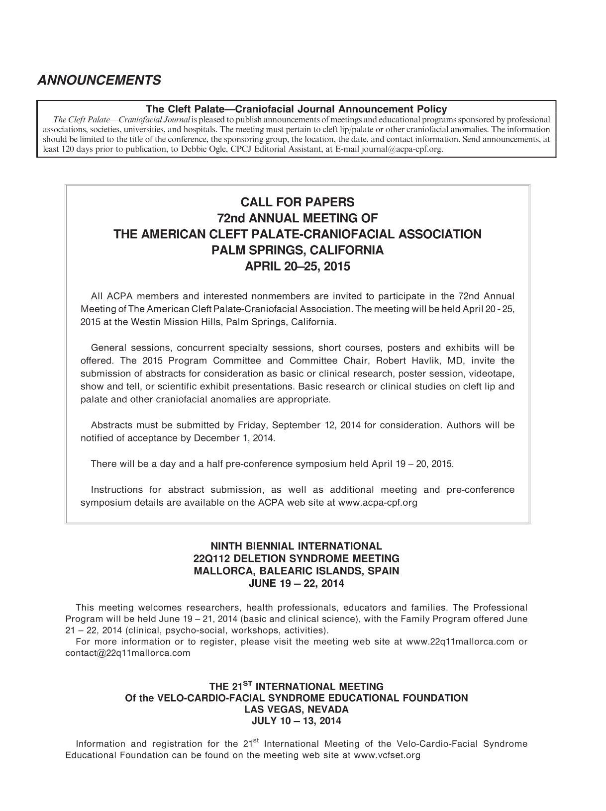
 Restricted accessAnnouncementFirst published May, 2014pp. 1-2

Select search scope: search across all journals or within the current journal


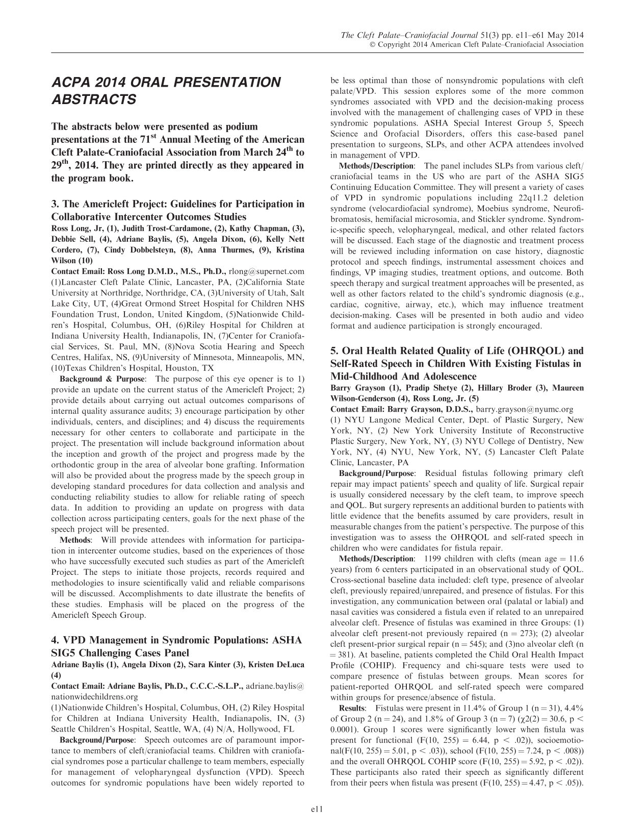
 Restricted accessAnnouncementFirst published May, 2014pp. 1-2Restricted accessAbstractFirst published May, 2014pp. 11-61
 Restricted accessEditorialFirst published May, 2014pp. 253-256
To celebrate the 50th year of the
Restricted accessOtherFirst published May, 2014pp. 257-263(1) To investigate the effect of probiotic bacterium
Double-blinded, randomized, placebo-controlled study.
A total of 30 orthodontically treated nonsyndromic cleft lip and palate patients (aged 19.22 ± 3.66 years): 15 in the intervention group (A) and 15 in the control group (B).
Milk with or without
Salivary mutans streptococci and lactobacilli were enumerated using a quantitative differential culture at baseline and once a week after the end of the administration period for 4 weeks. The persistence of
A statistically significant reduction in mutans streptococci counts occurred in group A, in contrast to group B (
The probiotic milk powder containing
Restricted accessOtherFirst published May, 2014pp. 264-269To investigate the reproducibility and repeatability of digital models of patients with a unilateral cleft lip and palate (UCLP) using the GOSLON yardstick.
Reproducibility and repeatability study.
Two examiners used the GOSLON yardstick to assess the intermaxillary dentoalveolar relationship of 30 consecutive UCLP patients by analyzing their 9-year (±3 months) dental study casts and digital study models. The records were rated 1 week apart to avoid bias. The process was repeated 1 month later as a measure of reproducibility. Reliability was assessed by comparing the GOSLON score achieved between the two modalities. Patient dental study casts were sent to 3M Unitek Australia to be scanned using the 3M Unitek Lava™ system to produce digital study models. The accuracy of the dental study cast occlusal registration was assessed by both raters prior to sending the study models for scanning.
The Linear Weighted Kappa statistic and Kendall's Coefficient of Concordance statistic were used to determine the levels of agreement within and between raters.
The linear weighted Kappa statistic for intrarater repeatability of digital study models scores were very high (0.89 and 0.97). This compared favorably to the intrarater repeatability of study model casts scores (0.86 and 0.97). There was very good agreement for interrater digital study model scores (0.80 and 0.87) and also for the interrater study model casts scores (0.80 and 0.90). Kendall's Coefficient of Concordance statistic (0.99) and Correlation Coefficient (0.86) support the weighted Kappa results of the digital study model scores.
Digital models can be used for GOSLON scoring with a high degree of reproducibility and repeatability.
Restricted accessOtherFirst published May, 2014pp. 270-273To establish for the first time the prevalence of fistula symptoms and the effectiveness of secondary alveolar bone grafting to treat these symptoms in a single surgeon cohort in Bristol, United Kingdom.
Direct questioning of 233 consecutive patients with cleft before and after secondary alveolar bone grafting as to the presence of fistula symptoms.
Southwest and South Wales Cleft Centre, Frenchay Hospital, Bristol, United Kingdom.
Consecutive patients with cleft who were being treated for secondary alveolar bone grafting.
Patients reporting presence of fistula symptoms. Data collection on cleft type (unilateral, bilateral), date of birth, and age at secondary alveolar bone grafting.
Of the 167 unilateral patients with cleft lip and palate and 66 patients with bilateral cleft lip and palate, 45% had symptoms of a fistula before alveolar bone grafting and 10% had symptoms of a fistula after surgery. There were no statistically significant differences between the presence of symptoms before or after secondary alveolar bone grafting between cleft types or by age at secondary alveolar bone grafting.
This is the first study examining the rates of fistula symptoms before and after secondary alveolar bone grafting in the United Kingdom. This study used the patient-centered outcome of the presence of symptoms as a way of measuring the presence of fistulas in this group. Secondary alveolar bone grafting reduces the incidence of symptomatic fistula in this setting.
Restricted accessOtherFirst published May, 2014pp. 274-282To describe and compare speech and phonology at age 3 years in children born with unilateral complete cleft lip and palate treated with three different methods for primary palatal surgery.
Prospective study.
Primary care university hospitals.
Twenty-eight Swedish-speaking children born with nonsyndromic unilateral complete cleft lip and palate.
Three methods for primary palatal surgery: two-stage closure with soft palate closure between 3.4 and 6.4 months and hard palate closure at mean age 12.3 months (n = 9) or 36.2 months (n = 9) or one-stage closure at mean age 13.6 months (n = 10).
Based on independent judgments performed by two speech-language pathologists from standardized video recordings: percent correct consonants adjusted for age, percent active cleft speech characteristics, total number of phonological processes, number of different phonological processes, hypernasality, and audible nasal air leakage. The hard palate was unrepaired in nine of the children treated with two-stage closure.
The group treated with one-stage closure showed significantly better results than the group with an unoperated hard palate regarding percent active cleft speech characteristics and total number of phonological processes.
Early primary palatal surgery in one or two stages did not result in any significant differences in speech production at age 3 years. However, children with an unoperated hard palate had significantly poorer speech and phonology than peers who had been treated with one-stage palatal closure at about 13 months of age.
Restricted accessOtherFirst published May, 2014pp. 283-289To report survey results from American Cleft Palate–Craniofacial Association members on the practice patterns of airway obstruction management in patients with Pierre Robin sequence.
A 10-question online survey was sent and the data were reviewed.
Online survey of members of the American Cleft Palate–Craniofacial Association.
Surveys assessed management patterns of patients with Pierre Robin sequence whom a surgeon member of the American Cleft Palate–Craniofacial Association treated for airway obstruction.
The survey comprised data on management strategies for airway obstruction in Pierre Robin sequence, including tracheostomy, tongue-lip adhesion, mandibular distraction, and treatments that falls in the “other” category.
A total of 87 American Cleft Palate–Craniofacial Association members completed the survey. Respondents' results were analyzed as a whole and by individual subspecialty: plastic surgery (n = 33), oromaxillofacial surgery (n = 21), and otolaryngology (n = 29). Although most of the surgeons were trained to manage airway obstruction in Pierre Robin sequence patients using tracheostomy (47%, n = 39) and tongue-lip adhesion (31%, n = 26), 48% reported a current preference for mandibular distraction (n = 40). Of surgeons who preferred to manage Pierre Robin sequence with tongue-lip adhesion (n = 23), 65% were trained to do so (n = 15). Surgeons preferring mandibular distraction (n = 40) and tracheostomy (n = 14) more often reported they were trained to manage Pierre Robin sequence with tracheostomy.
Currently there are various practice patterns for the management of airway obstruction in Pierre Robin sequence. Training habits and subspecialty category may influence a surgeon's preference in patients who fail conservative therapy. Treatment guidelines are lacking and may require significant collaboration among centers and subspecialties to develop a more standardized approach to a challenging clinical entity.
Restricted accessOtherFirst published May, 2014pp. 290-299To evaluate self-esteem, coping styles, and health-related quality of life and their relationships in Polish adolescents and young adults with unilateral complete cleft lip and palate and related sex differences.
Self-report questionnaires measuring self-esteem (Multidimensional Self-Esteem Inventory), coping styles (Coping Inventory for Stressful Situations), and health-related quality of life (WHOQOL-BREF) were completed by 48 participants with cleft lip and palate (age, 16 to 23 years; 31 males, 17 females) and 48 controls without cleft lip and palate (age, 16 to 23 years; 28 males, 20 females) matched for age, place of residence, and socioeconomic status.
Regarding self-esteem, individuals with cleft lip and palate scored higher on body functioning (
Late adolescents and young adults with and without cleft lip and palate differed little in terms of psychological adjustment measures. The higher scores in defensive self-enhancement of individuals with cleft lip and palate suggest the need for instruments measuring social approval in psychosocial adjustment research involving this group.
Restricted accessOtherFirst published May, 2014pp. 300-307To learn about (1) mothers' perspectives on their children's social experiences and (2) how mothers interpret social situations and provide guidance to their children in challenging situations.
This was a qualitative study analyzing narratives. Mothers participated in a semistructured interview; mothers and their children participated in a social coaching task involving responses to hypothetical situations. Transcripts of audio recordings were used to create thematic coding categories, and transcripts were reviewed and coded.
Children were patients at a reconstructive plastic surgery center in an urban hospital and medical school; families were recruited from a regional support group associated with the hospital.
Mothers of nine children with congenital craniofacial anomalies, aged 9 to 14 years.
Thematic narrative coding categories, focusing on mothers' perspectives on children's actual social experiences (from the interview) and mothers' advice and interpretations regarding challenging hypothetical social tasks (from the coaching task).
In the interviews, mothers reported positive and negative social experiences for their children. Multiple approaches were used by mothers to interpret social interactions experienced by children (interview) and hypothetical social situations (coaching task). These included consideration of situational factors, motivations of others, and factors within own child. Mothers' hypothetical advice was often prosocial, including concrete strategies to resolve conflict, to plan ways to avoid problems, to foster self-reliance, and to avoid hurtful situations.
Mothers showed active interest and concern in their children's peer relationships and were thoughtful in devising strategies to successfully manage potential social challenges.
Restricted accessOtherFirst published May, 2014pp. 308-313The purpose of this cross-sectional study was to assess the prevalence of dental caries in children with cleft lip, alveolus, and/or cleft palate living in central Germany between 1996 and 2010.
A total of 295 children 1 to 6 years of age from three birth cohorts (BC) with clefts from central Germany were included in the study. They were compared with 548 1- to 6-year-old cleft-free children from the same region.
Children with clefts underwent a dental examination in an outpatient dental clinic at the University of Leipzig. The first BC was examined between 1996 and 1998, the second between 2002 and 2004, and the third between 2008 and 2010. Controls were examined at day-care centers in Leipzig during the same periods.
The standard dental caries index for the primary dentition (dmf/t3-4) was used for clinical assessment.
Over the entire study period, the mean prevalence of dental caries in deciduous teeth was significantly higher (1.32 dmf/t3-4) in children with clefts compared with cleft-free children. However, a decline in caries (approximately 1 dmf/t3-4) and an increase in the proportion of children with healthy primary dentition were observed in both groups. These results represent a caries decline of 61% in children with clefts.
Caries rates for children 1 to 6 years of age with clefts from central Germany showed a considerable decline over the last years. The caries rates for clefts patients in the third BC (2008 to 2010) was similar to that of cleft-free children in the first BC (1996 to 1998).
Restricted accessOtherFirst published May, 2014pp. 314-319Experimental and clinical findings suggest that the vomer is involved in facial development and might contribute to the short and retrusive maxilla in cleft patients. The aim of this study was to investigate the relationship between vomer development and maxillary growth in unoperated submucous cleft palate (SMCP) patients.
Retrospective cohort study.
Thirty unoperated SMCP patients were included. The criteria for clinical diagnosis were: bifid uvula, a translucent zone in the midline of the soft palate, and a touchable “V” notch on the posterior border of the bony palate. There were 19 female and 11 male patients, with an age range from 3 to 25 years.
All patients were imaged using spiral computed tomography (CT) while in centric occlusion before the surgeries.
Three-dimensional (3D) reconstruction models were created, and dentoalveolar relationships were rated by three experienced doctors according to the GOSLON score principles. The patients then were divided into three groups: 1 - normal occlusion, 2 - edge-to-edge bite, and 3 - crossbite. The vomer-palate fusion rate was calculated on 3D CT images and represented the vomer development.
The sagittal extent of the palatal cleft and the malformation of vomer in SMCP were greatly varied. The vomer-palate fusion rate in the crossbite group (occlusal score = 3) was significantly lower than that in the normal occlusion group (
Our findings suggest that correlation exists between vomer development and sagittal maxillary growth in unoperated SMCP patients.
Restricted accessOtherFirst published May, 2014pp. 320-325Orofacial clefts are the most common malformations of the head and neck. In Africa, orofacial clefts are underascertained, with little or no surveillance system in most parts for clefts and other birth defects. A Nigerian craniofacial anomalies study, NigeriaCRAN, was established in 2006 to support cleft research specifically for epidemiological studies, treatment outcomes, and studies into etiology and prevention.
We pooled data from seven of the largest Smile Train treatment centers in the six geopolitical zones in Nigeria. Data from September 2006 to June 2011 were analyzed and clefts compared between sides and genders using the Fisher exact test.
A total of 2197 cases were identified during the study period, with an estimated prevalence rate of 0.5 per 1000. Of the total number of orofacial clefts, 54.4% occur in males and 45.6% in females. There was a significant difference (
The significant difference between unilateral clefts and the gender differences in the proportion of cleft palate only are consistent with the literature. The present study emphasizes the need for birth defects registries in developing countries in order to estimate the exact prevalence of birth defects including orofacial clefts.
Restricted accessOtherFirst published May, 2014pp. 326-333The purpose of this study was to evaluate the effect of different mechanical forces on the expansion of the palatine suture using transsutural distraction osteogenesis.
A total of 48 dogs were used in this study. The experimental groups were treated with a custom-designed internal distractor. Bone regeneration was determined with x-rays and histology. The computed values underwent statistical analyses using analysis of variance.
The maxillary complex was most noticeably advanced with an applied mechanical force of 600 g (20.15 ± 1.36 mm), compared with forces of 400 g (19.88 ± 1.41 mm) and 800 g (2.24 ± 0.93 mm). Immunohistochemical staining showed that the expression of bone morphogenetic protein-2 and bone morphogenetic protein-4 fluctuated with different mechanical forces. These changes were statistically significant when 600 g of force was applied within 30 days of distraction (
Transsutural distraction osteogenesis in the growing dog should be safe and well tolerated in inducing bony lengthening of the maxilla, and the optimal force is 600 × g. Bone morphogenetic protein-2 and bone morphogenetic protein-4 may play an important roles in the signaling pathways that link mechanical forces and biological responses.
Restricted accessOtherFirst published May, 2014pp. 334-343Patients with cleft lip and palate (CLP) and maxillary retrognathia are usually treated with maxillary advancement (Le Fort I osteotomy). The aim of this study was to investigate the impact of maxillary advancement on the velopharyngeal function (VPF) and symptoms of velopharyngeal incompetence in patients with CLP.
Retrospective group study before and after treatment.
All patients who had undergone Le Fort I osteotomy due to maxillary retrognathia from 2007 to 2010 at Karolinska University Hospital, Sweden (n = 13). Pre- and postoperatively standardized examinations were used. Blinded audio recordings were independently assessed by three experienced speech pathologists. Acoustical data (nasometry) and information on VPF (videoradiography and nasoendoscopy) were collected from the medical records. Two patients with additional malformations were considered outliers, and group data were based on a sample size of n = 11.
Perceptual and acoustic symptoms of velopharyngeal incompetence and overall assessment of VPF.
No assessment method showed a significant deterioration of the VPF postoperatively. Individual data revealed that 6 of the 13 patients had no or only one symptom pre- and/or postoperatively. The two patients with additional malformations had most symptoms pre- and postoperatively and did not change. Three patients had an increased number of symptoms postoperatively by at least two symptoms. No associations between the outcome and possible prognostic factors were found.
Maxillary advancement did not have a significant impact on the VPF at the group level, but three individuals had a somewhat deteriorated VPF postoperatively.
Restricted accessCase reportFirst published May, 2014pp. 344-349This article describes a new method that enables vector control during alveolar distraction osteogenesis in the treatment of a cleft palate patient. The patient presented with unilateral complete cleft lip and palate, and the alveolar part of the defect was covered by a mobile buccal flap. The distraction was performed by sliding the surgically released tooth segment with the help of an intraoral distractor over 1.5-mm stainless steel archwires held by metal crowns. This vector-controlled method enabled new bone and attached gingiva formation in harmony with the proper alveolar shape.
Restricted accessCase reportFirst published May, 2014pp. 350-353Epignathus teratomas are rare tumors that originate in the region of the palate or pharynx and are known as Rathke pouch. They may be associated with other malformations such as a cleft palate and bifid tongue or nose. The prenatal diagnosis can be established by two-dimensional ultrasonography showing a heterogeneous mass protruding through the mouth of the fetus. The three-dimensional ultrasonography in rendering mode permits better understanding of this anomaly by the parents, facilitating the counseling. The importance of prenatal diagnosis lies in the fact that this tumor may obstruct the airways, thus leading to death at birth due to respiratory insufficiency. We report a case of prenatal diagnosis of epignathus teratoma in the 30th week of pregnancy by two-dimensional ultrasonography. We focus on the importance of three-dimensional ultrasonography in rendering mode for demonstrating the spatial relationships of the tumor with the oral cavity and provide correlations between the ultrasound images and the anatomopathological findings.
Restricted accessCase reportFirst published May, 2014pp. 354-360The purpose of this case report is to report the orthodontic surgical treatment and subsequent dental rehabilitation in two patients with Axenfeld-Rieger syndrome. Axenfeld-Rieger syndrome is a rare autosomal dominant condition characterized by ocular, dental, craniofacial, and periumbilical abnormalities. The treatment of two patients with various anomalies in Axenfeld-Rieger syndrome is described and discussed. Early recognition of the syndrome and referral during childhood to a specialized oral and maxillofacial surgery, special dental care, and orthodontic unit provide the optimal starting point for this complex treatment.
Restricted accessOtherFirst published May, 2014pp. 361-364Pediatric patients with skull defects larger than available sources for splitting bicortical bone have limited options for autogenous cortical bone cranioplasty. Piezoelectric instruments allow donor bone to be chosen based on the best possible contour rather than the presence of bicortical bone. We present the use of piezoelectric technology to split thin unicortical calvarium for autogenous cranioplasty in a series of pediatric patients.
Retrospective review of a series of pediatric patients requiring reconstruction for skull defects.
Our series included a 2-year-old with a parietal skull tumor and resultant 3 × 3-cm defect after craniectomy, a 2-year-old with a 3 × 3-cm defect after excision of an occipital skull tumor, a 10-year-old with a 4 × 5-cm skull defect after excision of an occipital skull tumor, and a 13-year-old who suffered a gunshot to the forehead with a 12 × 7-cm frontal skull defect. We used a piezoelectric saw to precisely and safely split unicortical and bicortical cranium that ranged from 1 to 3 mm in thickness. The inner layer was used to reconstruct the donor site; whereas, the outer layer was used for the craniectomy defect.
The piezoelectric saw allows unicortical bone to be split and used for cortical bone cranioplasty. This technology allows choice of donor site based on the best contour rather than the presence of bicortical bone. This technique expands the possibilities of autogenous cranioplasty and enables primary repair of cranial defects that would otherwise require secondary cranioplasty with remote donor sites, foreign materials, or unstable particulate cranioplasty.
Restricted accessLetterFirst published May, 2014pp. 365-366 Restricted accessLetterFirst published May, 2014pp. 367-368
 Restricted accessLetterFirst published May, 2014pp. 369-370
 Restricted accessBook reviewFirst published May, 2014pp. 371-371