Editorial
Select search scope: search across all journals or within the current journal

It is important to know the range of normal vertebral artery (VA) flow volume. Diminished VA flow volume is associated with vertebrobasilar insufficiency. The study aim was to investigate the relationship between the posterior communicating artery (PCoA) calibration and the VA flow parameters, in patients with no underlying cerebrovascular disease, below 50 years of age.
The study population consisted of 102 cases below 50 years of age. The basilar artery (BA), internal carotid artery (ICA), and the PCoA calibrations were measured with magnetic resonance angiography. The VA flow parameters (maximum systolic velocity, end-diastolic velocity, mean velocity, VA flow volume, and VA calibration) were measured with duplex sonography.
There was no statistically significant difference between these two imaging groups, in terms of VA Vmax, VA flow volumes, VA calibrations, and BA calibrations. However, the mean right ICA and mean left ICA calibrations were significantly higher in group 2 than group 1 (
The findings indicate that the VA flow rate may be relatively maintained, regardless of PCoA measurements in this study.

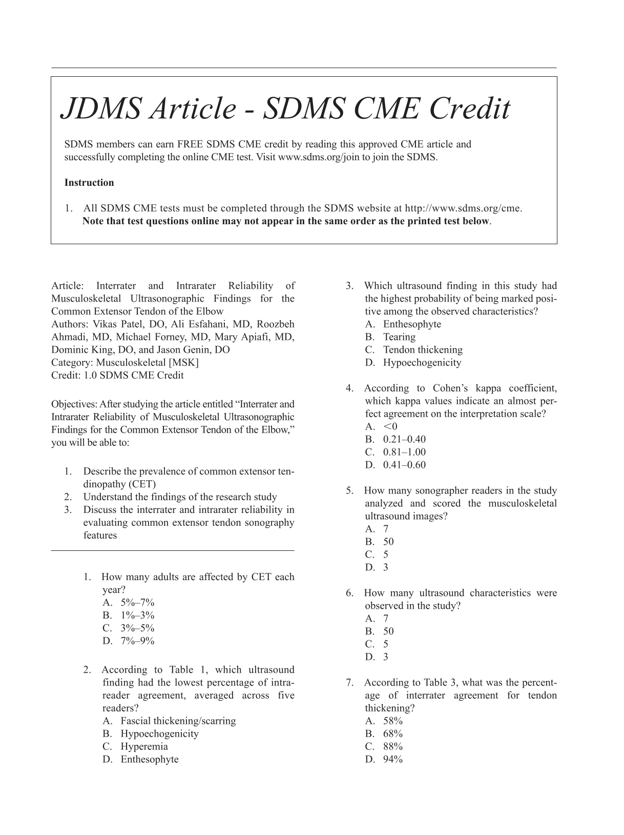
This retrospective case study evaluated the interrater and intrarater reliability of seven common extensor tendon pathologic features on musculoskeletal ultrasonography (MSK-US).
A cohort of 50 patients were imaged due to presenting with atraumatic nonradicular lateral elbow pain. Three experienced and two novice readers rated the images on two separate occasions, and AC1 and kappa coefficients were calculated for each feature.
The interrater reliability was fair with respect to fascial thickening/scarring (AC1 = 0.26), tearing (AC1 = 0.35), tendon thickening (AC1 = 0.38), and intratendinous calcification (AC1 = 0.33); substantial for enthesophytes (AC1 = 0.80); and near complete for hyperemia (AC1 = 0.83) and hypoechogenicity (AC1 = 0.92). Intrarater reliability was moderate for fascial thickening/scarring (κ = 0.48), tearing (κ = 0.41), tendon thickening (0.47), intratendinous calcification (κ = 0.56), and hypoechogenicity (κ = 0.47); substantial for hyperemia (κ = 0.71); and almost perfect for enthesophytes (κ = 0.86).
MSK-US may be a reliable tool to determine soft tissue changes in common extensor tendon pathology.

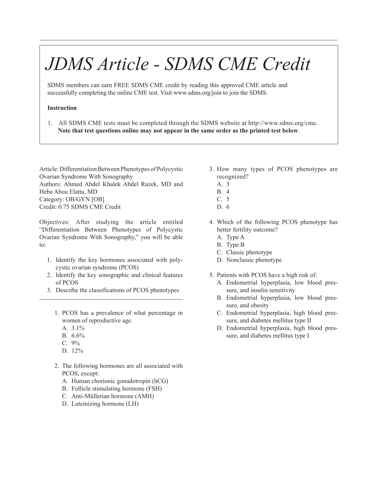
To differentiate between the classic (A/B) from the nonclassic (C/D) phenotypes of the polycystic ovarian syndrome (PCOS) with sonography.
Retrospective analysis was performed upon 72 female patients with PCOS. All patients underwent transvaginal sonography. The patients were classified into classic and nonclassic phenotypes of PCOS. Image analysis was performed for the ovarian volume, follicle counts, follicle size, and endometrial plate thickness.
There were significant differences in the ovarian volume (
Sonography findings can differentiate classic from nonclassic phenotypes of PCOS.

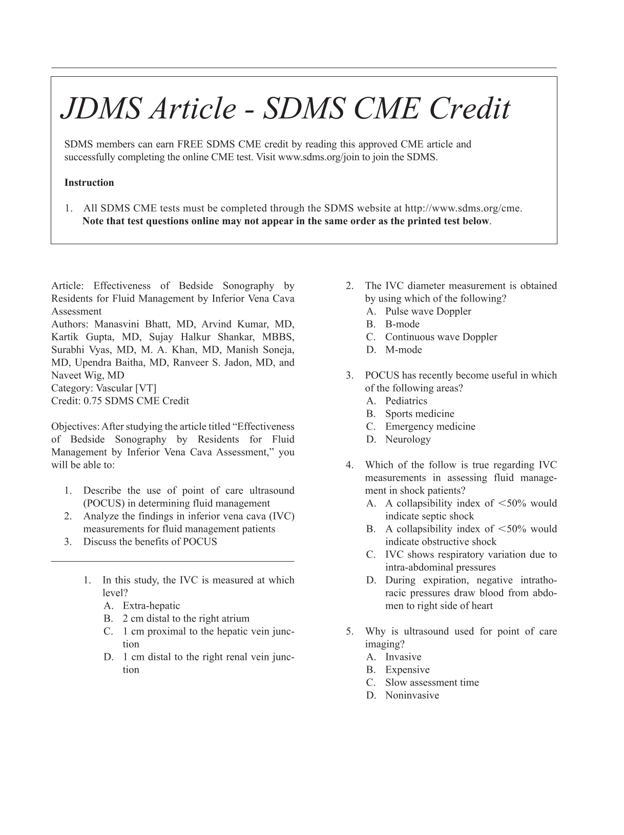
To study the utility of point of care ultrasound (POCUS) in the diagnosis and management of shock when performed by medicine residents.
A retrospective, case-control study was carried out in the medical intensive care unit at a tertiary care hospital. Consecutive patients who had received bolus fluid for hypotension during the hospital stay were screened from the case sheets and recorded.
A total of 57 patients were recruited into the study. Baseline parameters were comparable in both groups. The median amount of fluid given by the physician was 1000 milliliters (500, 1500) in the inferior vena cava (IVC) group as compared with 1000 milliliters (500, 1000) in the clinical group which was similar between the groups (
In this cohort, there was a significant increase in MAP, reduction in the APACHE II score, and heart rate in the IVC group when compared with the clinical group.

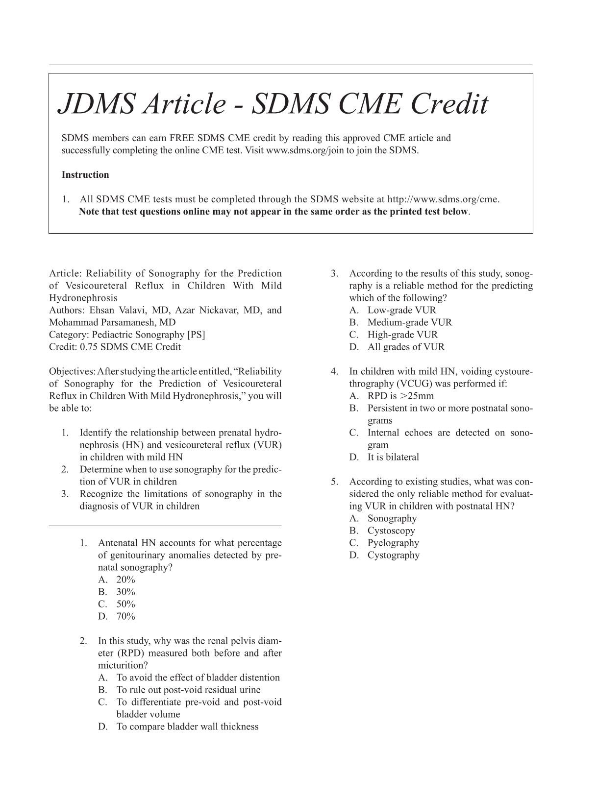
Postnatal evaluation of vesicoureteral reflux (VUR) remains controversial in patients with antenatal hydronephrosis (HN). The objective of this study was to identify the significance of mild postnatal HN as a marker of VUR and its severity.
Sonographic findings of 248 children (351 kidneys) with persistent postnatal HN were evaluated for the incidence and severity of primary VUR.
The majority of patients had mild (67.8%) HN, followed by moderate (27.6%) and severe (4.6%) HN. VUR was identified in 14.7% of patients with mild HN, 18.5% of patients with moderate HN, and 18.7% of patients with severe HN. About 11.44% of patients with mild HN had low-grade VUR, followed by moderate (45.71 %) and severe grades (42.85%).
A large number (89%) of patients with mild HN had moderate to severe VUR. In this cohort, renal sonography was not a reliable method for the prediction of VUR and its severity in patients with mild postnatal HN. However, it was reliable for high grades of VUR.
These results are limited due to the study design and the inability to generalize these findings. These results should be replicated across other multiple clinics, thereby recruiting more variety of patients, to validate these study recommendations.

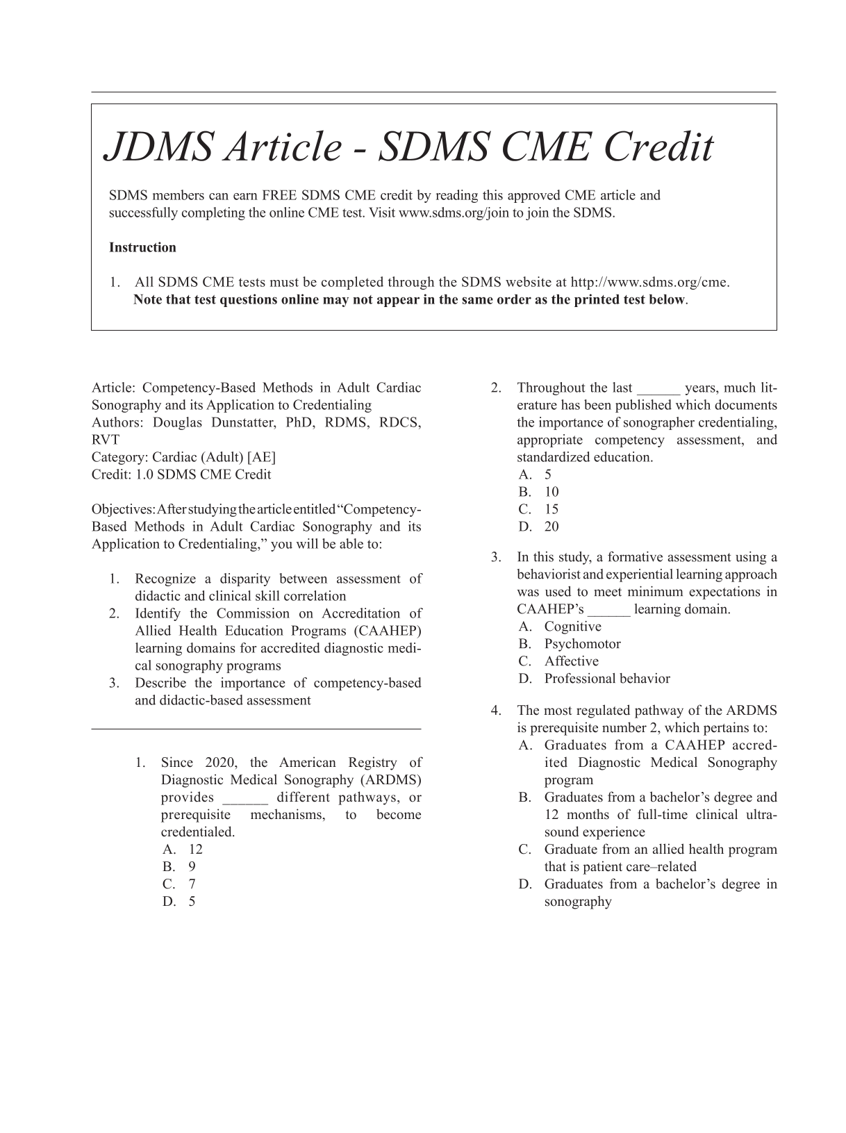
This research examined the effects of multiple combined competency-based methods in an adult cardiac Commission on Accreditation of Allied Health Education Programs (CAAHEP) accredited diagnostic medical sonography program on clinical and didactic scores.
A quasi-experimental nonequivalent group research design was used to evaluate students enrolled in an adult cardiac CAAHEP accredited echocardiography curriculum. Their cumulative final examination and clinical competency evaluation scores from multiple cohorts (pre- and post-intervention) were used for statistical analyses.
There were no significant differences between multiple competency-based assessments and didactic or clinical scores but unveiled reasons that coincide with the literature, such as nonvalid and subjective clinical assessment.
These results suggest further evaluation of the credentialing process to ensure clinical competency.

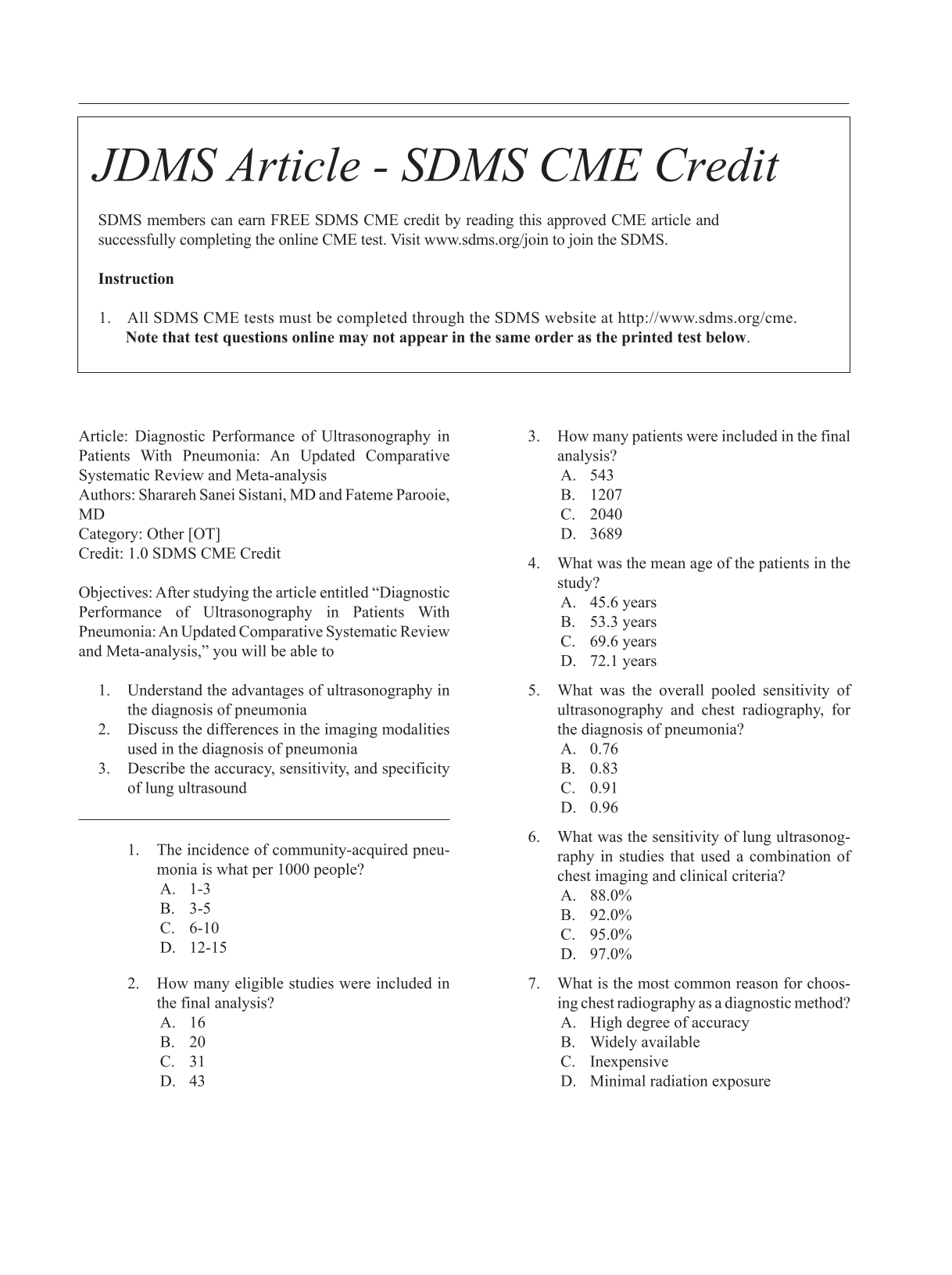
A chest radiograph (CXR) is still the preferred diagnostic method when pneumonia is suspected, although the sensitivity is relatively low. The aim of this study was to compare the diagnostic sensitivity, specificity, and accuracy of ultrasonography (US) for the diagnosis of community-acquired pneumonia (CAP), compared with CXR.
A principled search was conducted to identify original English articles using PubMed, EMBASE, Web of Science, Scopus, and the Cochrane library, with the end date of October 2020. A combination of keywords, such as “ultrasound” or “ultrasonography,” “pneumonia,” “sensitivity,” and “specificity,” was used. Methodologic quality was assessed using Quality Assessment of Diagnostic Accuracy Studies 2 criteria. Statistical analysis was completed on the resulting study data.
The search produced 16 eligible articles that reported on 2040 patients. The overall pooled sensitivity for US and CXR, to diagnose pneumonia, was 0.96 and 0.65, respectively. The overall pooled specificity for US and CXR was 0.85 and 0.81, respectively. The overall pooled positive likelihood ratio for US and CXR was 9.74 and 3.67, respectively. The negative likelihood ratio for US and CXR was 0.05 and 0.42, respectively. In addition, summary receiver operative characteristics areas under the curve were 0.98 for US and 0.77 for CXR.
This review demonstrated that lung US is a useful technique for the diagnosis of pneumonia. This diagnostic method can be used by emergency physicians with high accuracy, sensitivity, and specificity. Among an elderly population, this diagnostic method may be a better choice than CXR. The rapid performance of lung US may facilitate a quick, cost-effective, and safe diagnosis of this potentially fatal disease.

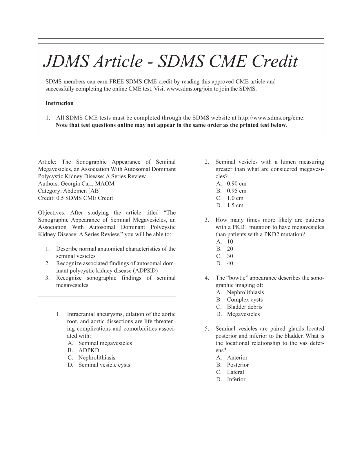
Megavesicles are an uncommon diagnostic finding during sonography of the bladder, especially when the examination is performed transabdominally. Although megavesicles are more likely seen with transrectal ultrasonography, computed tomogram (CT), vesiculography, or magnetic resonance imaging (MRI), it has been noted during transabdominal sonography for patients suffering from autosomal dominant polycystic kidney disease (ADPKD). When seminal vesicles become dilated, they are often visualized during transabdominal sonography. Two patient cases are provided of seminal megavesicles, associated with ADPKD and have documented sonographic findings. Both patient cases of megavesicles were discovered incidentally during the course of a renal sonogram. The importance of these diagnostic findings and the possible pathogenesis are provided.

A bursa is a thin sac filled with viscous fluid to facilitate friction and gliding. In the human body, there are two types of bursae: synovial bursae in well-defined positions and adventitial bursae, which develop in sites where friction or pressure increases. This case study is on a young woman with an old distal radial extremity fracture, with occupational exposure to pressure on distal ulnar forearm. She noticed a small and painful mass, with some mobility on the above-mentioned site. Sonography was used to detect a small fluid accumulation with features of adventitial bursitis. This case presents a rare localization of an adventitial bursitis. It is believed to have developed over the distal ulnar epiphysis, with a small bone malalignment, after an old and consolidated fracture, of the distal radial extremity.

Bilateral tubal pregnancy (BTP) is a very rare form of ectopic twin gestation. Many times, they occur after the use of assisted reproductive therapy. Most cases of BTP are diagnosed during laparoscopy. This case report demonstrates a rare preoperative, sonography diagnosis of a spontaneous BTP which occurred after a tubal ligation. The sonogram revealed two corpus luteal cysts, both located on the right ovary, which suggests that the left tubal pregnancy most likely occurred as a result of ovum transmigration. This case demonstrates the important role that sonography plays in the early diagnosis of ectopic pregnancies as well as the importance of thoroughly examining the entire pelvis during a pelvic sonogram.

Plain radiography is often considered a first-line diagnostic tool and gold standard for bony fractures. This case series and brief literature review highlight the need for increased training in musculoskeletal sonography. A case is presented of a distal radial fracture that was not detected on radiography but diagnosed with sonography on the same day. Similarly, two fractures of the distal fibula were diagnosed using sonography after negative radiographs at follow-up appointments, led to misdiagnoses and mismanagement. A fourth case is presented to show an acute rib fracture diagnosed by sonography. These cases emphasize the importance of musculoskeletal sonography training in the diagnosis of acute fractures and its underutilization at the bedside. Collaboration between sonographers and physicians may increase utilization of musculoskeletal sonography which in turn may improve patient care and outcomes.

Given the significant morbidity and mortality associated with tubo-ovarian abscess (TOA), prompt diagnosis and initiation of treatment is critical. If a diagnosis is made early, medical management remains an option thereby improving the chance of conservation of reproductive organs. TOA is a sequalae of pelvic inflammatory disease (PID) and the diagnosis remains difficult due to the wide inclusion criteria for this condition, which should exclude other causes of pelvic pain. This case study presents two atypical cases of TOA in which neither patient met the diagnostic criteria for PID, on initial presentation and delayed diagnosis. Sonographic findings of enlarging, complex hyper-vascular masses and markedly elevated CA-125 levels instead raised concerns for malignancy influencing management decisions. In retrospect, utilizing these two predictive diagnostic tools may facilitate an earlier diagnosis of TOA therefore even without overt PID. This becomes more important when the clinical signs support a potential diagnosis of malignancy.