Editorial
Select search scope: search across all journals or within the current journal

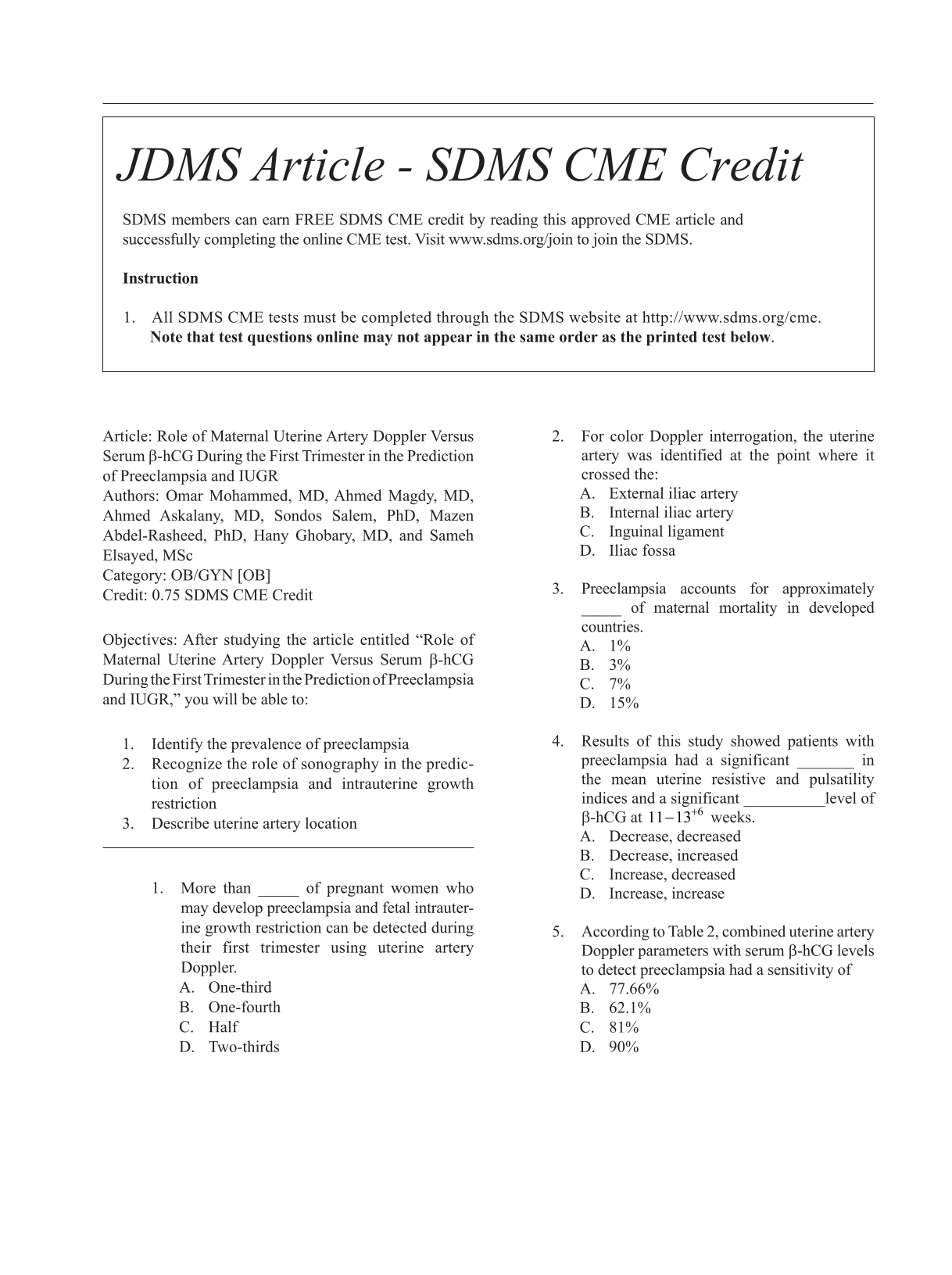
Preeclampsia accounts for 15% of maternal deaths and may cause fetal morbidity and mortality. The aim of this research was to evaluate the efficacy of maternal uterine artery Doppler versus serum beta-human chorionic gonadotropin (β-hCG), during the first trimester, in predicting preeclampsia and intrauterine growth restriction (IUGR).
In a convenient sample of 388 pregnant women, uterine artery resistive index (RI) and pulsatility index (PI) were assessed, and serum β-hCG level was measured at 11 to 13 weeks of gestation. The patients’ maternal blood pressure and fetal growth were monitored.
The patients with preeclampsia (n = 58) showed a significant uterine RI and PI increase with a significant β-hCG decrease compared with the normotensive patients (n = 330). The specificity of uterine PI and RI to predict preeclampsia and IUGR is higher than that of β-hCG. However, the sensitivity of combined diagnostic tools is higher than the singular use of these diagnostic tests.
Uterine artery Doppler may be better than serum β-hCG in predicting preeclampsia and IUGR. However, combined diagnostic techniques may be better to screen at-risk patients.

The objective of this study was to evaluate the association between reduced fetal thymus size and intrauterine growth restriction (IUGR). This study was devised to determine the association between thymus size and any abnormal Doppler indices within the fetal umbilical artery (UA), as well as the middle cerebral artery (MCA).
Forty-six pregnancies between 20 and 38 weeks of gestation with IUGR and 46 normal pregnancies within similar gestational age (GA) range were included. The transverse diameter of fetal thymus was measured. In the IUGR group, the fetal umbilical artery (UA) and middle cerebral artery (MCA) Doppler flow velocities were recorded.
The mean GA of fetuses with IUGR (33.5 weeks) was higher than control group (30.3 weeks). To adjust for the effect of GA, analysis of covariance (ANCOVA) was performed. The adjusted mean thymus diameters were 19.02 mm in IUGR and 21.25 within the control group (mean difference = 2.23 mm;
IUGR may be associated with reduced fetal thymus size, especially when coupled with abnormal Doppler findings. The thymus size in a group of IUGR fetuses, with abnormal Doppler findings, was smaller than IUGR fetuses, with normal Doppler findings.

The Thyroid Imaging Reporting and Data System (TIRADS) has been proposed to reduce the number of unnecessary fine needle aspirations (FNA) from thyroid nodules.
An individual radiologist provided sonographic examinations and FNA on a collection of 188 thyroid nodules. The recommendations based on the TIRADS system, for each nodule, was determined and evaluated against the cytology results.
The American College of Radiology (ACR), artificial intelligence (AI), European (EU), and Korean (K) scoring systems reduced FNAs by 53%, 56%, 48%, and 28%, respectively. Among those lesions without a recommendation for immediate FNA, The ACR would have missed four malignant nodules, the AI would have missed four malignant nodules, and K TIRADS would have missed three malignant nodules but with a recommended follow-up imaging. The ACR would have missed three malignant nodules, the AI would have missed four malignant nodules, and EU TIRADS would have missed four malignant nodules, without a recommended follow-up examination. The highest and lowest kappa interrelated agreements were between ACR and AI (0.902) and AI and K (0.448).
The ACR and AI TIRADS could substantially decrease the number of FNAs but rely on follow-up imaging. The EU TIRADS reduced the number of FNAs, the least however this system had less dependence on follow-up imaging. The K TIRADS was the most conservative method and the least dependent on follow-up diagnostics.

Sonographic estimated fetal weight (EFW) has an influence on the management of a pregnancy. The Hadlock 4 regression model (Hadlock-4), based on fetal biometry, is widely used. There are significant discrepancies noted between EFW, using Hadlock-4, compared to the actual infant birth weights (ABW) in the author’s clinical practice. The research objective was to compare the EFW, using Hadlock-4, with ABW and determine minor arithmetic modifications needed for this population.
A prospective observational study was done enrolling women in the third trimester, who underwent sonography and delivered within a week of the examination. The sonographic cases were divided into class intervals by gestational age. The EFW were compared with the ABW, using a Pearson coefficient and mean percentage errors (MPE). The EFW values were increased or decreased, by a certain percentage, to keep the mean percentage error in an acceptable range.
The strength of association between the EFW and ABW was 0.69 (
A simple 3 and 8% reduction of EFWs, using the Hadlock-4, with those sonographic examinations at 36-40 weeks and beyond 40 weeks gestation respectively, is proposed to increase reliable in this Indian patient practice.

The objective of this study was to determine the hemodynamic changes in the umbilical artery (UA) and middle cerebral artery (MCA) with oligohydramnios, during third trimester of pregnancy.
A cross-sectional descriptive study was done in a perinatal ultrasound center, over a 9-month period. The participant sample size was 64, and all of the women were conveniently consented and selected for the study. All individuals were referred for obstetrical sonography in the third trimester, with oligohydramnios. All fetal anomalies, such as a neural tube defect, dwarfism, an abdominal wall defect, were excluded from the study.
The mean systolic to diastolic (S/D) ratio for the UA was 3.01, within a range of 1.49 to 5.60 (± 0.79 SD). The mean pulsatility index (PI), of the UA, was 1.11, within a range of 0.41 to 5.51 cm (± 0.64 SD). The mean resistive index (RI) of the UA was 0.66, within a range of 0.33 to 1.25 cm (± 0.13 SD). The mean S/D ratio of the MCA was 5.68, within a range of 2.05 to 26.10 (± 3.10 SD). The mean PI of the MCA was 1.67 within a range of 0.75 to 2.55 cm (± 0.45 SD). The mean resistive index (RI), of the UA, was 0.82 within a range of 0.51 to 1.77 cm (± 0.82 SD).
The Doppler indices may rise as pregnancy advances, especially in fetuses with oligohydramnios. In these types of patients, the flow within the MCA may decrease, with an increase in the gestational age. Consequently, it appears that oligohydramnios may lead to vascular remodeling of the UA and MCA.

Female infertility may be a commonly encountered problem that presently accounts for a great percentage of women seeking gynecologic services. A systematic review was preformed to evaluate the most common cause of infertility, using sonography.
A search was executed with Google Scholar, PubMed, NCBI, and Medscape databases, from 2001 to 2020. Two investigators independently reviewed and assessed those studies for eligibility. The data were tabulated in a Microsoft Excel sheet. The Statistical Package for the Social Sciences (SPSS), version 24 software was used to evaluate the data.
Out of 70 studies, the contributing factors, detected with sonography, for infertility were as follows: polycystic ovarian syndrome (PCOS), 44.9%; fibroids, 43.6%; endometriosis, 33.3%; polyps, 29.5%; adhesions, 29.5%; pelvic inflammatory disease, 23.1%; ovarian cysts, 23.1%; congenital anomalies, 20.5%; and adenomyosis, 11.5%.
The most common cause of infertility, detected with sonography, was PCOS, and the least contributor to infertility was adenomyosis.

The prevalence of segmental testicular infarction is extremely uncommon and very few cases have been reported in literature. Clinical and sonographic presentation of this condition can mimic testicular neoplasms or testicular torsion. Therefore, accurate diagnosis of segmental testicular infarction is imperative in the treatment process. This case study presents the sonographic diagnosis of testicular infarction in a 49-year-old man who reported mild testicular tenderness. A conservative treatment approach was used, saving the patient unnecessary surgical intervention.
Non-Hodgkin lymphoma (NHL) is a cancer of the immune system, and accounts for 90% of all lymphomas. When lymphoma is discovered outside of the immune system, it is considered extranodal lymphoma. Any organ can be the primary site of NHL, with the gastrointestinal tract being the most common site of involvement. This case study explores the sonographic discovery of an uncommon presentation of NHL with an extranodal mass originating from the pancreatic body and tail, and further extending into the splenic hilum.

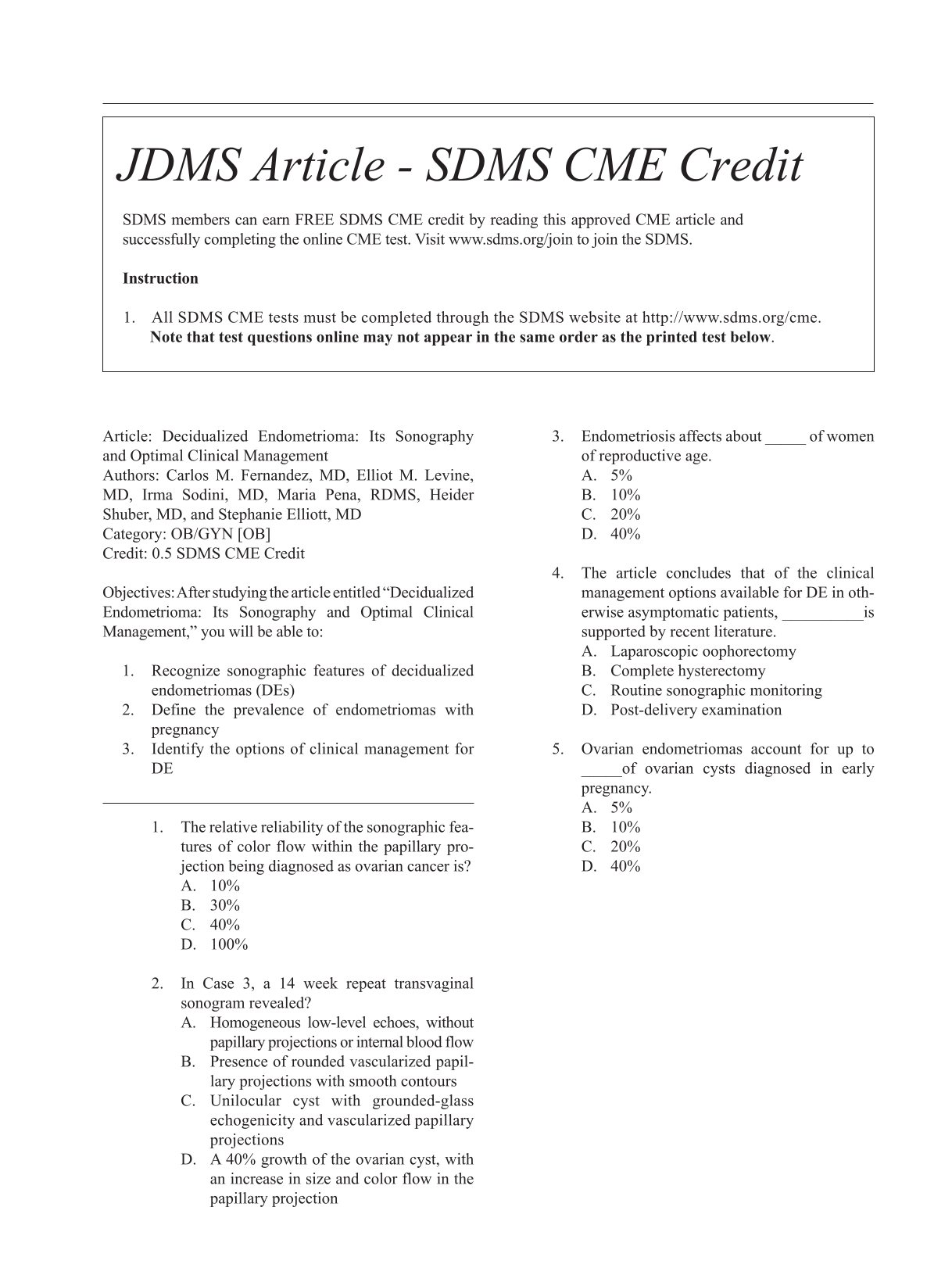
Endometriosis is a condition which affects many reproductive age women. During pregnancy, an ovarian endometrioma may undergo decidualization, which may take on the sonographic appearance suggestive of malignancy. In this instance, a surgical approach for treatment may be deemed necessary. Three cases are presented with different outcomes, worthy of viewing the contrasting management. In case 1, a patient was diagnosed with a left ovarian cyst at her routine 20-week gestation sonogram. This sonographic examination was suggestive of an ovarian malignancy. The patient underwent laparoscopy with left salpingo-oophorectomy at 23 weeks gestational age, without complications. The pathologic diagnosis of the removed mass was a decidualized endometrioma. In case 2, a female patient presented with bilateral ovarian decidualized endometriomas that were demonstrated sonographically and had the appearance of malignancy. This patient was treated conservatively with serial follow-up sonograms during her pregnancy. At the 2-month post-partum follow-up sonogram demonstrated persistent bilateral endometriomas, without decidualization. However, in case 3, possible decidualization of an endometrioma was seen, and a follow-up sonogram revealed the presence of ovarian carcinoma. There is previously published support for

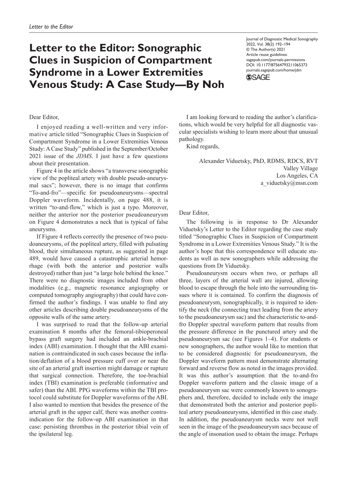
Sonography is an essential imaging modality, known for its real-time capabilities, relative low cost, and multiple diagnostic applications. Although there are protocols which are well-established for specific examinations, there are not clear guidelines for sonographic examinations of soft tissues. Many sonographers have to establish their own imaging protocols when completing a soft tissue examination. This case series details five soft tissue examinations that represent just a few of the clinical requests generated in an Urgent Care (UC) facility. Since UC usage has increased over the past several years, it appears likely that any sonographer working in such outpatient settings will benefit from as much exposure to soft tissue examinations as possible.

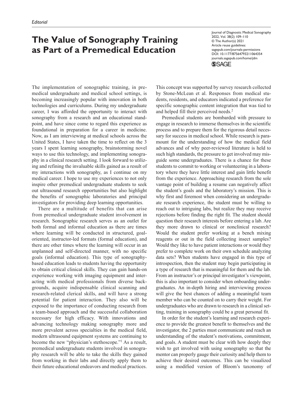
Sonographers experience a high cognitive load in hospital-based care. High ambient noise and frequent noise-based interruptions include knocking on the room door, questions from others in the room or through communication technology, alarms, alerts from personal devices, and carts and people passing in the hallway. In addition, other providers turning on the overhead light is distracting for exams that need to be conducted in reduced lighting conditions. This article suggests strategies to improve working conditions for sonographers conducting exams on a patient in the hospital room. Our strategies emerge from human factors methods and principles, which derive from communication principles and theory. These strategies are organized by reducing noise-based and light-based interruptions in the hospital room and hallway, primarily through changes to the built environment and communication technology settings and reducing the use of speech during cognitively challenging time periods through training. Most of the strategies are low-cost and can be implemented within the current built environment and communication technology infrastructure. We anticipate that these strategies could enhance patient outcomes, increase patient satisfaction, improve sonographers’ job satisfaction, protect provider health, and increase procedural efficiency.