Editorial
Select search scope: search across all journals or within the current journal

The purpose of this study was to examine the effects of course delivery methods on examination grades and student perceptions in a sonography course.
The participant included all sonography students (n = 103), enrolled at a Midwestern university, during the academic years (AY) of 2010–2021. A retrospective, convergent mixed-methods design was used to collect and analyze data, related to the course delivery method.
The highest overall mean examination score and course satisfaction rating resulted from the blended learning format and the lowest mean examination scores and course satisfaction resulted from the flipped learning format.
Health professions students are a diverse group of learners. Pedagogical practices should include course design and delivery methods which educate all learners. Courses that balance both face-to-face learning with opportunities for self-directed learning improve student satisfaction which could lead to improved student outcomes and provide the foundation for students to become competent health care professionals.

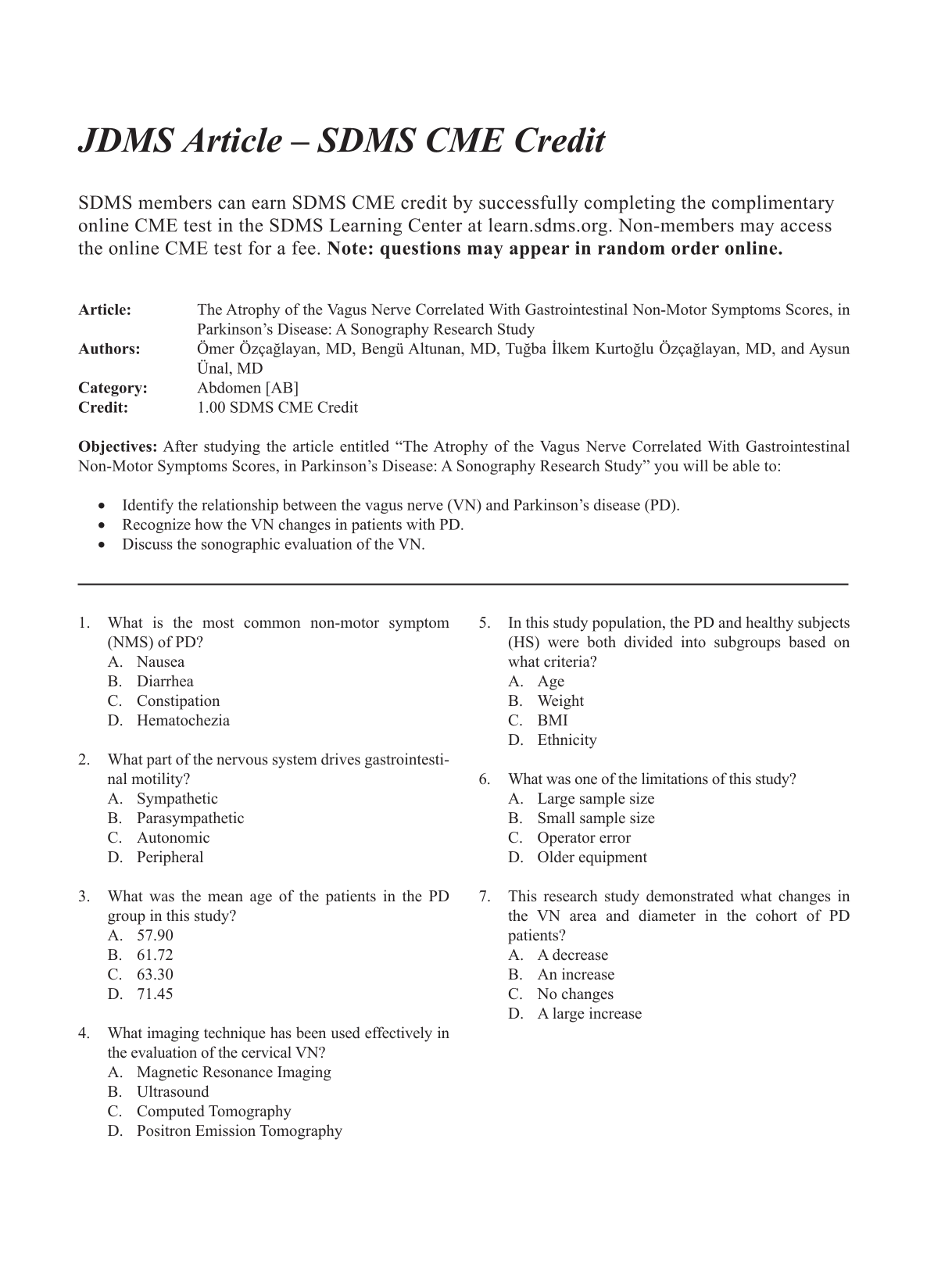
To investigate the vagus nerve (VN) dimensional changes with Parkinson’s disease (PD), compared with healthy subjects. Additionally, it is important to investigate whether there is any relationships between these changes and patient’s motor and non-motor symptoms (NMS) of PD.
A cohort of 43 patients with PD formed a group that was compared with 44 patients without PD, denoted as the healthy subject (HS) group. The diameter and areas of VN of study groups were measured using ultrasonography (US). The study groups were further divided into <65 and ≥65 subgroups, to evaluate the possible effect of age on the VN and evaluated relationships of VN dimensions, between subgroups. In the PD group, a correlational analysis was conducted between the diameter and area of the VN and the motor and NMS scores.
There was statistically significant difference in right (
The US demonstrated that VN dimensions may possibly reduce in those patients affected by PD. In this cohort, it appears that an increase in gastrointestinal NMS scores may be explained by atrophy of the VN.

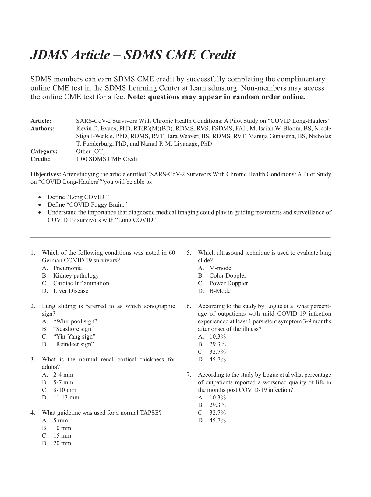
As the global pandemic resulting from this virus continues, surviving patients continue to report chronic symptoms long after a diagnosis of coronavirus disease (COVID-19). A pilot study was conducted with a convenient sample to ascertain the proper diagnostic testing for detecting chronic disease.
A convenient group of nine patients were directed by their primary care physician to be evaluated based on a variety of symptoms being reported by those surviving the acute phase of severe acute respiratory syndrome coronavirus 2 (SARS-CoV-2). The patients who volunteered provided sonographic imaging data of the lungs, kidney, and heart. This modified sonographic assessment was completed in a Family Practice office, with a laptop ultrasound equipment system, a blood draw, as well as completion of the EuroQol (EQ-5D) questionnaire. The data were collected as part of the patients 3- to 4-month follow-up primary care visit.
In this cohort, six patients were diagnosed and were able to recover at home. The other three patients chose to undergo monoclonal antibody therapy. The two subgroups’ data are provided and describe the types of residual effects that some individuals continue to suffer.
Those patients with residual symptoms of SARS-CoV-2 have been dubbed “COVID long-haulers.” This combination of diagnostic tests, specifically renal sonography, flow cytometry, and dynamic sonographic assessment, may hold promise for guiding the treatment and surveillance of these patients.

This research was designed to investigate the effectiveness of programmatic accreditation, when performing a virtual site visit, compared with a traditional on-site visit.
This study used a quasi-experimental, mixed-methods design with the virtual site visits as the independent variable and the site visit outcomes (number of citations, level of the citation, and recommendation for continuing accreditation) as the dependent variable. For this study, there were 28 diagnostic medical sonography programs throughout the United States, applying to the Commission on Accreditation of Allied Health Education Programs (CAAHEP), for reaccreditation status. Those programs that experienced a virtual site visit were compared with their counterpart programs (N = 109), which had a traditional on-site visit.
Based on these comparisons, there was no significant difference in diagnostic medical sonography programmatic accreditation effectiveness in the United States when performing a virtual site visit.
These study results would highly suggest that there was no significant difference in the level of the citations or the accreditation outcomes, based on a virtual site visit compared with those programs having an on-site visit. This research points to the effectiveness of the accreditation process, when is it conducted through a virtual site visit.

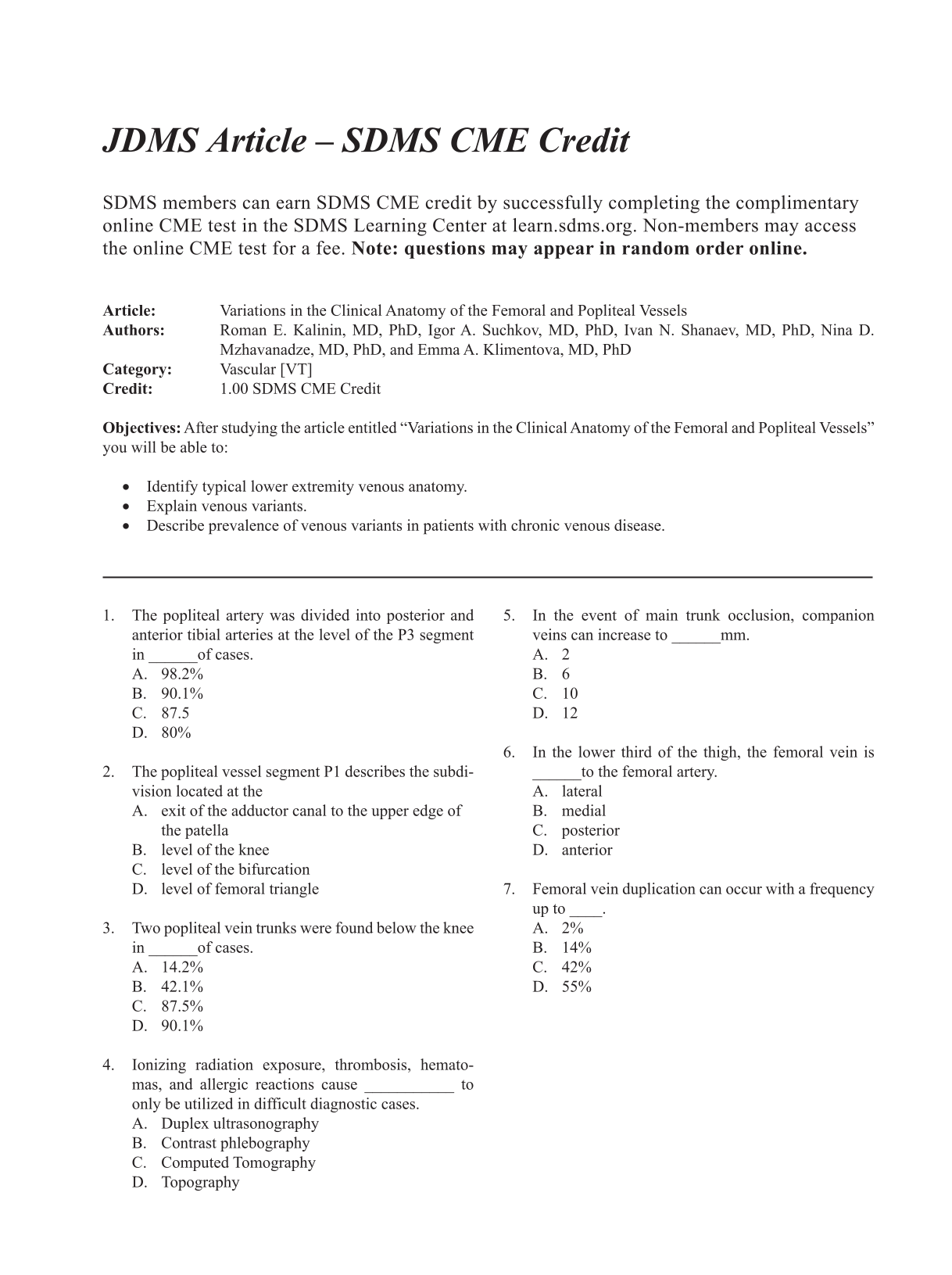
The aim of this study was to assess the topography of the femoral and popliteal vessels focusing mainly on the venous variants, in patients with chronic venous disease (CVD).
Duplex ultrasonography (DUS) was performed on 200 patients with varicose veins (VVs) and 253 patients with postthrombotic syndrome (PTS). In addition, an anatomical dissection was performed on 30 amputated lower extremities, without signs of CVD.
Duplication of the femoral vein (FV) was found in 14% of patients with VVs. Multiple FV trunks were identified in 42% of patients with PTS. One patient had a developmental anomaly with a hypoplastic FV and dilated deep FV. Two trunks of the popliteal vein (PV) below the knee level were detected in 83.3% of cases during anatomical dissection, in 87.5% of cases in patients with VVs, and in 90.1% of cases in PTS subjects during DUS. In 98.2% of cases, popliteal artery (PA) had a typical bifurcation. One patient was presented with a duplication of PA above the level of the knee. In 1.8% of cases, the PA was divided into an anterior tibial and peroneal artery.
This study found variant anatomy in the FV and PV, which is frequent, with major changes occurring in patients with PTS. Variant anatomy of the PA was also found and is considered uncommon, occurring in less than 2% of cases.

The present methods for the linear measurement of the liver with sonography are based on external reference lines. In addition, these measurements are subject to interrater variability due to relative mobility of liver and abdominal wall during breathing, change in position, and so on. A novel method using a plane with two internal references (plane of Y0) was developed as an alternative. The purpose of this study was to estimate the reliability of the novel method and compare with other commonly used methods.
A prospective, analytical, cross-sectional study was completed in a radiology department, including individuals referred for sonography and a contrast-enhanced computed tomography (CT) of abdomen. Three radiologists sonographically measured the craniocaudal dimension of liver in the “plane of Y0,” midclavicular, and anterior axillary lines for each participant. In 60 participants, the craniocaudal measurements obtained in the “plane of Y0,” in both contrast-enhanced CT and sonography, were compared for validity. Interrater correlation coefficients were calculated for each method.
Three hundred thirty-four participants underwent sonography of the liver. Sixty of them also underwent a contrast-enhanced CT. The craniocaudal measurements within the “plane of Y0” showed excellent reliability in comparison with other methods. It was found to be valid when compared with measurements completed on the CT image.
Measuring the liver in the “plane of Y0,” with two identifiable internal references, is reliable, valid, and recommended for possible use in daily clinical practice.
Using internal reference points for measurements of internal organs, with sonography, will reduce interoperator variability.
The “plane of Y0” passing through two internal reference points within the liver, the branching of right portal vein (Y shaped) and gallbladder (0 shaped lumen), can be easily acquired with sonography.
Measurement in the craniocaudal dimension of liver, in the “plane of Y0,” showed validity and high interrater reliability (0.91). The normal range of measurements in adults was found to be 10.5 to 15.1 cm.

Identify factors predictive of therapeutic success following ultrasound-guided sheath injection of the long head of the biceps tendon.
A total of 162 cases were divided into three groups based on clinical responses: complete, partial, or no relief. The presence or absence of clinical symptoms, glenohumeral joint structural pathology, and treatment method was compared.
Of the 115 injections with follow-up, 19.1% reported no clinical response while 53.0% had a complete response. Patients with fibromyalgia or chronic spine pain were strongly associated with a poor outcome (odds ratio [OR] = 5.7,
Ultrasound-guided intra-sheath injection is an effective method of non-surgical management for biceps tendinopathy.

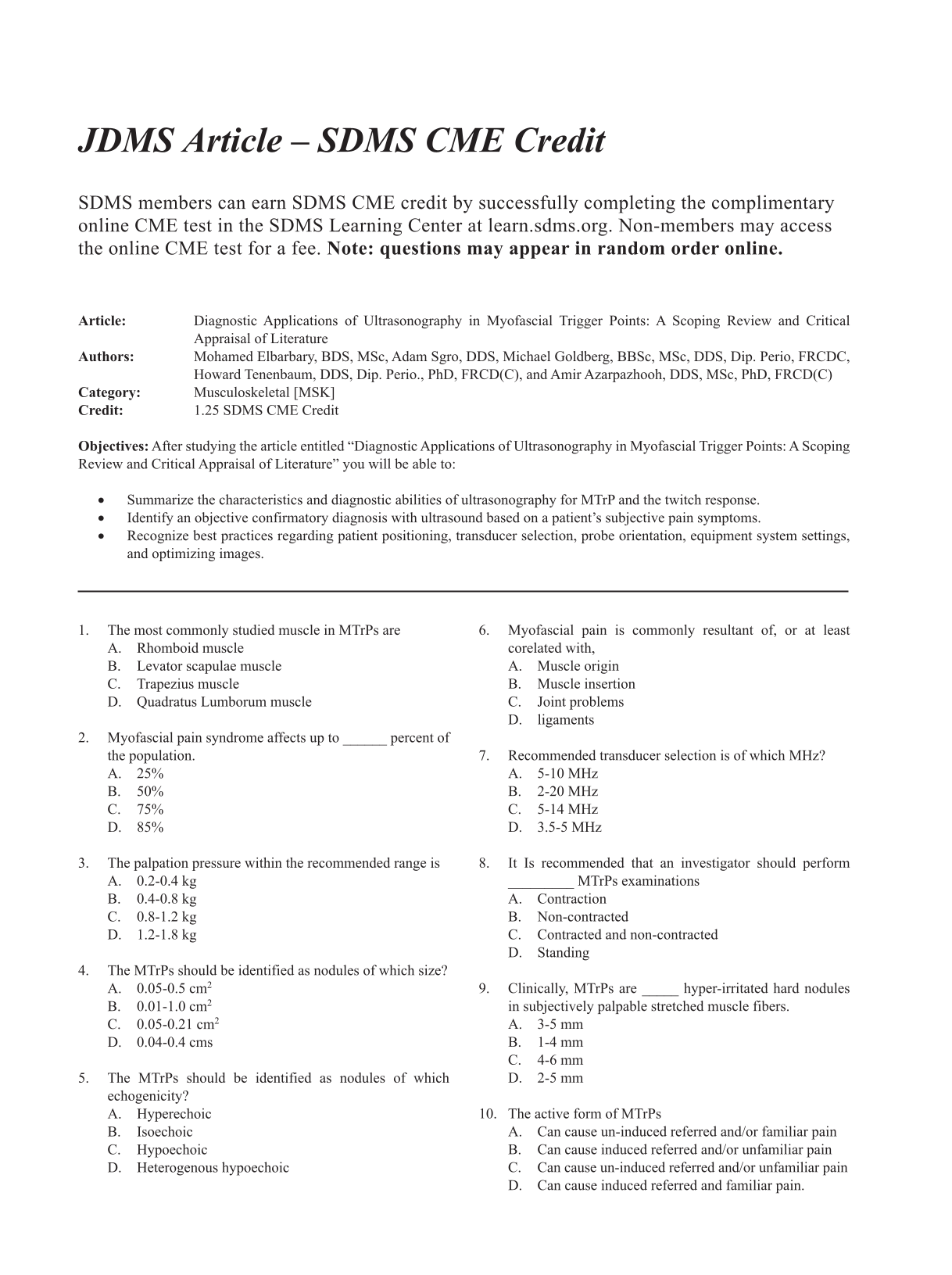
Myofascial trigger points (MTrPs) are pathognomonic of myofascial pain syndrome. The detection ability of MTrPs via ultrasonography is underreported and the characteristics of MTrPs are not sufficiently standardized. The objective was to summarize the characteristics and diagnostic abilities of ultrasonography for MTrP investigations.
A multi-database, and bibliography hand-search was implemented. Studies of ≥10 patients, published after 1980, appraising ultrasonography as a diagnostic aid for myofascial pain syndrome were included. The Quality Assessment of Diagnostic Accuracy Studies 2 (QUADAS-2) tool was used to evaluate the diagnostic accuracy of the included studies.
Six cross-sectional studies met the inclusion criteria. The back and trapezius muscles were the most studied locations. The diagnostic studies were of low-medium risk of bias. The studies reported a large range of diagnostic metrics (accuracy 58%–100%, sensitivity 33%–91%, specificity 75%–100%, positive predictive value 91%–100%, negative predictive value 47%–97%, positive likelihood ratio 3.6, and negative likelihood ratio 0.12–0.67).
This review found low-medium risk of bias evidence in support of ultrasonography for MTrP investigations. The clinical studies identified in the scoping review used gray-scale ultrasound equipment systems with a 5 to 14 MHz transducer to diagnose MTrPs and the local twitch response, and MTrPs were visualized mostly as hypoechoic nodules.

Renal transplant anastomotic pseudoaneurysms (RTAPs) are a rare vascular postoperative complication. The etiology of these pseudoaneurysms is attributed to infection or surgical vessel damage. RTAPs can lead to allograft dysfunction, allograft loss, or patient mortality due to rupture or sepsis. Because of these serious complications, expedient diagnosis is imperative for initiating surgical intervention and medical management. Sonography is a useful imaging modality for diagnosing and characterizing RTAPs. The sonographic findings, although not often described in detail in the literature, are definitive and unique. This case report describes the sonographic findings of a patient 22 days post renal autotransplant, who presented with excruciating abdominal pain. Sonographic examination identified multiple classic pseudoaneurysm findings and demonstrated intrarenal and extrarenal vascular abnormalities.

Although polyp size and stalk diameter are significant for the differential diagnosis of a gallbladder polyp, it is necessary to catch genuine polyp characteristics to differentiate a giant fibrous polyp from gallbladder carcinoma (GBC). This case report analyses the images and their correlation with the pathology. Transabdominal sonography revealed gallstones and a giant pedunculated gallbladder polyp measuring 32 × 18 × 14 mm with a thick stalk. The polyp lacked a hyperechoic surface line and was not delineated by either a deep hypoechoic area or a conically thickened outermost hyperechoic layer. Contrast-enhanced ultrasonography (CEUS) depicted a periphery-dominant enhancement, during the post vascular phase. A noncontrast computed tomogram (CT) did not demonstrate a gallbladder mass; meanwhile, a dynamic study revealed an enhanced polyp showing a periphery-dominant enhancement. Hence, without diagnostic findings suggestive of an obvious advanced GBC or excluding an early GBC, a full-thickness laparoscopic cholecystectomy was performed. The histopathology demonstrated a fibrous polyp with a thick stalk. In conclusion, a giant gallbladder polyp with a thick stalk showing a periphery-dominant contrast enhancement could suggest a fibrous polyp.

Pseudoaneurysms of the lateral plantar artery (LPA) are rare. In this case study, a very rare case of a pseudoaneurysm of the LPA, caused by a knife cut, is reported. A female adult patient presented with a growing lump in the arch of the left foot for 4 weeks. She had an accidental steak knife stab her on the arch of the left foot and had the cut sutured 5 weeks prior. The physical examination revealed a warm, pulsatile, and tender lump without a clear border deep to the scar in the lateral arch. Duplex ultrasonography (DUS) detected a cavity with the wall uneven in thickness and the outer wall undistinguished. Doppler demonstrated a typical Yin-yang sign inside the cavity and a neck-like structure connecting the cavity to an artery along the path of the LPA, suggesting that this was a LPA pseudoaneurysm, caused by penetration trauma. Later, the patient received priority surgery to have the pseudoaneurysm repaired.

A systemic framework for teaching vascular sonography that is centered on hemodynamics is useful to unify the initial test-by-test approach that developed with the pioneering and expansion of the field. Vascular sonography education is most powerful when taught with a system-level view of the circulation that connects how vascular anatomy (form) and physiology (function) are inextricably linked with hemodynamics (flow). Furthermore, understanding hemodynamic principles is key to using waveform morphology as a diagnostic tool. Teaching a system-based, hemodynamic-centered perspective aligns with the objectives of the recent Consensus Statement on Doppler Waveforms by the Society for Vascular Medicine and the Society for Vascular Ultrasound and equips the vascular sonography student to recognize, understand, and appropriately document vascular findings.