Editorial
Select search scope: search across all journals or within the current journal

The aim was to determine if the renal sinus fat-to-cortical thickness ratio was a better predictor of chronic kidney disease (CKD) than cortical thickness alone.
A total of 199 patients were included in the study. Renal fat, parenchyma, and cortical thicknesses were evaluated by sonography retrospectively. Same day serum glomerular filtration rate (GFR) values were obtained and correlated.
The study patients had an average fat-to-cortical thickness ratio <0.4 and had a 63% probability of developing renal failure. The probability increased almost linearly from 65% to 85% for patients with a ratio between 0.4 and 0.5, and then plateaued at 85% probability for a ratio >0.5. Additionally, the average fat-to-cortical thickness ratio was statistically significant (
The renal sinus fat-to-cortical thickness ratio has a significant negative correlation to GFR, specifically for the left kidney. The probability of developing renal failure increases with higher ratios. The renal sinus fat-to-cortical thickness ratio may also be better than cortical thickness alone in predicting progression toward CKD and it acts as an internal control for differences in body size.

To investigate the role of the uterine endometrial thickness and sonographic pattern as potential predictors for a pregnancy with an unknown location (PUL) and its possible outcomes.
A convenient sample of 330 symptomatic extra preposition female patients were enrolled in this study with a diagnosis of PUL. The clinical variables of endometrial stripe thickness and endometrial sonographic pattern were determined with transvaginal sonography. These sonographic examinations (it seems that examination in this context is a countable noun) were conducted during the first 24 hours of referral, and their predictive values for PUL outcomes (normal intrauterine pregnancy (IUP), ectopic pregnancy (EP), and pregnancy loss) were assessed after clinical follow-up sessions provided a definite outcome. The statistical significance was set a priori at a
The mean initial endometrial stripe thickness among participants, with a normal IUP, was more than those patients with an abnormal pregnancy outcome (
In this large cohort of patients, the endometrial stripe thickness of more than 11 mm, among those who were symptomatic and deemed as PUL, had the potential to predict ectopic pregnancy as an unlikely diagnosis.

The Centers for Disease Control and Prevention (CDC) has documented that the leading cause of death in the United States is cardiovascular disease (CVD). Specifically, CVD is responsible for the death of one out of every five women in the United States. Therefore, the research question was, “What specific diagnostic tests, including a novel approach to detecting abdominal aortic calcifications (AAC), could be added to screening women for CVD?”
A prospective cohort of 14 female volunteers, between the ages of 40 and 60 years, were consented to sonography of the aorta, collection of demographic data, as well as contributing biometric data. Each volunteer gave informed consent in compliance with the university’s Institutional Review Board. Volunteers also were provided with a Health Insurance Portability and Accountability Act (HIPAA) release that was signed prior to the collection of any data. All sonographic images were analyzed post-examination and aortic calcifications were graded using a modified scoring system derived from a dual-energy x-ray absorptiometry (DXA)-based grading system.
In this cohort of 14 females, their AAC category scores ranged from I to IIIB (grades: normal to multiple calcified atheroma). Descriptive statistics demonstrate that it is possible to document AAC on this cohort of women using sonography. Correlational statistics showed a statistically significant association between a volunteer’s posterior aortic-intimal media thickness (a-IMT) and their atherosclerotic cardiovascular disease (ASCVD) 10-year risk score.
This pilot study suggests the need for further research on screening techniques to determine women’s specific CVD risk in this age group. Although the preliminary results allude to sonographic measures of a-IMT being a possible CVD screening method in the future, more research data are needed.

Most health care programs have competitive entry and selective admission procedures. The purpose of this study was to investigate the relationship between cognitive variables such as the Test of Essential Academic Skills (TEAS) composite scores, the preprogram academic grade point average (GPA), as well as clinical GPA for diagnostic medical sonography (DMS) or radiography (RT) students.
The participants of this study were 26 imaging students. The convenient sample included 10 DMS and 16 RT students, enrolled in an Associate Degree in Applied Sciences program, within a community college. The student’s program success was examined over a period of one academic year.
The analysis of the findings determined that a significant relationship existed between some of the cognitive variables such as the TEAS composite scores, academics, as well as the clinical success for the DMS students. However, these RT students did not show the same outcome. In addition, there was no significant relationship between preprogram GPA and academic as well as clinical success, for either educational program.
The results of this study provide empirical evidence that a strong relationship may exist between the TEAS composite scores, academics, as well as clinical success for the DMS students. This is an important finding that may support the use of the TEAS test, as part of the admission process, for other DMS programs.

Assess the correlation between the qualitative sonographic score for detecting hepatic steatosis (HS) and liver fat quantification, using proton magnetic resonance spectroscopy (MRS).
Sixty-six patients with known or suspected nonalcoholic fatty liver disease (NAFLD) underwent ultrasonography (US) and magnetic resonance imaging (MRI). The qualitative sonographic score and fat quantification, measured by MRS, were the techniques used. A Kappa coefficient was used for agreement calculation, and a Fisher test was used to assess the normality of the variables. The MRS results were the gold standard for US quality assessment.
The agreement between MRS and US was 50% (Kappa 0.35). Ultrasonography results were more severe in 42.4% of the cases, and 66.7% of the patients had a body mass index greater than 30 (
Ultrasonography is a reliable exam for detecting HS, although not for grading purposes. Therefore, US is not a good predictor of HS severity for the management and follow-up of NAFLD.

Community-acquired pneumonia (CAP) is a common respiratory infection, and diagnosis is frequently performed using a chest radiography (CXR). Sonography is an available method with less radiation exposure, but has not been confirmed for diagnosis of CAP. The objective was to compare the diagnostic accuracy of sonography.
In this cross-sectional study, 90 adult patients (aged >18 years) were admitted to the emergency department of two university-affiliated hospitals in Southwest Iran, from July to December 2019, with a confirmed diagnosis of CAP. The patient symptoms and CXR results were included as part of this study. Within 24 hours after obtaining a CXR, a lung ultrasonogram (LUS) was performed. The diagnostic accuracy of semiquantitative LUS (SQLUS) was compared with CXR results using the Pearson chi-square test and Fisher’s exact test.
The mean age of participants was 52.98 ± 16.77 years. 51 were men (56.7%). 28 patients (31.1%), who had abnormal SQLUS results, were not associated with CXR findings (
This study results could not confirm LUS as an accurate method for diagnosing CAP in adult patients; although due to the convenient sample of adults and clinical-based diagnosis of CAP, any generalization of the results should be made with caution.

A reproduction management strategy is an essential component of any cattle production operation. Options include palpation per rectum, transrectal sonography, or biochemical blood tests. To determine the best method, veterinary costs must be weighed against operational costs of maintaining open cattle.
An online literature search using PubMed, Medline, CINAHL, EBSCO, and Google Scholar was completed to find relevant articles regarding pregnancy detection in cattle, costs of reproductive management in cattle, and pregnancy loss in cattle. Priority was given to original research articles pertaining to palpation per rectum, transrectal ultrasound, fetal gender, and pregnancy loss.
Transrectal sonography can detect a pregnancy up to 15 days earlier than palpation per rectum and has sensitivities and specificities as high as 97% when performed between days 21 and 35 post artificial insemination.
Cattle producers can be confident that transrectal sonography is a useful tool in the beef and dairy cattle industry. Its use facilitates reproductive management decisions, such as re-breeding or culling, thus reducing expenses, and increasing profitability.

This case series is presented to illustrate the clinical value of identifying, sonographically, the type of cesarean scar ectopic pregnancy that may present, as this has become more frequently encountered. The choice of surgical treatment appears to be dependent on the sonographic typing, of this specific type of ectopic pregnancy. This case study illustrates the importance of this topic, given the rising incidence of cesarean deliveries coupled with the increase in first trimester sonograms being more commonly performed. The value of proper diagnostic detection is important, given the contemporary management of this new type of ectopic pregnancy.

Pentalogy of Cantrell (POC) is a rare syndrome, with an omphalocele and ectopia cordis (EC) being pathognomonic anomalies. In this case report, a 12-week fetus, for nuchal translucency (NT) assessment, displayed a sac protruding through the midline of the fetal abdomen and lower thorax. The fetus’ cardiac activity was detected within the sac. These sonographic appearances are consistent with omphalocele and EC, respectively. A thickened NT and cystic hygroma (CH) were also identified. An important finding was a 1.8 mm maxillary gap (MG), which is a new marker for possible early diagnosis of cleft lip and palate (CLP), in the first trimester. The coexistence of a MG with POC is unique.

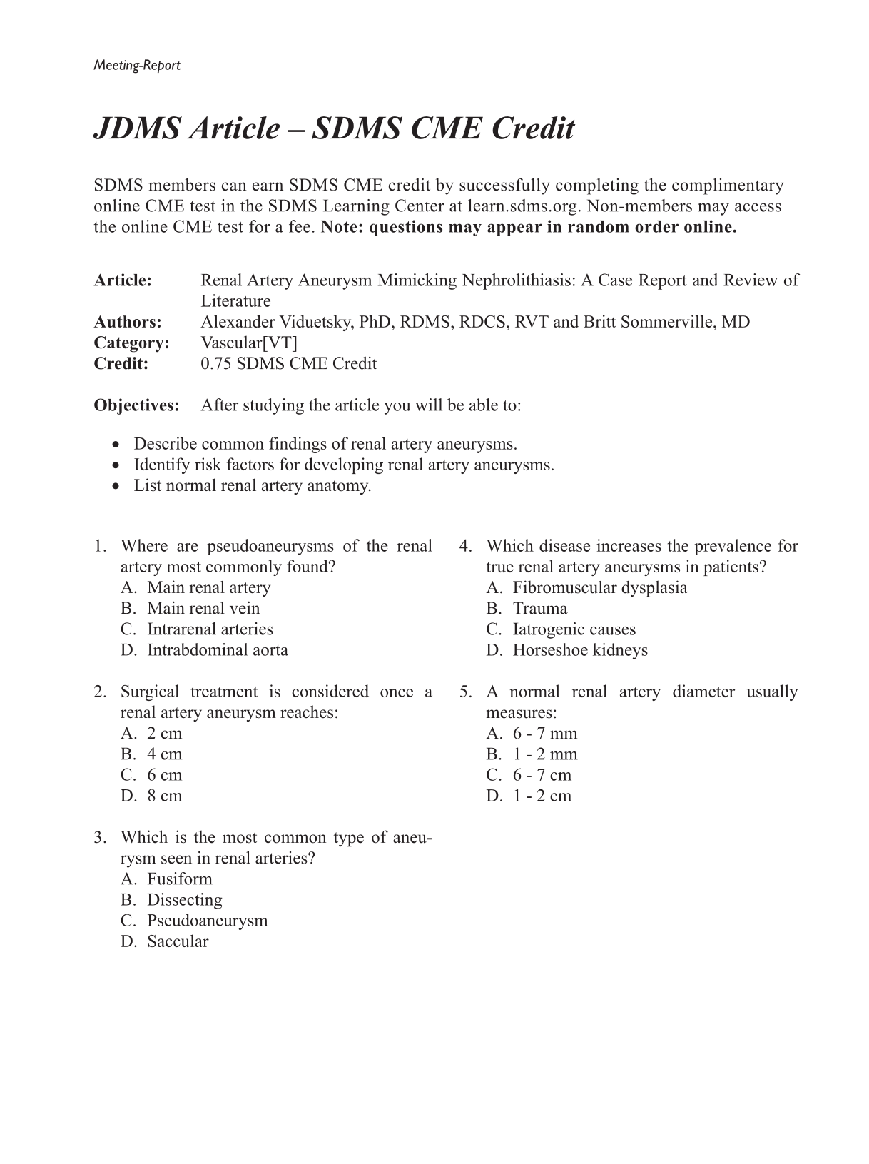
While originally described in the 18th century, the incidental detection of renal artery aneurysms (RAAs) has increased significantly with the advent and increased usage of modern cross-sectional imaging. Although the incidence remains low, RAAs are clinically important, especially if measuring greater than 2 cm in diameter. Therefore, ultrasonographers and radiologists alike should be aware of its features on multiple imaging modalities as well as imaging pitfalls. Renal duplex sonography is widely used for the evaluation of renal arteries stenosis, fibromuscular dysplasia, renal veins patency, and for the status of renal transplants. RAA is another, albeit rare, indication for renal arteries duplex examination. Furthermore, RAAs may be an incidental finding on abdominal or renal ultrasound. Therefore, familiarity with the sonographic presentation of a RAA is important. Diagnosing RAAs with duplex ultrasound may become exceptionally difficult when the aneurysm has peripheral calcification, even more so when the aneurysm is situated by the renal hilum. Those calcified arterial walls may produce strong acoustic shadowing, mimicking a renal calculus.

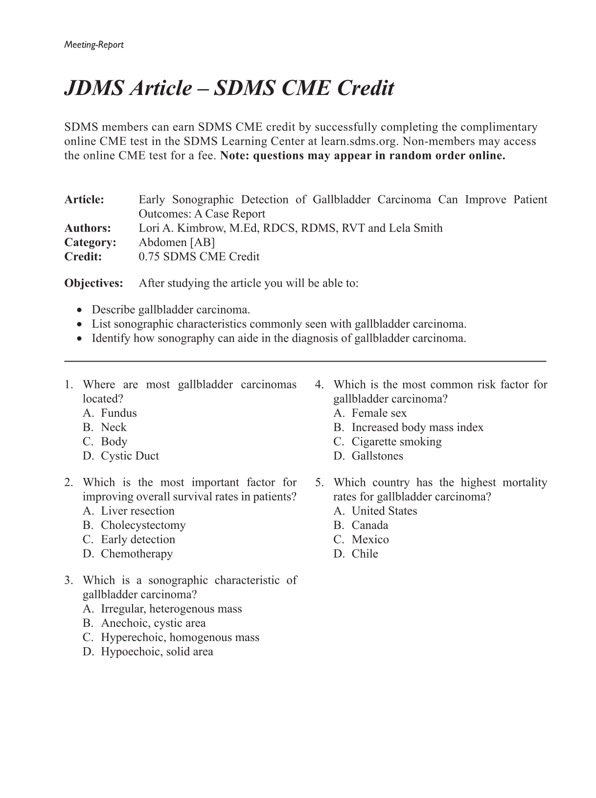
Gallbladder carcinoma (GBC) is a lethal and rare disease despite it being the most common malignancy of the biliary tract. Most cases of GBC are also associated with cholelithiasis and inflammation. The poor prognosis of GBC is mainly due to a lack of symptoms in the early stages of the disease. Early detection and cholecystectomy can lead to a higher survival rate when the cancer is confined to the gallbladder mucosa. This case study describes a man in his late 50s who presented with elevated liver function tests and was diagnosed incidentally with GBC with sonography.

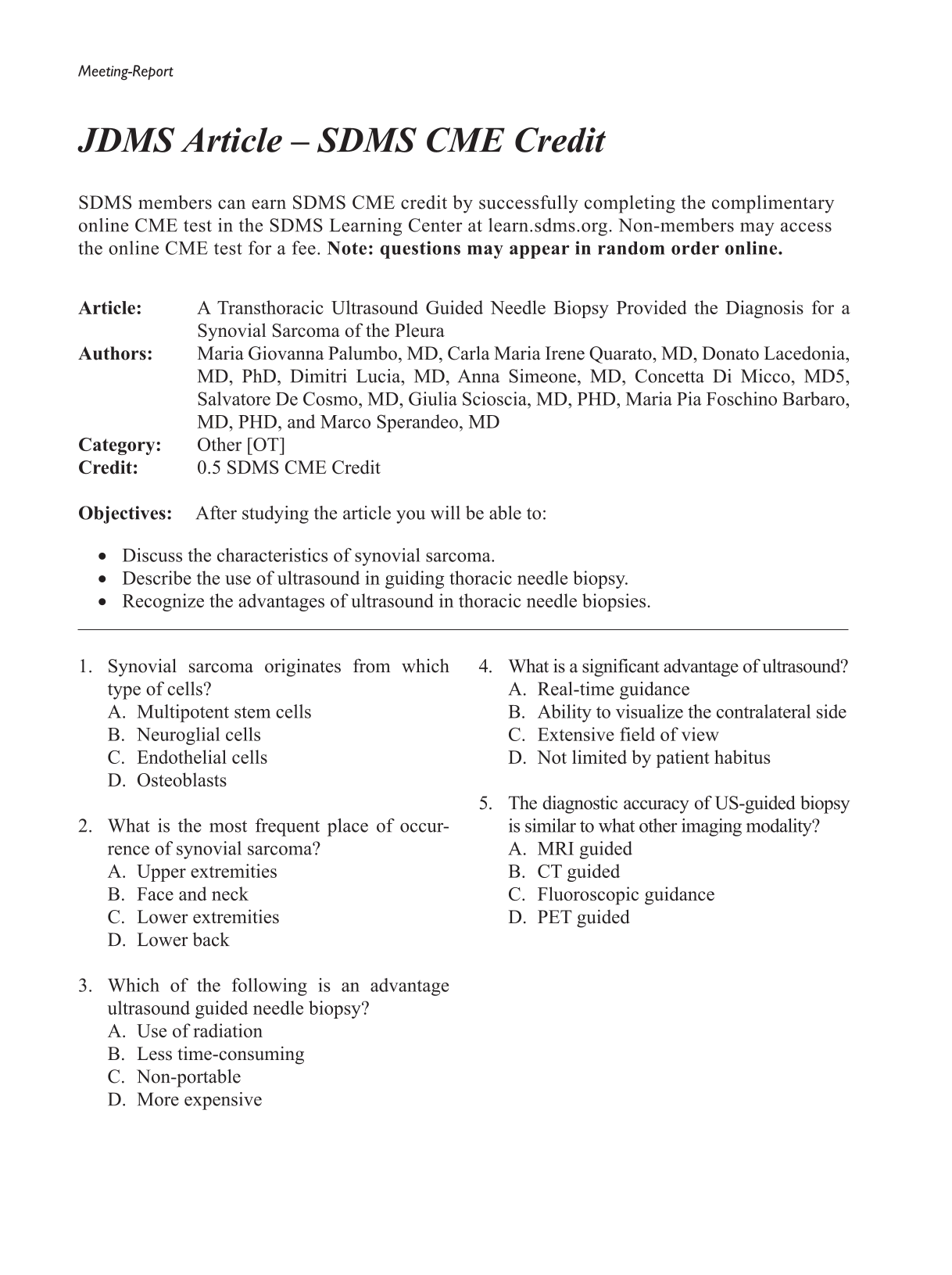
A case report is presented of a 72-year-old woman with monophasic synovial sarcoma of the buttocks, diagnosed four years before. The tumor was treated with surgical removal followed by radiotherapy. The patient regularly underwent follow-up examinations and the last chest computed tomogram (CT) showed a left-sided pleural mass and ipsilateral effusion. The patient was admitted in hospital to obtain a specific histological diagnosis before treatment of chest pathology was initiated. An ultrasound (US)-guided fine needle aspiration biopsy (FNAB) of the pleural lesion and US-guided thoracentesis with drainage of blood serum pleural fluid were performed. The pathological examination of the lesion revealed a definitive diagnosis of synovial sarcoma of the pleura. For pleural-based lesions, the diagnostic accuracy of US-guided biopsy is similar to that of CT-guided biopsy, with a lower complication rate, the absence of radiation exposure to patients, and a significantly reduced procedural time. The presence of pleural effusion creates a favorable acoustic window for US-guided biopsy of pleural lesion, which is useful for the diagnosis.