Editorial
 Free accessEditorialFirst published September, 1998pp. 285-285

Select search scope: search across all journals or within the current journal


 Free accessEditorialFirst published September, 1998pp. 285-285
 Restricted accessResearch articleFirst published September, 1998pp. 287-297
Vertebral bone strength is determined by several factors: cortical thickness, bone size, trabecular bone density, and microarchitecture. All these factors change with age as a result of the two dynamic processes: remodelling and modelling. When the changes become pronounced, osteoporotic fractures occur. There is a different aging pattern for men and women:
1. Men achieve a higher peak bone mass than women (mainly because of a larger cross-sectional area of their bones);
2. Men have no accelerated bone loss in middle age; and
3. Men seem to be able to compensate for their loss of cancellous bone strength by increasing their vertebral cross-sectional area with age. The general pattern, for both men and women is, though, that of an extreme (70–80%) decline in whole vertebral body strength during normal aging. The accompanying decline in bone density is much less pronounced (35–45%). This clearly illustrates the power relationship between bone density and strength. However, the role of changes in trabecular bone microarchitecture for vertebral bone strength during aging still needs to be determined.
Restricted accessResearch articleFirst published September, 1998pp. 299-306The stiffness and strength of cancellous bone depends on the amount of bone mineral (BMD) and on the three-dimensional distribution of the mineral (architecture). The relationship between mechanical properties and architecture, excluding confounding effects due to BMD can be studied using computer models of cancellous bone. It was shown that adaptation to mechanical deformation energy leads to an architecture which is an optimal or semi-optimal configuration with respect to maximal stiffness and minimal mass. Thus, the stiffness of the cancellous bone relative to the amount of bone (the bone density) can be considered as an optimality criterion. Based on these findings we assumed that the status of osteoporosis – or better fracture risk – could be related to how close this optimality criterion was met. In other words, we assumed that a higher fracture risk is simply related to a less optimal structure. This was tested for cancellous bone samples taken from post mortem vertebral bodies from two groups of subjects: one group with high fracture incidence during their lives and one group of “healthy” controls. It was found that the specimen from the high fracture incidence group had an architecture leading to a slightly stiffer structure relative to the BMD value. The conclusion is therefore that vertebral bone specimen from subjects with high fracture incidence are better optimized which was contradictory to what we expected. This finding indicates that bone specimen from the “healthy” control subjects had bone matrix at locations which are relatively unloaded. This tissue can be considered as not mechanically efficient or functional. A possible explanation of the present findings is that bone from subjects with increased fracture incidence is better adapted to mechanical stress, because it needs all bone material to carry the load. This stronger adaptation might be related to a compromised safety factor against bone loss, or diminished intrinsic matrix properties (e.g., microdamage).
Restricted accessResearch articleFirst published September, 1998pp. 307-320This article reviews recent progress in magnetic resonance microimaging of cancellous bone in vitro and in vivo from the perspective of the authors’ laboratory. It is shown that in particular in vivo the key technical prerequisites to satisfy are: (i) achieving sufficient signal-to-noise ratio (SNR) to allow for adequate spatial resolution; (ii) the image processing algorithms have to be robust enough to provide accurate structural information in the limited spatial resolution regime, i.e., in the presence of inevitable partial volume blurring and noise. The practical lower limit of voxel size in vivo was found to be about
Restricted accessResearch articleFirst published September, 1998pp. 321-327Bone mineral density and three-dimensional trabecular structure play a significant role in predicting bone strength and biomechanical properties. MR is a non-invasive technique for determining trabecular architecture both in vivo and in vitro. In this paper we review the use of magnetic resonance imaging to obtain high resolution images of trabecular bone structure and quantify the three-dimensional architecture of the trabecular bone network. Studies assessing the anisotropy of the trabecular architecture in human cadaveric specimens from the distal and proximal femur, and the thoracic and lumbar vertebrae, are reviewed. The contributions of the MR derived measures of 3D trabecular bone structure to the biomechanical strength of the specimen are presented. In vivo, the relationship between the high resolution MR derived trabecular bone structure parameters in the distal radius and calcaneus in patients with hip fractures, are compared to age matched normal controls. MR derived measures are compared to measures of trabecular bone mineral density (BMD) in the hip using dual X-ray absorptiometry (DXA).
Restricted accessResearch articleFirst published September, 1998pp. 329-337In vivo examinations of bone microarchitecture have become available recently through high resolution computed tomography (3D-QCT) and magnetic resonance imaging. The spatial resolution of the resulting images, however, is not sufficient to depict individual trabeculae in their true shape. Nevertheless, structural indices such as relative bone volume, trabecular number, mean thickness and mean separation can be extracted with the help of a ridge detection algorithm. Precision of the procedure is of the order of 1%, accuracy is ascertained using a micro-CT based calibration.
In this work we report first results of time serial examinations. Eighteen healthy postmenopausal women (no HRT) were measured at months 0, 6, and 12, and the temporal changes were analyzed. Examination site was the distal radius. The above mentioned structural indices, the average densities and the thickness of the cortical shell were determined. Of the 18 women 6 showed no significant bone loss of any kind, 5 lost primarily cancellous bone, 4 lost primarily cortical bone, and 3 had a substantial loss of cortical as well as cancellous bone. We conclude that even in a homogenous group such as postmenopausal women, there are considerable differences in the reason why bone is weakend and that high resolution 3D-QCT allows to differentiate between various types of bone loss.
Restricted accessResearch articleFirst published September, 1998pp. 339-350This paper describes the application of synchrotron radiation microtomography to osteoporosis research. By taking advantage of the high intensity, collimation, and monochromaticity of synchrotron radiation, we have been able to image the three-dimensional trabecular bone structure in living rats, thus providing serial data on the earliest architectural changes that occur with estrogen loss. Results from these in vivo animal experiments demonstrate that one of the earliest manifestations of estrogen loss, in addition to a decrease in the amount of trabecular bone, is decreased connectivity. We demonstrate that estrogen replacement therapy, when initiated soon after significant changes have occurred, restores bone mass to baseline levels but does not recover the trabecular connectivity. Even without an associated recovery in trabecular connectivity, finite element calculations on the three-dimensional images suggest that estrogen recovers the original structural modulus of elasticity. We believe the recovery of the elastic properties is due to an increase in trabecular thickness above baseline values.
Restricted accessResearch articleFirst published September, 1998pp. 351-359Improved methods for evaluation and quantification of the three-dimensional (3D) architecture of bone are needed in order to more fully understand the role of trabecular architecture in bone strength. Computed tomography (μCT) is capable of examining bone at resolutions below 30 μm (isotropic), with collection of a three-dimensional data set which can then be subjected to image analysis. In this paper, we discuss automated methods for important steps in this analysis, including methods for (1) segmenting the image into bone and background; (2) defining the volume of interest for determination of structural parameters; and (3) segmenting the bone into trabecular and cortical components. Evaluation of bone structure using these techniques provides new information about the 3D architecture of bone tissue, and may be useful for evaluation of structural changes in bone caused by aging, disease, or drug treatment.
Restricted accessResearch articleFirst published September, 1998pp. 361-372There is a tremendous unmet therapeutic need for the treatment of osteoporosis and osteoarthritis. The ovariectomized rat and the guinea pig are widely used animal models for the evaluation of new therapeutics for osteoporosis and osteoarthritis, respectively.
We have utilized X-ray micro-CT techniques to quantitatively evaluate the differences in trabecular bone in the rat proximal tibia following ovariectomy and treatment with estrogen (17-B-estradiol). Results demonstrate a loss of trabecular bone and architecture following ovariectomy (
Micro-CT and MR images were also obtained to study age related changes in the stifle joint of the guinea pig. Significant boney changes can be seen in the tibia and femur from the animals at various ages. Changes in cartilage and joint space can also be visualized in the images.
The utility of micro-CT imaging in evaluating the mouse skeletal system is illustrated by obtaining morphological and architectural details from high resolution images of the mouse hind limb and proximal tibia, respectively.
The results demonstrate the advantages that multi-dimensional imaging techniques can offer in evaluating bone and joint related changes in animal models of osteoporosis and osteoarthritis.
Restricted accessResearch articleFirst published September, 1998pp. 373-390The problem of quantifying the structure of cancellous bone has been addressed in the past by histomorphometry and more recently by imaging techniques using X-ray attenuation. The current approaches compute and describe parts of the construction of the trabecular net. We developed a new technique which quantifies cancellous bone of human lumbar vertebrae as a whole. The interactions, transactions, and interrelationships of all parts of the structural composition of the trabeculae are accounted for and quantified. The method is based on the concept of structural complexity within the framework of nonlinear dynamics.
The methodology was developed by using axial high resolution computed tomography images. The technique was transferred to quantitative computed tomography images and is based on the non-invasive assessment of 50 human L3 specimens.
The value of Houndsfield units per pixel representing trabecular bone of the vertebrae was transformed into color-encoded and alphabet-encoded symbols. The procedure of transformation of the X-ray attenuation pixels into symbols was necessary as a basis on which measures of complexity were introduced to assess the composition of symbols within the images. The development of a generalization of symbolic dynamics, a mathematical method, to work with two-dimensional images was a prerequisite.
The results of this study demonstrate that the structural composition of cancellous bone declines more rapidly than bone mineral density during the loss of bone. This outcome strongly suggests an exponential relationship between bone mineral density and the architectural composition of cancellous bone.
Normal trabecular bone has a complex ordered structure. The structural composition during the osteopenic phase of bone loss is characterized by lower structural complexity and a significantly higher level of architectural disorder. A high grade of osteoporosis leads again to an ordered structure, although its structural complexity is minimal.
Restricted accessResearch articleFirst published September, 1998pp. 391-401Tomographic techniques are attractive for the investigation of trabecular bone architecture. Using either conventional X-ray sources or synchrotron sources currently allows the acquisition of 3D images in a wide range of spatial resolution that may be as small as a few micrometers. Since it is technically possible to examine trabecular architecture at different scales, a question is to know what type of information it is possible to get at each scale. For this purpose, a series of ten vertebrae samples from healthy females of different ages (33 to 90) was imaged at various resolutions on three different micro-CT systems (cubic voxel size respectively 14, 6.7 and 1.4
Restricted accessResearch articleFirst published September, 1998pp. 403-412We are exploring methods of quantitating the 3D microstructure of bone in a way that will provide quantitative information about the functional status of the bone. The basic strategy is to image the spatial distribution of a selected, local, marker of function (e.g., material properties or new bone formation) and relate this to the simultaneously imaged 3D anatomic microstructure. Many of these approaches are extensions of well-established 2D imaging techniques (e.g., use of fluorophores and autoradiography) to 3D micro-CT. Local stresses throughout the microstructure can be estimated from the 3D geometry (and change in that geometry in response to stress applied to the outside of the bones) and correlated to the local function.
In addition to study of bone, we are also exploring calcification of arterial walls, both within the bone and outside the bone, such as coronary arteries. Arterial calcification in ovariectomised rats has been observed.
Restricted accessResearch articleFirst published September, 1998pp. 413-420Recently, new micro-finite element (micro-FE) techniques have been introduced to calculate cancellous bone mechanical properties directly from high-resolution images of its internal architecture. Also recently, new peripheral quantitative computed tomography (pQCT) and magnetic resonance (MR) imaging techniques have been developed that can create images of whole bones in vivo with enough detail to visualize the internal cancellous bone architecture. In this study we aim to investigate if the calculation of cancellous bone mechanical properties from micro-FE models based on such new pQCT and MR images is feasible. Three bone specimens were imaged with the pQCT scanning system and the MR-imaging system. The specimens were scanned a second time using a micro-CT scanner with a much higher resolution. Digitized reconstructions were made based on each set of images and converted to micro-FE models from which the bone elastic properties were calculated. It was found that the results of both the pQCT and the MR-based FE-models compared well to those of the more accurate micro-CT based models in a qualitative sense, but correction factors will be needed to get accurate values.
Restricted accessResearch articleFirst published September, 1998pp. 421-427Prevention of osteoporotic fractures requires accurate methods to detect the increase in bone fragility at an early disease stage as well as effective therapies to reduce the risk of bone fractures. Presently the prediction of the patient-specific bone fracture risk is primarily based on bone density, since this is the only parameter which can routinely be measured in vivo. However, these predictions might not always be precise because the fracture risk is also determined by the bone microarchitecture and the bone’s loading conditions. The aim of this paper is to introduce and evaluate new methods which could contribute to a better quantification of bone fracture risk.
Recently, a new approach, combining computational engineering methods (finite element (FE) method) and 3D high-resolution imaging techniques, has been introduced which can account not only for bone density but also for microarchitecture and loading conditions. High-resolution imaging techniques allow acquisition of 3D images of the bone microarchitecture, whereas FE methods applied to these images allow very precise calculation of the mechanical properties of bone. However, such a detailed FE analysis was not feasible for bone in vivo mainly because the resolution was not sufficient to measure the bone microarchitecture. It is shown here, from preliminary results, that the FE approach based on high-resolution images from a new CT scanner now allows prediction of the mechanical behavior of peripheral bones in vivo. It is expected that, eventually, the FE approach will lead to a better patient-specific fracture risk prediction than earlier methods based on bone density alone. Hence, with this new approach, it might be possible to detect the increase in bone fragility at an early stage of osteoporosis and it might also be possible to evaluate treatments more accurately.
Restricted accessResearch articleFirst published September, 1998pp. 429-432The elastic modulus and hardness of embedded and rewet trabecular bone lamellae of the proximal femur have been quantified in four males and four females by nanoindentation. The average elastic moduli ranged from 6.9 to 15.9 GPa and were found to be significantly different among individuals. Hardness correlated with elastic modulus and followed similar trends.
Restricted accessResearch articleFirst published September, 1998pp. 433-444Many bones within the axial and appendicular skeleton are subjected to repetitive, cyclic loading during the course of ordinary daily activities. If this repetitive loading is of sufficient magnitude or duration, fatigue failure of the bone tissue may result. In clinical orthopedics, trabecular fatigue fractures are observed as compressive stress fractures in the proximal femur, vertebrae, calcaneus and tibia, and are often preceded by buckling and bending of microstructural elements. However, the relative importance of bone density and architecture in the etiology of these fractures is poorly understood. The aim of the study was to investigate failure mechanisms of 3D trabecular bone using micro-computed tomography (μCT). Because of its nondestructive nature, μCT represents an ideal approach for performing not only static measurements of bone architecture but also dynamic measurements of failure initiation and propagation as well as damage accumulation. For the purpose of the study, a novel micro-compression device was devised to measure loaded trabecular bone specimens directly in a micro-tomographic system. The measurement window in the device was made of a radiolucent, highly stiff plastic to enable X-rays to penetrate the material. The micro-compressor has an outer diameter of 19 mm and a total length of 65 mm. The internal load chamber fits wet or dry bone specimens with maximal diameters of 9 mm and maximal lengths of 22 mm. For the actual measurement, first, the unloaded bone is measured in the μCT. Second, a load-displacement curve is recorded where the load is measured with an integrated mini-button load cell and the displacement is computed directly from the μCT scout-view. For each load case, a 3D snap-shot of the structure under load is taken providing 34 μm nominal resolution. Initial measurements included specimens from bovine tibiae and whale spine to investigate the influence of the structure type on the failure mechanism. In a rod-like type of architecture as seen in the whale spine, structural failure was described by an initial buckling and bending of structural elements followed by a collapse of the overloaded trabeculae. In the more plate-like bovine tibial architecture, buckling and bending could not be observed. Failure rather seemed to occur instantaneously. In conclusion, micro-compression in combination with 3D μCT allows visualization of failure initiation and propagation and monitoring of damage accumulation in a nondestructive way. We expect these findings to improve our understanding of the relative importance of density, architecture and load in the etiology of spontaneous fractures of the hip and the spine. Eventually, this improved understanding may lead to more successful approaches to the prevention of age-related fractures.
Free accessOtherFirst published September, 1998pp. 445-449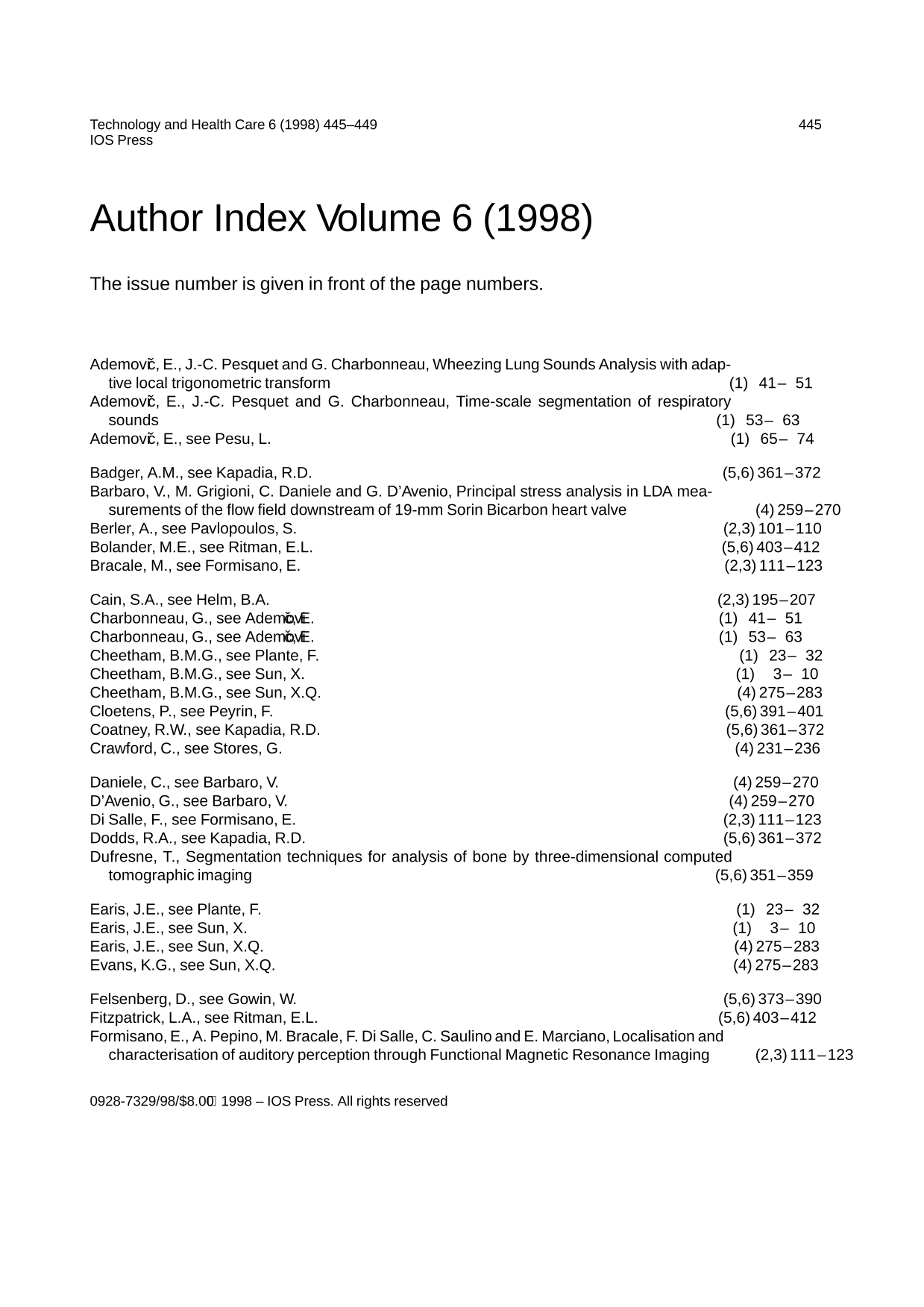 Free accessOtherFirst published September, 1998pp. 451-452